Case Report
Epstein-Barr virus-associated NK-cell
lymphoproliferative disorder treated with
haploidentical hematopoietic stem cell transplantation
Dan Luo1,2, Xiangrong Hu1, Linan Deng1, Peiyuan Dong1, Hanying Sun1, Miao Zheng1
1Department of Hematology, Tongji Hospital, Tongji Medical College, Huazhong University of Science and Technol-ogy, Wuhan 430030, China; 2Taikang Tongji (Wuhan) Hospital, Wuhan 430050, China
Received March 26, 2018; Accepted July 14, 2018; Epub October 15, 2018; Published October 30, 2018
Abstract: Epstein-Barr virus (EBV) is a ubiquitous herpes virus whose infection is usually asymptomatic and persists for a lifetime. It often causes symptomatic diseases, such as infectious mononucleosis (IM), but rarely leads to EBV associated-NK-cell lymphoproliferative disorder (EBV-NK-LPD) in non-immunocompromised individuals. No studies have described EBV-NK-LPD treated with haploidentical hematopoietic stem cell transplantation (haplo-HSCT). The present study reports a case of a 29-year-old man with recurrent fever and cervical lymph node enlargement. The result of sorting lymphocytes with immunomagnetic beads was diagnosis of EBV-NK-LPD.To stop progression of this disease, haplo-HSCT was employed, with a good prognosis. The patient has survived for one year with no signs of recurrence.
Keywords: Chronic active Epstein-Barr virus infection, Epstein-Barr virus-associated NK-cell lymphoproliferative disorder, EBV target cell sorting, haploidentical hematopoietic stem cell transplantation (haplo-HSCT)
Introduction
Epstein-Barr virus (EBV) is one of eight human ubiquitous herpes viruses, often causing symp-tomatic diseases, such as infectious mononu-cleosis (IM) and lymphoproliferative disorder (LPD), in immunocompromised individuals [1]. In addition, EBV has been linked with some human malignancies, including Burkitt lympho-ma (BL), nasopharyngeal carcinolympho-ma, and gas-tric carcinoma. A small minority of individuals develop chronic EBV infections with persistent IM-like symptoms, without apparent
immuno-deficiency. These patients have high EBV-DNA
loads in the peripheral blood and monoclonal T-cells or natural killer (NK) cells. Because of life-threatening complications, such as malig-nant lymphomas, hemophagocytic lymphohis-tiocytosis, and organ failure, they invariably
have poor prognosis. To be unified, this disease
has been generally named “chronic active EBV infection” (CAEBV) [2]. The number of reported cases with this entity have significantly increased in the last three decades. Fur- thermore, recent development of diagnostic
procedures has enabled confirmation of certain
diseases, especially malignant ones. Okano et.al proposed guidelines for diagnosing CAEBV, in 2005, to clarify this enigmatic disorder [3]. However, due to various clinical hallmarks, out-comes, and underlying diseases, including LPD derived from T-cell or NK-cell lineages, confu-sion persists concerning diagnosis of CAEBV [4-7]. Moreover, it should be emphasized that Epstein-Barr virus-associated NK-cell lymphop-roliferative disorder (EBV-NK-LPD) is a very dan-gerous disease, with a high rate of early mortal-ity, high misdiagnosis rate, and limited thera- peutic regimen. Therefore, early diagnosis and treatment are of the utmost importance for these patients [8]. The present study presents an adult case of EBV-NK-LPD, with a timely diagnosis and good outcome.
Case report
patient began to suffer from irregular fever with no obvious cause. The highest temperature was 39.8°C during the entire course of the dis-ease. Afterward, multiple swelling and painful lymph nodes were found in his cervical region.
Thanks to treatment with antibiotics, the amount of enlarged lymph nodes was reduced and the pain was alleviated. However, the dis-ease recurred within a short time. He was admitted to the hospital for further examina-tion and treatment. The patient had no previ-ous medical history or family history. He had a splenomegaly, with 3 cm under the costal region. Blood tests revealed that his leukocyte count was 1.52×10⁹/L,with a hemoglobin con-centration of 106 g/L, and a thrombocyte count of 83×109/L. Elevated transaminase (ALT 136 U/L, AST 220 U/L), bilirubin (TB 30.9 μmol/L), and decreased albumin (23.2 g/L) indicated liver dysfunction. The EBV genome load in his peripheral blood was notably high (6.27×10⁵ copies/mL).
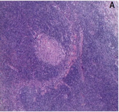
[image:3.612.92.523.76.339.2]To identify whether there were malignant hema-tological diseases, he received bone marrow aspiration and cervical lymph node biopsy. The bone marrow smear showed that the cyte percentage was low and immature lympho-cytes occasionally appeared. Bone marrow flow cytometry results revealed that lymphocytes accounted for about 13.4% of total karyocytes. These lymphocytes included 86.8% CD3 + T Figure 1. Bone marrow flow cytometry results revealed that 12% CD56 + NK cells, which presented as purple, were identified with CD2, CD7, CD45RA expression, and CD8 partial expression, without CD57, CD5, CD4, CD45RO, and CD16 expression.
[image:3.612.90.289.397.583.2]lymphocytes, 12% CD56 + NK cells, and 1.2% CD19 + CD20 + B lymphocytes. T lymphocytes expressed CD2, CD5, and CD7, with partial expression of CD57 and CD56. NK cells were identified with CD2, CD7, and CD45RA expres -sion, with CD8 partial expression and without CD57, CD5, CD4, CD45RO, and CD16 expres-sion. There were no monoclonal abnormal B lymphocytes in the bone marrow (Figure 1). As results of the cervical lymph node biopsy did not identify the disease, T lymphocytes, B lym-phocytes, and NK cells were sorted with immu-nomagnetic beads to clarify the target cells of EBV infection. Next, real- time probe method was conducted to quantify expression of EBV nucleic acid amounts in each type of lympho-cytes. Eventually,quantitative test results could be represented by the ratio of EBV copy number and cell count. Test reports suggested that infected T lymphocytes, B lymphocytes, and NK cells amounts were 2.58×10⁵, 1.69×10⁵, and 1.95×10⁷ (/2×105), respectively, indicating NK cells as the main target cells. Finally, lymph node biopsy pathology showed parts of the nor-mal structures were reserved while T lympho-cytes hyperplasia was found in the paracortex zone, with slightly bigger lymphoid cells among them (Figure 2). Different shapes of multifocal red dye necrotic areas were also found in the lymph nodes. There were a group of infiltrated
cells expressing CD3, CD56, Grb, and CD5, while the ratio of Ki-67-positive cells was esti-mated at 40%, according to immunohistochem-istry (IHC). In situ hybridization targeted EBV-encoded RNAs (EBERs) of these cells were also positive. Combined with these results, it was
qod, twice)/Bu/cy/ATG regimen for preprocess-ing, on September 22. Ten days later, he was successfully injected with his father’s bone marrow and peripheral blood stem cells. Bone marrow chimeric analysis on November 3rd showed that donor cells accounted for 96.94%, characterized by complete chimerism. Bone marrow flow cytometry results after haplo-HSCT showed that the percentage of lympho-cytes was low and CD4/CD8 ratio decreased without antigen loss. This assay did not show obvious abnormal expression of perforin and granular enzyme in NK cells. Subsequently, this patient suffered from graft-versus-host disease (GVHD), mainly in the intestinal tract with cyto-megalovirus infection. Thanks to active general treatment with glucocorticoids, immunosup-pressors, basiliximab, and antivirals, his symp-toms were well alleviated. On May 12, 2017, the EBV genome load was decreased to 2.32×10³ copies/mL in peripheral blood mono-nuclear cells (PBMC) and was negative in the plasma, compared to previous results. The patient has remained in good condition for two years, with no evidence of recurrence.
Discussion
EBV is a ubiquitous virus infecting >90% of the adult population, worldwide. B lymphocytes are often the target cells, resulting in infectious mononucleosis. Active EBV infections are rarely prolonged, with abnormal expansion of poly-clonal, oligopoly-clonal, and monoclonal T or NK
cells. These conditions were defined as
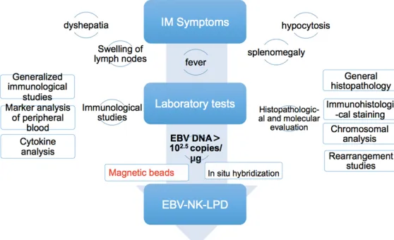
[image:4.612.91.370.74.244.2]EBV-associated T/NK lymphoproliferative diseases Figure 3. Diagnostic flow chart of EBV-NK-LPD.
considered that the patient suffered from chronic active Epstein-Barr virus disease-natural killer-cell lymphoprolif-erative disorder (CAEBV-N- K-LPD).
After the chemotherapy GEM- OX regimen, symptoms were
briefly controlled, but still
that belong to CAEBV. In 2008, Ohshima et al. proposed a clinicopathological categorization of EBV-T/NK-LPD as category A1 (polymorphic LPD without clonal proliferation of EBV-infected cells), category A2 (polymorphic LPD with clon-ality), category A3 (monomorphic LPD with clonality), and category B (monomorphic LPD with clonality and fulminant course). It was based on pathological evaluations and molecu-lar data, providing a guide for better under-standing of this disorder [9].
Although diagnostic criteria and classifications have been articulated, it remains difficult to
diagnose EBV-NK-LPD in clinical practice. Thus, the appropriate approach to diagnose this dis-ease is necessary for clinicians. A detailed diagnostic process was shown in Figure 3. Currently, molecular techniques, such as poly-merase chain reaction (PCR) and in situ hybrid-ization, target EBV-encoded RNAs (EBERs), make it possible to detect EBV genomes. Thanks to these techniques, malignant patho-logical abnormalities in affected tissues are no more essential in patients with CAEBV at the time of diagnosis [3]. Furthermore, it has been demonstrated that patients with CAEBV have viral loads of more than 102.5 copies/mg DNA in the mononuclear cells of peripheral blood [10, 11]. The most important thing in diagnosing CAEBV is to identify target cells in time, thus further examinations are necessary. It is now possible to use techniques, such as double-staining, in situ/immuno-histochemistry, or the magnetic beads procedure to differentiate tar-get cells from human B lymphocytes or epithe-lial cells [12, 13]. As the pathology is often dif-ficult to obtain, this has usually been acco- mplished with immunobead sorting of PBMC
into lymphocyte subsets, followed by quantifi -cation EBV-DNA in each subset with PCR [14]. In the present case, the patient presented with repeated fever, organ enlargement, lymphade-nopathy, and pancytopenia. These symptoms
are typical but with low specificity for diagnos -ing EBV-NK LPD. To discover the kinds of this disease, PCR was used to detect EBV DNA loads in peripheral blood to screen for EBV infection. This patient showed unexpected high levels of EBV DNA load (6.27×10⁵ copies/mL), prompting the diagnosis of CAEBV. Subse- quently, routine bone marrow examinations and immunomagnetic beads procedure were
used to sort lymphocytes. With these tests, NK cells were found to be the main target cells,
eventually confirmed by pathology. Thus, this case was considered a rare adult EBV-NK-LPD category A2.
In the absence of HSCT, therapy for EBV-NK-LPD is often unsatisfactory, while momentarily delaying progression of disease. Antiviral thera-py and immunomodulatory agents are usually invalid. Corticosteroids and other immunosup-pressive agents only alleviate symptoms. Moreover, patients could develop progressive
immunodeficiency, become refractory to thera -py, and submit to occasional infections with extended time. Use of cytotoxic chemotherapy
and autologous EBV specific CTLs have also
been unsuccessful. Although these regimens may induce sustained complete remission, in some exceptional cases[15, 16], HSCT is still the only curative therapy for CAEBV [17]. Moreover, allogeneic HSCT has been success-fully performed in several cases in Japan [18-20]. In 2011, Kawa et al. reported excellent results with HSCT, following a non-destructive pretreatment of reduced-intensity hematopoi-etic stem cell transplantation (RIST). There were 18 pediatric patients with CAEBV treated with RIST in the study. The 3-year event-free survival rate was 85.0 ± 8.0% and 3-year over-all survival rate was 95.0 ± 4.9% [20]. In addi-tion, if a patient had no family donor, unrelated cord blood could be an alternative source for RIST [21]. HSCT is, therefore, the best choice for CAEBV. However, it is still accompanied with high risk of transplantation-related complica-tions [22]. Therefore, hope remains of develop-ing novel therapies that are better than HSCT. Bortezomib [23] and valproic acid [24] are two candidate drugs for CAEBV that have been included in recent preclinical studies.
In the present case, the patient was initially treated with chemotherapy including glucocor-ticoids. Some symptoms, such as fever and lymphadenectasis, were briefly controlled, but there were no significant changes in EBV DNA
loads. To achieve the purpose of a cure, it was suggested that he receive allogeneic HSCT as soon as possible. Since he had no HLA identi-cal suited donors, his father was chosen as a donor to implement haplo-HSCT. In recent
years, Haplo-HSCT has been confirmed to be a
malignancies in China [25, 26], though no liter-ature reports of EBV-NK-LPD treated with Haplo-HSCT have been published. It has been reported that the number of life-threatening complications and plasma viral load may be useful factors in predicting outcomes of HSCT [2]. Thus, this study added VP-16 into pretreat-ment before haplo-HSCT to reduce side effects after transplantation. Although this patient experienced mild GVHD, cytomegalovirus infec-tion, and liver damage after suitable treatment, he fortunately recovered. The EBV genome load decreased to 2.32×10³ copies/mL in PBMC and was negative in plasma. The most gratify-ing thgratify-ing is that he has survived for more than one year with no signs of recurrence. This patient has had a relatively good outcome, until now, but long-term follow up is still necessary. Acknowledgements
The work was supported by the National Natural Science Foundation of China (No. 81300387)
and the Scientific Research Foundation for
Returned Scholars of Tongji Hospital. Disclosure of conflict of interest
None.
Address correspondence to: Miao Zheng, Depart- ment of Hematology, Tongji Hospital, Tongji Medical College, Huazhong University of Science and Tech- nology, Wuhan 430030, China. Tel: 13871286124; E-mail: zmzk@sina.com
References
[1] Macsween KF and Crawford DH. Epstein-Barr virus-recent advances. Lancet Infect Dis 2003; 3: 131-140.
[2] Fujiwara S, Kimura H, Imadome K, Arai A, Ko-dama E, Morio T, Shimizu N and Wakiguchi H. Current research on chronic active Epstein-Barr virus infection in Japan. Pediatr Int 2014; 56: 159-166.
[3] Okano M, Kawa K, Kimura H, Yachie A, Wakigu-chi H, Maeda A, Imai S, Ohga S, Kanegane H, Tsuchiya S, Morio T, Mori M, Yokota S and Imashuku S. Proposed guidelines for diagnos-ing chronic active Epstein-Barr virus infection. Am J Hematol 2005; 80: 64-69.
[4] Kawa K, Okamura T, Yasui M, Sato E and Inoue M. Allogeneic hematopoietic stem cell trans-plantation for Epstein-Barr virus-associated T/ NK-cell lymphoproliferative disease. Crit Rev Oncol Hematol 2002; 44: 251-257.
[5] Yachie A, Kanegane H and Kasahara Y. Ep-stein-Barr virus-associated T-/natural killer cell lymphoproliferative diseases. Semin Hematol 2003; 40: 124-132.
[6] Suzuki K, Ohshima K, Karube K, Suzumiya J, Ohga S, Ishihara S, Tamura K and Kikuchi M. Clinicopathological states of Epstein-Barr vi-rus-associated T/NK-cell lymphoproliferative disorders (severe chronic active EBV infection) of children and young adults. Int J Oncol 2004; 24: 1165-1174.
[7] Kimura H, Hoshino Y, Kanegane H, Tsuge I, Okamura T, Kawa K and Morishima T. Clinical and virologic characteristics of chronic active Epstein-Barr virus infection. Blood 2001; 98: 280-286.
[8] Cho EY, Kim KH, Kim WS, Yoo KH, Koo HH and Ko YH. The spectrum of Epstein-Barr virus-as-sociated lymphoproliferative disease in Korea: incidence of disease entities by age groups. J Korean Med Sci 2008; 23: 185-192.
[9] Ohshima K, Kimura H, Yoshino T, Kim CW, Ko YH, Lee SS, Peh SC, Chan JK; CAEBV Study Group. Proposed categorization of pathological states of EBV-associated T/natural killer-cell lymphoproliferative disorder (LPD) in children and young adults: overlap with chronic active EBV infection and infantile fulminant EBV T-LPD. Pathol Int 2008; 58: 209-217.
[10] Kimura H, Morita M, Yabuta Y, Kuzushima K, Kato K, Kojima S, Matsuyama T and Morishi-ma T. Quantitative analysis of Epstein-Barr vi-rus load by using a real-time PCR assay. J Clin Microbiol 1999; 37: 132-136.
[11] Maeda A, Wakiguchi H, Yokoyama W, Hisaka-wa H, Tomoda T and Kurashige T. Persistently high Epstein-Barr virus (EBV) loads in periph-eral blood lymphocytes from patients with chronic active EBV infection. J Infect Dis 1999; 179: 1012-1015.
[12] Okano M. Haematological associations of Ep-stein-Barr virus infection. Baillieres Best Pract Res Clin Haematol 2000; 13: 199-214. [13] Kasahara Y, Yachie A, Takei K, Kanegane C,
Okada K, Ohta K, Seki H, Igarashi N, Maru-hashi K, Katayama K, Katoh E, Terao G, Saki-yama Y and Koizumi S. Differential cellular tar-gets of Epstein-Barr virus (EBV) infection between acute EBV-associated hemophago-cytic lymphohistiocytosis and chronic active EBV infection. Blood 2001; 98: 1882-1888. [14] Cohen JI, Jaffe ES, Dale JK, Pittaluga S, Heslop
[15] Koyama M, Takeshita Y, Sakata A, Sawada A, Yasui M, Okamura T, Inoue M and Kawa K. Cy-totoxic chemotherapy successfully induces du-rable complete remission in 2 patients with mosquito allergy resulting from Epstein-Barr virus-associated T-/natural killer cell lymphop-roliferative disease. Int J Hematol 2005; 82: 437-440.
[16] Zhang X, Wang Z, Wang L and Yao H. An adult case of systemic Epstein-Barr virus-positive T/ natural killer-cell lymphoproliferative disorder with good outcome. Int J Clin Exp Pathol 2013; 6: 2620-2624.
[17] Okamura T, Hatsukawa Y, Arai H, Inoue M and Kawa K. Blood stem-cell transplantation for chronic active Epstein-Barr virus with lymphop-roliferation. Lancet 2000; 356: 223-224. [18] Gotoh K, Ito Y, Shibata-Watanabe Y, Kawada J,
Takahashi Y, Yagasaki H, Kojima S, Nishiyama Y and Kimura H. Clinical and virological charac-teristics of 15 patients with chronic active Ep-stein-Barr virus infection treated with hemato-poietic stem cell transplantation. Clin Infect Dis 2008; 46: 1525-1534.
[19] Sato E, Ohga S, Kuroda H, Yoshiba F, Nishimu-ra M, Nagasawa M, Inoue M and Kawa K. Allo-geneic hematopoietic stem cell transplanta-tion for Epstein-Barr virus-associated T/natural killer-cell lymphoproliferative disease in Japan. Am J Hematol 2008; 83: 721-727.
[20] Kawa K, Sawada A, Sato M, Okamura T, Sakata N, Kondo O, Kimoto T, Yamada K, Tokimasa S, Yasui M and Inoue M. Excellent outcome of al-logeneic hematopoietic SCT with reduced-in-tensity conditioning for the treatment of chron-ic active EBV infection. Bone Marrow Transplant 2011; 46: 77-83.
[21] Sawada A, Inoue M, Koyama-Sato M, Kondo O, Yamada K, Shimizu M, Isaka K, Kimoto T, Kiku-chi H, Tokimasa S, Yasui M and Kawa K. Um-bilical cord blood as an alternative source of reduced-intensity hematopoietic stem cell transplantation for chronic Epstein-Barr virus-associated T or natural killer cell lymphoprolif-erative diseases. Biol Blood Marrow Transplant 2014; 20: 214-221.
[22] Kimura H, Morishima T, Kanegane H, Ohga S, Hoshino Y, Maeda A, Imai S, Okano M, Morio T, Yokota S, Tsuchiya S, Yachie A, Imashuku S, Kawa K, Wakiguchi H; Japanese Association for Research on Epstein-Barr Virus and Related Diseases. Prognostic factors for chronic active Epstein-Barr virus infection. J Infect Dis 2003; 187: 527-533.
[23] Iwata S, Yano S, Ito Y, Ushijima Y, Gotoh K, Kawada J, Fujiwara S, Sugimoto K, Isobe Y, Nishiyama Y and Kimura H. Bortezomib induc-es apoptosis in T lymphoma cells and natural killer lymphoma cells independent of Epstein-Barr virus infection. Int J Cancer 2011; 129: 2263-2273.
[24] Iwata S, Saito T, Ito Y, Kamakura M, Gotoh K, Kawada J, Nishiyama Y and Kimura H. Antitu-mor activities of valproic acid on Epstein-Barr virus-associated T and natural killer lymphoma cells. Cancer Sci 2012; 103: 375-381. [25] Mo XD, Xu LP, Liu DH, Zhang XH, Chen H, Chen
YH, Han W, Wang Y, Wang FR, Wang JZ, Liu KY and Huang XJ. Nonmalignant late effects in survivors of partially matched donor hemato-poietic stem cell transplantation. Biol Blood Marrow Transplant 2013; 19: 777-783. [26] Xu T, Chen J, Jin ZM, Miao M, Fu CC, Qiu HY,