Original Article
Study on new classification and treatment of vascular
malformations in the extremities
Xuejian Liu, Hailin Yang, Yangzhang Wen, Zhenfeng Zhu, Wenchuan Yang, Yuhua Li, Xia Wu
Department of Oncology, People’s Hospital of Linyi Economic and Technological Development Zone (Linyi Third People’s Hospital), Linyi 276023, Shandong Province, China
Received January 3, 2018; Accepted February 4, 2018; Epub April 15, 2018; Published April 30, 2018
Abstract: Objective: To propose a new clinical classification of vascular malformations (VMs) in the extremities, and to investigate the correlation between the new classification and the effectiveness of treatment. Methods: We ret-rospectively analyzed the clinical data of 256 patients who were treated in our hospital for VMs in the extremities, including their clinical characteristics, imaging data, conventional classification, treatment process and prognosis. Based on the extent of lesion determined by the imaging test, the cases were classified into six categories, which were superficial type, mono-localized type, poly-localized type, extensive type, nerve type, and diffuse type. The treatment process and treatment outcome in all the patients were analyzed, and the correlation between the new classification and the treatment effectiveness were investigated. Results: According to the conventional classifica-tion, the lesions (256 cases) could be divided into high-flow VM (arteriovenous malformaclassifica-tion, 77 cases) and low-flow VM (venous malformation, 179 cases), while according to the new classification, they could be classified into six types, which were superficial type (84 cases), mono-localized type (56 cases), poly-localized type (23 cases), exten-sive type (74 cases), nerve type (9 cases), and diffuse type (10 cases). Treatment methods were as follows: most of the patients with superficial type received laser treatment (80/84); most of the patients with mono-localized type or poly localized type underwent surgical excision only (25/56, 12/23); among patients with extensive type, 21 of them underwent surgical excision only (21/74), while 34 received surgical excision plus muscle transfer (34/74); patients with nerve type underwent surgical excision only (2/9) and embolization of draining vein and sclerotherapy (7/9); more than half of the patients with diffuse type received embolization of draining vein and sclerotherapy (6/10, the other 4 patients didn’t received any treatment). Patients were followed up for 1-10 years (average 6.5 years). Under the conventional classification based on the hemodynamics, the improvement rates in patients with high-flow type and low-flow rate were 84.42% and 94.97% respectively, whereas under the new classification based on the extent of lesion, the improvement rates in groups of superficial type, mono-localized type, poly-localized type, extensive type, nerve type and diffuse type were 95.24%, 98.21%, 95.65%, 87.84%, 55.56% and 80.00% respectively (P<0.001). Logistic regression analysis found significant difference in the prognosis between these two classifications (P=0.02). Conclusion: The new classification proposed in this study was found to be closely related to the prognosis of VMs in extremities, and treatments received by patients within the same group under the new classification tended to be the same. This finding suggests that the new classification can be applied clinically for assisting in the clinical diagnosis and treatment.
Keywords: Extremities, vascular malformations, therapeutics
Introduction
Vascular malformation (VM) is a common be- nign vascular lesion in soft tissues of extremi-ties, which accounts for about 7% of the benign soft tissue tumors. It is mainly an abnormality in blood vessel structure, with no abnormal cell [1]. VMs in extremities are often manifested as extensive lesions, which can bring about seve- re complications, and even threaten the lives of
prognosis, and provides very limited assistance in making treatment plans [6]. Due to the de- velopment of imaging and interventional tech-nologies, new classifications are expected to be established. Therefore, based on patients’ case histories and imaging results, the present study proposed a new type of classification, and investigated its correlation with the prog-nosis of the disease.
Materials and methods
Case selection
The clinical data of 256 cases of VMs (with complete medical records) were collected for the study. The study was approved by the Eth- ics Committee of People’s Hospital of Linyi Economic and Technological Development Zo- ne and informed consents were obtained from patients.
Inclusion criteria were as follows: patient met the diagnostic criteria of VM (test of X-ray and CT found phleboliths in soft tissues of extre- mities; vascular anomaly in extremities was detected by color ultrasound; abnormality in feeding artery was identified by DSA); patient aged between 18 and 70 years (with no restric-tion on gender); the treatment process was complete and patient was not lost to follow-up (follow-up period was 2 years).
Exclusion criteria: patients were found to have open blood vessel injuries of the extremities; patient had evident heart, liver, lung, or kidney failure.
Data extraction
The following information was collected for the study, which included patients’ basic informa-tion (age, gender, underlying diseases), symp-toms and physical signs, clear diagnosis, con-ventional classification, imaging results (DX, CT, MRI, ultrasound), pathological feature, com-plete treatment process with no missing infor-mation, follow-up period, and prognosis. Information regarding the treatment methods was collected, which included parameters of laser treatment (such as laser type and power), and data related to the surgical excision (such as signs, excision area, whether or not there was any muscle transfer, embolization, and
sclerotherapy). Based on these information, the treatment methods in patients were classi-fied into five groups: 1) Laser treatment; 2) Surgical excision; 3) Surgical excision + muscle transfer; 4) Embolization of feeding artery + excision of tumor; 5) Embolization of draining vein + sclerotherapy.
The effectiveness of treatment was defined by using following criteria: 1) Cured: VM or vascu-lar tumor was removed completely; patient could get back to normal life and experienced no recurrence during a more than one-year fol-low-up; 2) Markedly improved: the lesions were almost completely removed, or the tumor size was reduced by over 80%; the tumor was stable or grew slowly during follow-up period; patient had no sign of pain; the affected limb could function properly; 3) Improved: the main tumor in the lesion was removed, or the tumor size was reduced by over 30%; the pain was relieved and the function of the limb improved, which wouldn’t keep patient from living normal life; 4) Not improved: the tumor was not noticeably reduced; patient’s symptoms were not evident-ly improved and patient still couldn’t get back to normal life [7].
Basis of the new classification
Lesions were classified into the following six types based on their extents: 1) Superficial type: the lesion was only in the superficial tis-sue, and didn’t extend to the muscle; 2) Mono-localized type: the lesion was only in one area or one muscle; 3) Poly-localized type: the lesions were in the single muscle group of mul-tiple areas, where less than 50% of the mus-cles were affected; 4) Extensive type: the lesions were in multiple areas or multiple cle groups, where more than 50% of the mus-cles were affected; 5) Nerve type: the tunica vaginalis and perineurium of the nerve trunk in extremities were affected; 6) Diffuse type: the lesions existed in multiple muscle groups and soft tissues of the entire limb.
Outcome measures
Table 2. Treatment of patients under conventional classification
Treatment method Case High-flow type Low-flow type
Case % Case %
Laser treatment 94 42 54.55 52 29.05
Surgical excision 64 12 15.58 52 29.05
Surgical excision + muscle transfer 34 4 5.19 30 16.76
Embolization of feeding artery + resection of tumor 34 14 18.18 20 11.17 Embolization of draining vein + sclerotherapy 21 - - 21 11.73
Amputation 5 3 3.90 2 1.12
No treatment 4 2 2.60 2 1.12
Total 256 77 100.00 179 100.00
Statistical analysis
SPSS software was applied for the statistical analysis. The count data were expressed as percentage (rate) and the measurement data were expressed as mean ± sd; comparison of the treatment effectiveness among individuals in different classification were performed by X2 test.
The association between the method of new and conventional classification and the prog- nosis was analyzed by multiple logistic regres-sion. The prognoses were divided to two gr- oups, which were group of improvement and group of non-improvement. The method of binary classification was adopted. The conven-tional classification was taken as variable 1, and the high-flow and low-flow type were coded 0 and 1 respectively. The new classification was taken as variable 2, and the dummy vari-ables were used in coding: the superficial type was taken as control X1=0, X2=0, X3=0, X4=0, X5=0; mono-localized type: X1=1, X2=0, X3=0, X4=0, X5=0; poly-localized type: X1=0, X2=1, X3=0, X4=0, X5=0; extensive type: X1=0, X2=0, X3=1, X4=0, X5=0; nerve type, X1=0, X2=0, X3=0, X4=1, X5=0; diffuse type: X1=0, X2=0, X3=0, X4=0, X5=1. Method of binary
classification was used in gender and treat-ment effectiveness, in which the male and female were coded 0 and 1 respectively, and the improvement and non-improvement were coded 0 and 1 respectively. Gender, age, and type of classification were entered into the regression analysis (Backward: Wald). The alpha-to-enter significance level was 0.10, while the alpha-to-remove significance level was 0.15. A value of P<0.05 was considered as statistically significant.
Results
Basic information
[image:3.612.90.523.226.359.2]Among the 256 patients from our hospital, there were 134 male patients and 122 female patients (the ratio of males to females was 1:1). The age distribution of the patients was as follows: 11 patients aged 18-20 years, 18 patients aged 21-25 years, 28 patients aged 26-30 years, 113 patients aged 31-40 years, 49 patients aged 41-50 years, 22 patients aged 51-60 years, 9 patients aged 61-70 years, and 6 patients aged over 70 years (mean age ± sd, 48.3±4.7). The average medical history was 8.9 years.
Table 1. Symptoms and physical signs
Symptoms Case Percentage (%)
Limb hypertrophy or presence of localized soft tissue masses 243 94.92
Blue skin 111 43.36
Red spots on skin 24 9.38
Increase in skin temperature 21 8.20
Pain in affected limbs 138 53.91
Affected limbs had different levels of deformities and malfunctions 38 14.84
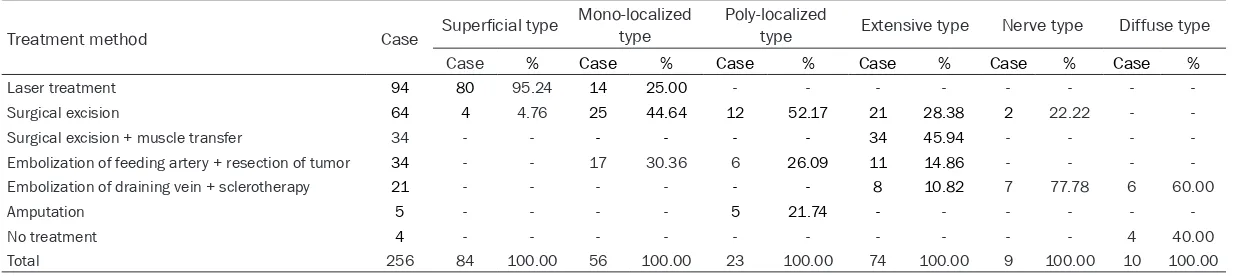
Table 3. Treatment of patients under new classification
Treatment method Case Superficial type Mono-localized type Poly-localized type Extensive type Nerve type Diffuse type
Case % Case % Case % Case % Case % Case %
Laser treatment 94 80 95.24 14 25.00 - - -
-Surgical excision 64 4 4.76 25 44.64 12 52.17 21 28.38 2 22.22 -
-Surgical excision + muscle transfer 34 - - - 34 45.94 - - -
-Embolization of feeding artery + resection of tumor 34 - - 17 30.36 6 26.09 11 14.86 - - - -Embolization of draining vein + sclerotherapy 21 - - - 8 10.82 7 77.78 6 60.00
Amputation 5 - - - - 5 21.74 - - -
-No treatment 4 - - - 4 40.00
Table 4. Follow up of patients under conven-tional classification
High-flow type Low-flow type
Follow-up 77 179
Healed 36 70
Markedly improved 25 59
Improved 4 41
Not improved 12 9
Improvement rate 84.42 94.97
X2 value 7.968
P value 0.005
Symptoms and physical signs
There were 243 cases (94.92%) in which patients’ affected limbs were hypertrophic or presented with localized soft tissue masses (the diameter of the largest mass was about 3-4 cm), 111 cases (43.36%) where skin color turned blue, 24 cases (9.38%) where there were red spots on skin, 21 cases (8.20%) where skin temperature increased, 138 cas- es (53.91%) where pain existed in affected limbs (among which 18 cases reported unbe- arable pain (7.03%), and one patient suffered from secondary gastrointestinal bleeding due to long-term use of painkillers), 38 (14.84%) cases where the affected limbs had differ- ent levels of deformities and malfunctions, 6 (2.34%) cases where the tumors were compli-cated by localized ulcers which later ruptured and bled (2 cases were arteriovenous malfor-mation (AVM) where blood spurted; 1 case was complicated by fingertip ischemia; 1 case was extensive AVM complicated by reduction in platelet count (30*109/L)). See Table 1. Treatment plans
According to the conventional classification based on the hemodynamics, there were 77 patients with high-flow type, among which 42 received laser treatment, 12 underwent surgi-cal excision, 4 received surgisurgi-cal excision plus muscle transfer, 14 received embolization of feeding artery and tumor resection, 3 under-went amputation, and 2 didn’t receive any treatment. There were 179 patients with low-flow type, among which 52 received laser tre- atment, 52 received surgical excision, 30 re- ceived surgical resection and muscle transfer, 20 received embolization of feeding artery and
tumor resection, 21 received embolization of draining vein and sclerotherapy, 2 received amputation, and 2 had no treatment. See Table 2.
Under the new classification based on the extent of lesion, cases were divided into six types, which were superficial type (84), mono-localized type (56), poly-mono-localized type (23), extensive type (74), nerve type (9), and diffuse type (10). Among the patients with superficial type, 80 received laser treatment and 4 re- ceived surgical resection; for patients with mono-localized type, 14 received laser treat-ment, 25 received surgical resection only, and 17 received embolization of feeding artery and tumor resection; among patients with poly-localized type, 12 received surgical resection only, 6 received embolization of feeding artery and tumor resection, 5 underwent amputa- tion; among patients with extensive type, 21 received surgical resection only, 34 received surgical resection and muscle transfer, 11 received embolization of feeding artery and tumor resection, 8 received embolization of draining vein and sclerotherapy; 2 patients wi- th nerve type received surgical excision only, while another 7 patients with this type received embolization of draining vein and sclerothera-py; 6 patients with diffuse type received em- bolization of draining vein and sclerotherapy, and another 4 with this type received no treat-ment (Table 3).
Treatment effectiveness
differences had statistical significance (P< 0.001, Tables 4 and 5).
Correlation between treatment effectiveness
and classification
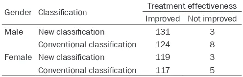
The logistic regression analysis was performed to investigate the association between the two classifications and the improvement rates in patients with VMs in extremities. The result showed significant difference in the prognosis between two classifications (P=0.02), while gender had no impact on the prognosis (P=0.175). See Tables 6 and 7.
The result of the logistic regression analysis found that there was no significant difference in the improvement rates between high-flow type and low-flow type under conventional clas-sification after treatment (P=0.05, OR=1.2), whereas there was great difference in impro- vement rates among various types under new classification. The improvement rates in mono-localized, poly-mono-localized, and extensive type after treatment were similar to that in super- ficial type (P=0.236, P=0.998, P=0.144). As compared to control group, patients with nerve and diffuse type experienced evident improve-ment following treatimprove-ment (P<0.001, OR=1.23; P=0.007, OR=2.43).
[image:6.612.90.527.85.215.2] [image:6.612.88.336.263.342.2]these diseases lasted for 8.9 years on avera- ge. The ratio of males to females was 1:1. These data were consistent with other studies [8, 9].
Classification and treatment plan
Currently, the treatment plan was usually ma- de based on the impact of the lesion on pa- tient’s quality of life. In most cases, the conser-vative treatment was applied. However, in the cases where patients experienced clinical com-plications, percutaneous puncture or interven-tional treatment would then be necessary. According to lesion’s location, severity and le- vel of deformity, sclerosants and embolic ag- ents used in artery may need to be used in combination, such as ethanol, bleomycin, 3% sodium tetradecyl sulfate (STS), polidocanol and various coils and polymer microspheres [10, 11]. Ethanol sclerotherapy has been suc-cessfully applied clinically in some cases of low-flow VMs. It can be used as either a single treatment or a treatment before the operation [12, 13]. When treating high-flow VM, the aim of the treatment is to cut off arteriovenous fis-tula by ethanol. However, the sclerotherapy cannot solve this issue well, which may be part-ly due to the fact that ethanol can make the infused or injected drug flow out rapidly. So far, Table 5. Follow up of patients under new classification
Superficial
type Mono-localized type Poly-localized type Extensive type Nerve type Diffuse type
Follow-up 84 56 23 74 9 10
Healed 62 44 - - -
-Markedly improved 10 4 14 47 5 4
Improved 8 7 8 18 - 4
Not improved 4 1 1 9 4 2
Improvement rate 95.24% 98.21% 95.65% 87.84% 55.56% 80.00%
X2 value 23.920
P value <0.001
Table 6. Gender and effectiveness of treatment under two classifications
Gender Classification Treatment effectiveness Improved Not improved
Male New classification 131 3
Conventional classification 124 8
Female New classification 119 3
Conventional classification 117 5
Discussion
Disease occurrence
Table 7. Impacts of the two classifications and gender on treatment effectiveness
B S.E Wals df Sig. Exp (B)
Step 1a Variable 1 8.235 5 0.144
Variable 1 (1) -0.482 0.407 1.404 1 0.236 0.617
Variable 1 (2) -18.786 7,882.490 0.000 1 0.998 0.000
Variable 1 (3) -1.124 0.770 2.132 1 0.144 0.325
Variable 1 (4) 23.620 28,420.722 0.000 1 0.999 1.812E10
Variable 1 (5) 1.319 0.723 3.329 1 0.068 3.738
Constant -2.417 0.279 75.102 1 0.000 0.089
Step 2b Variable 1 13.437 5 0.020
Variable 1 (1) -1.129 0.420 7.217 1 0.007 0.323
Variable 1 (2) -0.303 8,540.342 0.000 1 1.000 0.739
Variable 1 (3) 17.359 3,286.914 0.000 1 0.996 3.460E7
Variable 1 (4) 42.103 28,610.160 0.000 1 0.999 1.928E18
Variable 1 (5) 19.802 3,286.914 0.000 1 0.995 3.979E8
Classification -19.396 3,286.914 0.000 1 0.995 0.000
Constant -1.504 0.295 25.913 1 0.000 0.222
Step 3c Variable 1 6.221 5 0.285
Variable 1 (1) -18.997 3,748.382 0.000 1 0.996 0.000
Variable 1 (2) -18.774 9,282.271 0.000 1 0.998 0.000
Variable 1 (3) -1.112 4,901.725 0.000 1 1.000 0.329
Variable 1 (4) 23.632 28,840.325 0.000 1 0.999 1.834E10
Variable 1 (5) 1.331 4901.725 0.000 1 1.000 3.784
Classification -19.211 3,158.566 0.000 1 0.995 0.000
Gender 18.286 3,748.382 0.000 1 0.996 8.737E7
Constant -1.504 0.295 25.913 1 0.000 0.222
Note: a, key in variable (variable 1) in step 1; b, key in variable (classification) in step 2; c, key in variable (gender) in step 3.
there has been no consensus on any ideal treatment method for more complicated VMs, while some studies proposed the idea of mul- tidisciplinary approach [10, 11, 14]. For pati- ents with high-flow type, surgeons should pay much attention to the preoperative preparation and intraoperative procedure in order to pre-vent any severe uncontrollable bleeding during surgery, especially if they are going to operate on areas where bleeding can occur easily, su- ch as hip and groin. After embolization of the artery, the dilated malformed vascular mass and venous pool would shrink and the tension would decrease, the borders between tumors and normal tissues would become clear, which can make the resection of the tumor easier, and reduce the volume of blood loss [15, 16]. In an effort to find ways to provide better ass- istance in making suitable treatment plans, we used the new classification to divide VMs in extremities into six groups, which were super- ficial type, mono-localized type, poly-localized
type, extensive type, nerve type and diffuse type [17, 18].
on patients with high-flow type which is adjunc-tive to the surgery [19]. In the laser treatment, oxyhemoglobin in blood vessel can selectively absorb the color radical in light energy and gen-erate heat locally, thus damaging the affected vessel and removing the lesion [20]. The treat-ment methods for patients with superficial or mono-localized type in the study were as fol-lows, 80 patients with superficial type and 14 with mono-localized type received laser treat-ment, 4 with superficial type and 25 with mo- no-localized type received surgical excision, 17 with mono-localized type received embolizati- on of feeding artery and resection of tumor. Patients were followed up after surgery, which found that improvements in the 84 cases of superficial type (95.24% of improvement rate) and 53 cases with mono-localized type (98.21% of improvement rate). For patients with poly-localized or extensive type, surgeries should be performed if lesions impair the limb func-tion. The type of vascular tumor, and area and depth of the lesion should be determined prior to the operation. Good knowledge of anatomy and tissue structure is required for maximum possible resection of tumor. In cases where muscle transfer can be conducted, the affect-ed muscle should be removaffect-ed as much as pos-sible; whereas the resection of muscle shoul- dn’t exceed 50% in cases where muscle trans-fer cannot be performed, so as to keep the function of extremities. A total of 23 patients with poly-localized type and 74 with extensive type were followed up (95.65% and 87.84% of improvement rate, respectively), while 1 case in poly-localized type and 9 cases in extensive type had no improvement. For nerve type in which nerve is affected, the nerve compression should be eliminated, meanwhile blood supply should be maintained in order to avoid nerve ischemia. There were 9 patients with nerve type and 10 with diffuse type, who were fol-lowed up after surgery. The improvement rate of nerve type was 55.56% and 4 cases had no improvement; the improvement rate of diffuse type was 80.00% and 2 cases had no improve-ment. The treatment of VM in diffuse type is still quite difficult. Normally patients with this type are treated with embolization of draining vein by anhydrous ethanol plus sclerotherapy, which is mainly to control the development of disease, and to relieve clinical symptoms. Im- aging examination is taken after the surgery immediately, while some scholars suggest
tak-ing this test 3 days after surgery [14]. Color Doppler ultrasound can be used selectively fol-lowed by CT angiography, but it is believed that MRI is still the best way for evaluating medium- and long-term management of the disease [14]. Disclosure of conflict of interest
None.
Address correspondence to: Xia Wu, Department of Oncology, People’s Hospital of Linyi Economic and Technological Development Zone (Linyi Third People’s Hospital), No.117 Huaxia Street, Linyi Economic and Technological Development Zone, Linyi 276023, Shandong Province, China. Tel: +86-0539-8769202; E-mail: wuxia2837@163.com
References
[1] Ly JQ, Sanders TG, Mulloy JP, Soares GM, Beall DP, Parsons TW and Slabaugh MA. Osseous change adjacent to soft-tissue hemangiomas of the extremities: correlation with lesion size and proximity to bone. AJR Am J Roentgenol 2003; 180: 1695-1700.
[2] Madani H, Farrant J, Chhaya N, Anwar I, Marmery H, Platts A and Holloway B. Peripher-al limb vascular mPeripher-alformations: an update of appropriate imaging and treatment options of a challenging condition. Br J Radiol 2015; 88: 20140406.
[3] Johnson JB, Cogswell PM, McKusick MA, Binkovitz LA, Riederer SJ and Young PM. Pre-treatment imaging of peripheral vascular mal-formations. J Vasc Diagn 2014; 2014: 121-126.
[4] Al-Shahi R and Warlow C. A systematic review of the frequency and prognosis of arteriove-nous malformations of the brain in adults. Brain 2001; 124: 1900-1926.
[5] Buckmiller LM, Richter GT and Suen JY. Diag-nosis and management of hemangiomas and vascular malformations of the head and neck. Oral Dis 2010; 16: 405-418.
[6] Redondo P, Aguado L and Martinez-Cuesta A. Diagnosis and management of extensive vas-cular malformations of the lower limb: part I. Clinical diagnosis. J Am Acad Dermatol 2011; 65: 893-906; quiz 907-898.
[7] McCafferty I. Management of low-flow vascular malformations: clinical presentation, classifi-cation, patient selection, imaging and treat-ment. Cardiovasc Intervent Radiol 2015; 38: 1082-1104.
[9] Azizkhan RG. Complex vascular anomalies. Pe-diatr Surg Int 2013; 29: 1023-1038.
[10] McCafferty IJ and Jones RG. Imaging and man-agement of vascular malformations. Clin Radi-ol 2011; 66: 1208-1218.
[11] Legiehn GM and Heran MK. A step-by-step practical approach to imaging diagnosis and interventional radiologic therapy in vascular malformations. Semin Intervent Radiol 2010; 27: 209-231.
[12] Hyodoh H, Hori M, Akiba H, Tamakawa M, Hy-odoh K and Hareyama M. Peripheral vascular malformations: imaging, treatment approach-es, and therapeutic issues. Radiographics 2005; 25 Suppl 1: S159-171.
[13] Goyal M, Causer PA and Armstrong D. Venous vascular malformations in pediatric patients: comparison of results of alcohol sclerotherapy with proposed MR imaging classification. Radi-ology 2002; 223: 639-644.
[14] Vogelzang RL, Atassi R, Vouche M, Resnick S and Salem R. Ethanol embolotherapy of vascu-lar malformations: clinical outcomes at a sin-gle center. J Vasc Interv Radiol 2014; 25: 206-213; quiz 214.
[15] Maclellan RA, Chaudry G and Greene AK. Com-bined lymphedema and capillary malformation of the lower extremity. Plast Reconstr Surg Glob Open 2016; 4: e618.
[16] Michelini S and Cardone M. Veno-lymphatic vascular malformations: medical therapy. 2015.
[17] Huang JT and Liang MG. Vascular malforma-tions. Pediatr Clin North Am 2010; 57: 1091-1110.
[18] Fujino J, Ishimaru Y, Tahara K, Suzuki M, Hat-anaka M, Igarashi A, Hamajima A, Hasumi T and Ikeda H. Staged-surgery for vascular mal-formations of the extremities and the trunk. Journal of the Japanese Society of Pediatric Surgeons 2011; 47: 261-268.
[19] Uihlein LC, Liang MG, Fishman SJ, Alomari AI and Mulliken JB. Capillary-venous malforma-tion in the lower limb. Pediatr Dermatol 2013; 30: 541-548.