Current Status of Urinary/Cardiac Catheter
Indwelling Pathogens and their Biofilm Formation
Capabilities
Srushti T. Gamit
1, Dr. Manisha N. Shah
21 PG Student, 2Assistant Professor, Department of Microbiology, Shree Ramkrishna Institute of Computer Education and Applied
Sciences,
Abstract: A great threat to indwelling devices like urinary and cardiac catheters is by an accumulation of microorganisms embedded in an exopolysaccharide matrix. This may result into slow and persistent infections and thereby interfering with antibiotic therapy too. This study was aimed to find out the bacterial and fungal etiological agents that are the major cause of biofilm formation on tips of urinary and cardiac catheters along with their higher resistance towards antimicrobials. Total 36 urinary catheter tips and 4 cardiac catheter tips were collected from various clinics of Surat region, since December 2018 to March 2019 from the admitted patients. Out of these samples, many of them were polymicrobials and produced total of 63 isolates. Among these isolates, 93.66% were bacterial etiological agents and 6.34% were fungal etiological agents. Gram negative isolates predominated (55.56%) over gram positives. Antibiotics susceptibility patterns of isolates were determine by Kirby Bauer disc diffusion method. Prevalence of ESBLs producing gram negative isolates and determination of Oxacillin resistant gram Positive cocci (S.aureus) were carried out. Biofilm forming capabilities were observed in majority of the isolates when performed by Tube, Congo Red Agar (CRA), and Microtitre Plate (MTP) Methods. Partial molecular sequencing (18S r RNA) of screened isolate with high biofilm formation capabilities and its phylogenetic analysis is determined.
Keywords: Indwelling devices, Exopolysaccharide matrix, Urinary and Cardiac catheter tips, Antibiotic susceptibility test, ESBLs producers, Oxacillin resistant
I. INTRODUCTION
Bio-medical devices are important part of modern medical practices. Such devices increases the chances of survival and also improves the quality of life of patients. In medicine, a catheter is a thin tube made from medical grade materials serving broad range of functions. Catheters can be inserted into a body cavity, duct or vessel. Nowadays, Hospital Acquired Infections (HAIs) is a common public health problem caused due to colonization of microorganisms on to different medical devices. More than 60% of these infections worldwide are due to bacteria forming biofilm on medical devices. Many patients suffers from Hospital acquired infections caused due to frequent use of bio-medical devices such as indwelling catheters [1]. Microorganisms are more susceptible to antimicrobial agents when they are in free floating phase but interestingly becomes ‘1000 fold’ resistant to the same antimicrobial agents when present in a biofilm [2]. Almost 100 Million catheters are sold annually worldwide [3]. The most common urinary catheter in use is the Foley indwelling urethral catheter, a closed sterile system that is comprised of a tube inserted through the urethra and held in place by an inflatable balloon to allow urinary drainage of the bladder.
There are 3 main types of urinary catheters:
1) Indwelling Catheter
2) Condom Catheter
3) Intermittent Self-Catheter
II. MATERIALS AND METHODS
The present study was conducted over a period of three months, i.e., from December 2018- March 2019. Under strict aseptic conditions, a total of 36 urinary and 4 cardiac catheter tips were collected into sterile universal containers from the patients admitted into different wards of Hospitals.
A. Sample Processing
Urinary and cardiac catheter tips were cut into 2 pieces: First piece was immersed into sterile 3.5 ml of normal saline and referred as Non-Disinfected Catheter (NDC). While second piece was disinfected using 10 ml of Hydrogen Peroxide (H2O2) and immersed into
saline solution, it is refereed as Disinfected Catheter (DC) [8]. Both the sections of tips were properly vortex in order to remove biofilm and achieve good bacterial suspension. Suspension were streaked onto generalized media (Nutrient agar), differential and Selective media (MacConkey’s agar, Mannitol Salt agar and Sabouraud’s Dextrose agar – for fungi). Plates were incubated at 370C
for 24-48 hours.
B. Isolation and Identification
Significant difference in the growth of isolates between NDC and DC were observed and identification of isolates were carried out by standard microbiological procedures.
C. Antibiotic Susceptibility Test
The isolated and identified organisms were further tested for their antimicrobial susceptibility towards commonly used different groups of antibiotics discs with known concentration by KirbyBauer disc diffusion method. For antifungal test Muller-Hinton Agar supplemented with 2% dextrose and 0.5 mcg/ml methylene blue was used to improve the yeast/fungal growth and provide sharp zones of inhibition. The zone of inhibition were measured (diameter in mm) using ruler and interpreted according to the interpretation chart provided by the manufacturer.
D. Determination of MDR
Antimicrobial resistance shown by a species of microorganism to multiple groups of antimicrobial drugs. The etiological agents that confirm resistance against three and more than three groups of antibiotics were accounted as MDR.
E. Detection of ESBLs (Extended-Spectrum Beta Lactamases) Producers
ESBLs is an enzymes that confer resistance to most beta-lactam antibiotics. ESBL producer are most commonly associated with therapeutic failure and therefore poor outcome of infection were noted. All gram negative isolates were tested for ESBL. Production capabilities of Cefoperazone and Cefoperazone + Sulbactum were used to check ESBLs production. Disc containing 105 mcg concentration of Cefoperazone and Cefoperazone + sulbactum were used. A difference in zone of inhibition of ≥ 5 mm of Cefoperazone and Cefoperazone + sulbactum indicates production of ESBL.
F. Detection of Oxacillin Resistant Gram-Positive Cocci:
Oxacillin (trade name Bactocill) is a penicillinase-resistant beta-lactam. It is similar to methicillin, and has replaced methicillin in clinical use. Therefore we intended to detect ORSA. 0.5 MacFarland turbid inoculum was prepared of well isolated colony of Staphylococcus species and inoculated in M-H agar plates. After prediffusion time of 15 minutes the Oxacillin disc (30 mcg) were placed on the medium with the sterile forcep. Plates were incubated at 37oC for 24 hours. After incubation measure the diameter of
zone and zone diameter of <10 mm is considered as resistant (ORSA).
G. Detection of Biofilm Formation Capabilities
Three different methods were used to detect biofilm formation capabilities of isolates such as:
1) Tube Method [9]: 5 ml of Trypticase soy broth (TSB) with 1% glucose was inoculated with a loop full of test organism from overnight culture plates in test tubes. Incubated all the tubes at 370 C 24-48 hours. The contents were then gently decanted and
2) CRA Method (Freeman et al., 1989): Slime production by each isolates were determined by CRA method. CRA medium was prepared with brain heart infusion broth (BHI) supplemented with 5% sucrose and Congo red. Congo red was prepared as concentrated aqueous solution and autoclaved at 121°C for 15 minutes, separately from other medium constituents and was then added when the agar had cooled to 55°C. CRA plates were streaked with each bacterial isolates and incubated aerobically for 24 hours at 37o C. The appearance of dark-black colonies with a rough, dry and crystalline consistency was considered as
indicative of slimeproducers. Non-slime producers show pinkish red smooth colonies with a darkening at the center [10]. 3) MTP method [11]: Individual wells of sterile, propylene, 96 well Microtitre plates were filled with 100 μl of Trypticase Soya
Broth (Hi-media) with 1% glucose (10ml) was inoculated 50 μl of 24 hrs old microbial cultures to be tested and then incubated for 24 hours at 37°C. After incubation (24 h at 37°C), the microtiter plates content of each well was removed by tapping the bottom plates. 200 μl phosphate buffer saline is used to wash the wells as it removes planktonic bacteria. The plates were then inverted and blotted on paper towels and allowed to air dry for 15 min [12] and stained with saffranin (0.1% w/v) and allowed to incubate at room temperature for 15 min. After removing the crystal violet solution, wells were washed three times with 1 × PBS or deionised water to remove unbound dye. Finally, all wells were filled by 200 μl 33% glacial acetic acid or 95 % ethanol to release the dye from the cells. Optical density (OD) of stained adherent bacteria was determined with an Absorbance Microtitre Reader (model EL×800) / micro ELISA auto reader (model 680, Bio rad) at wavelength of 570 nm [13].
4) Microscopic Observation of Biofilm:
Microscopic observation of biofilm was performed using Microtiter Plate Method and matrix of organisms associated with biofilm were observed under oil immersion lens.
H. 18S r RNA Sequencing of Screened Isolates and Construction of Phylogenetic Analysis:
Molecular identification was done on the basis of 18S rRNA sequence analysis of most potent isolate with high resistance against antifungal agents and also strong biofilm producer was selected and identified
Phylogeny tree was constructed by taking the sequences obtained in the blast search. Sequence obtained from BLASTn (nucleotide blast) was obtained in FASTA format and relation between each sequence could be known by multiple sequence alignment using a software CLUSTAL algorithm.
III. RESULTS AND DISCUSSION A. Positivity of Sample
In present study as per sample size calculation 40 catheter tips were collected. Among 40 catheter tips, 37 were culture Positive for both bacterial and fungal etiological agents and 3 were culture Negative. In contrast to our study 37% positivity and 63% culture Negative was reported by Ventaka Hemalatha Neeli et al., 2016 [14].
B. Distribution of Samples
Fig.2 Distribution of samples
C. Isolation And Identification Of Etiological Agents
Fig.3 High growth on NDC compare to DC
Isolates were proceeded for various standard microbiological procedures that help to identify particular etiological agents. Colonial, growth and morphological characteristics were observed on generalized, selective /supportive/differential and specialized media along with their motility patterns, capsule staining and for fungi wet mount and confirmatory germ test were performed. Biochemical profiling of isolates were analysed thoroughly for their identification.
From 37 positive cases, 89.18% were urinary catheter tips whereas 10.82% were cardiac catheter tips.
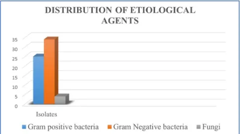
D. Distribution of Isolates
[image:5.595.51.294.140.276.2]E. Frequency wise Distribution of Isolates
Fig.5 Frequency wise distribution of isolates F. Antimicrobial Susceptibility Test
After phenotypic identification, all isolates were tested for their antibiotic susceptibility against commonly used antibiotics. Total 25 etiological agents were identified as gram-positive isolates and 34 etiological agents were identified as gram-negative isolates and 4 were fungal agents. Resistogram of these isolates were separately studied.
Fig.6 For gram-positive isolates Fig.7 For gram-negative isolates
Figure 4: Distribution of isolates
From the results of all presumptive & confirmatory identification tests the etiological agents were successfully identified. In the present study Gram negative isolates predominates (54%) over Gram Positive isolates (40%) followed by Fungi (6%). In accordance to our study, predominating gram negative bacteria (55.56%) followed by gram positive bacteria (40.74%) and yeast (3.70%) were observed by AL- Mjalwai [15].
[image:5.595.42.276.370.511.2] [image:5.595.69.523.603.726.2]Fig.8Resistogram for gram positive isolates
As represented, majority of isolates were resistant to Cefotaxime and Co-Trimoxazole while show susceptibility towards
Cloxacillin and Ciprofloxacin.
Fig.9 Resistogram for gram negative isolates
In present study, the most resistant antibiotic were Ofloxacin followed by Co-Trimoxazole and isolates were mostly susceptible to Amikacin followed by Ceftizoxime antibiotics.
Fig.10 For fungal isolates Fig.11 Resistogram for fungal isolates
As represented in graph, all Candida isolates were most resistant to Itraconazole. Whereas more susceptible to Fluconazole followed by Clotrimazole.
0 2 4 6 8 10 12 14 16
Resistant
Intermediate
Susceptible
0 0.5 1 1.5 2 2.5 3
Resistant Intermediate Susceptible
0 5 10 15 20 25 30
G. Detection of Multi Drug Resistance (MDR)
Fig.12 Prevalence of MDR
H. Detection of ESBLs producer and prevalence of ORSA
Fig.13 ESBLs test
In our study, 11 gram-negative isolates were observed as ESBLs producers. So, 32.35% positivity is the striking outcome of our study.
Fig.14 Oxacillin resistant test
In present study 13 gram positive cocci were identified as Staphylococcus species and were tested for their sensitivity towards Oxacillin. In our study, 46.15% isolates were Oxacillin Resistant.
I. Determination Of Prevalence Of Biofilm Producers
A biofilm is an aggregation of microorganisms in which cells are frequently embedded within a self-produced matrix of extracellular polymeric substances (EPS). Biofilm EPS, Which is also referred as slime is a polymeric conglomeration generally composed of extracellular DNA, Polysaccharides and Proteins.
In present study majority of isolates are MDR. Total 69% isolates were MDR. In accordance to our study, 68.69% isolates were MDR observed by Mahabubul Islam Majumder [17].
0 5 10 15 20 25
Fig.15 Biofilm test by Tube method Fig.16 Biofilm Detection by CRA Method
Fig.17 Biofilm test by MTP method Fig.18 Evaluation of different method for biofilm
formation
J. 18s RRNA Sequencing and Phylogenetic Tree Construction Of U5 Isolates
From the antibiotic susceptibility patterns and biofilm production capabilities isolate no. U5 was screened for partial sequencing (18S rRNA sequencing) and therefore phylogenetic analysis was carried out.
In partial molecular identification of isolate U5 was compared for homology with sequences contained within large database using BLAST tool of NCBI. Partial sequence of the isolate showed 100% identity with 18S rRNA partial sequence and identified as Candida albicans. The partial sequence of 18S rRNA of isolate U5 was deposited in NCBI database and the Accession no is MK729537.
Fig.19 Phylogenetic Tree Analysis of Isolate U5 Moderate producer
Strong producer
IV. CONCLUSIONS
In our study, a total of 63 clinical procure isolates of Gram positive cocci, gram negative rods and yeasts from various urinary and cardiac catheter tips were collected from Hospitals of Surat, Gujarat. Among these gram negative isolates 34 (54%) predominates over gram positive isolates 25 (40%) and yeast 4 (6%). Most isolates showed biofilm positive production by three conventional methods: Tube method, Congo Red Agar Method & Microtiter Plate Method.
In our Study, Out of 69 isolates, 29 (46.03%) isolates found as strong biofilm producers, 13 (20.63%) as moderate biofilm producers and 21 (33.33%) as non-producers of biofilm, by Tube Method & 12 (19.04%) isolates found as strong biofilm producers, 5 (7.95%) as moderate biofilm producers and 46 (73.01%) as non-producers biofilm, by Congo Red Agar Method. OD values < 0.40 was considered non biofilm producer, 0.40 – 0.70 was moderate and > 0.70 was considered as strong biofilm producers.
Biofilm producing bacteria are responsible for many recalcitrant infections and are notoriously difficult to eradicate. The information on the capacity of a clinical isolate to produce biofilm would help a clinician to evaluate the measure of its virulence and devise an appropriate treatment plan for the patient.
Our study shows TCP is the better screening test for biofilm production than CRA and TM. The test is easy to perform and assess both qualitatively and quantitatively. Detection of biofilm formation can help prevent potentially fatal and persistent infections. Tissue Culture Plate method (TCP or MTP) can be recommended as a general screening method for the detection of biofilm producing bacteria in laboratories.
REFERENCES
[1] Jansen, A. M., Lockatell, C. V., Johnson, D. E., & Mobley, H. L. (2003). Visualization of Proteus mirabilis morphotypes in the urinary tract: the elongated swarmer cell is rarely observed in ascending urinary tract infection. Infection and immunity, 71(6), 3607-3613.
[2] Hassan, A., Usman, J., Kaleem, F., Omair, M., Khalid, A., & Iqbal, M. (2011). Evaluation of different detection methods of biofilm formation in the clinical isolates. The Brazilian Journal of Infectious Diseases, 15(4), 305-311.
[3] Chakravarti, A., Gangodawila, S., Long, M. J., Morris, N. S., Blacklock, A. R. E., & Stickler, D. J. (2005). An electrified catheter to resist encrustation by Proteus mirabilis biofilm. The Journal of urology, 174(3), 1129-1132.
[4] Jacobsen, S. M., Stickler, D. J., Mobley, H. L. T. and Shirtliff, M. E., (2008). Complicated Catheter-Associated Urinary Tract Infections Due to Escherichia coli and Proteus mirabilis. Clinical microbiology review, 21(1), Pp: 26-59.
[5] Mah TC and O’Toole GA, (2001) ‘Mechanisms of biofilm resistance to Antimicrobial agents TRENDS in Microbiology; 9(1):34-39.
[6] Geng V, Boekhorst HC, Farrell J, Gea-Sánchez M, Pearce I, Schwennesen T, Vahr S, Vandewinkel C, (2012) ‘Catheterization Indwelling catheters in adults. Evidence based guideline for best practices in urological health care European Union of urology nurses’ 3-102
[7] Deka, N. (2014). Comparison of Tissue Culture plate method, Tube Method and Congo Red Agar Method for the detection of biofilm formation by Coagulase Negative Staphylococcus isolated from Non-clinical Isolates. International journal of current microbiology and applied sciences, 3(10), 810-815.
[8] Essomba, C. N., Leme, L., Esiene, A., Abong, T., Etoundi, O., Gweth, M. N. & Bilong, C. F. (2013). Identification and quantification of bacteria associated with indwelling urinary catheterization. Int. J. Curr. Microbiol. App. Sci, 2(5), 168-177.
[9] Christensen, G.D., Simpson, W.A., Younger, J.A., Baddour, L.M., Barrett, F.F. and Melton, D.M., (1982). J. Clinical Microbial, 22 Pp: 996-1006. [10] Samant Sharvari, A. (2012). Evaluation of different detection methods of biofilm formation in clinical isolates of staphylococci.
[11] Christensen, G. D., Simpson, W. A., Younger, J. J., Baddour, L. M., Barrett, F. F., Melton, D. M., & Beachey, E. H. (1985). Adherence of coagulase-negative staphylococci to plastic tissue culture plates: a quantitative model for the adherence of staphylococci to medical devices. Journal of clinical microbiology, 22(6), 996-1006.
[12] Broschat, S. L., Call, D. R., Kuhn, E. A., & Loge, F. J. (2005). Comparison of the reflectance and Crystal Violet assays for measurement of biofilm formation by Enterococcus. Biofilms, 2(3), 177-181.
[13] Ghellai, L., Hassaine, H., Klouche, N., Khadir, A., Aissaoui, N., Nas, F., & ZINGG, W. (2014). Detection of biofilm formation of a collection of fifty strains of Staphylococcus aureus isolated in Algeria at the University Hospital of Tlemcen. Journal of Bacteriology Research, 6(1), 1-6.
[14] Ventaka Hemalatha Neeli, T.Parvathi and Perala Balamurali Krishna., (2016) ‘Study of biofilm production and Anti-microbial susceptibility pattern of bacterial and fungal isolates from urinary catheters.
[15] AL-Mjalawi, B. S. A., AL-Hamil, A. R. H., & Ridha, H. A. (2016). Isolate and Identification some of the pathogens of cardiac catheterization patients in the city of Karbala. Journal of Kerbala University, 14(2), 1-9.
[16] Murugan, K., Selvanayaki, K., & Al-Sohaibani, S. (2016). Urinary catheter indwelling clinical pathogen biofilm formation, exopolysaccharide characterization and their growth influencing parameters. Saudi journal of biological sciences, 23(1), 150-159.
[17] Majumder, M. I., Ahmed, T., Hossain, D., Ali, M., Islam, B., & Chowdhury, N. H. (2014). Bacteriology and antibiotic sensitivity patterns of urine and biofilm in patients with indwelling urinary catheter in a tertiary hospital in Bangladesh. Mymensingh Med J, 23, 99-104.