Abstract— The state-of-the-art in monolith with proteins as chiral selectors for enantiomer separations is comprehensively reviewed, focused not only on those few literature specific to monoliths with proteins as chiral selectors, but also on the papers related to it. Proteins or glycoproteins , through different ways, combined on monolithic columns comprising in situ organic polymer monoliths, silica monoliths or molecularly imprinted polymer monoliths are discussed herein. Thus, we can conclude monolithic columns with proteins as chiral selectors present a considerable promising future for enantiomer separations.
Index Terms—monoliths, proteins, chiral selectors, separation
I. INTRODUCTION
A lot of chemical compounds used in pharmaceutical formulations feature one or more chiral centers, responsible for optical activity, can strongly impact their pharmacological and pharmacokinetic properties. Therefore, development of new approaches for the separation of chiral compounds is a source of global research efforts and innovative incentives.
The broad applicability of proteins as chiral selectors is evident in the large number of racemates separated so far, and is further expressed by simple adsorption or immobilization to the stationary phase for inducing a reversible change of the selector conformation, thus obtaining different enantioselective properties of the same protein. Because of their chiral nature and the variety of functional groups present at their surface, proteins can interact with chiral entities by forming not only relatively weak and non-specific bonds, but also stronger and more specific interactions [1],[2], which will be discussed specifically.
In the past decade, monolithic separation media [3] have become popular and different types of monolithic materials, that is polymer monoliths, silica monolith, and molecularly imprinted polymer monoliths, have been developed, which will be elaborated later in the field of enantio-separation using proteins as chiral selectors.
Yan Zheng is with the Department of Analytical Chemistry, China
Pharmaceutical University, 24 Tongjia Xiang, Nanjing 210009, China.
(e-mail: ayumi19880302@163.com).
Yibing Ji is with the Department of Analytical Chemistry and State Key Laboratory of Bioactive Natrual Products and Function, China
Pharmaceutical University, 24 Tongjia Xiang, Nanjing 210009, China.
(Corresponding author to provide phone: +86 25 83271310, fax: +86 25 83271310. e-mail: jiyibing@jlonline.com).
II. PROTEINSELECTORS A. General View
A protein and glycoprotein, both of which are chiral, have the possibility to discriminate a chiral molecule. However, only a limited number of proteins have been investigated as chiral selectors, including albumins and glycoproteins. The most extensively investigated protein ligands in chiral monoliths are bovine serum albumin (BSA), human serum albumin (HSA), and α-acid glycoprotein (AGP).
Due to the structural complexity of these macromolecular protein selectors, their chiral recognition mechanisms at the molecular level remained unknown for a long time. However, with the advent of modern techniques, like protein NMR [4], X-ray crystallography [5] and docking studies [6], the mysterious mechanisms for chiral recognition are likely to be unveiled. The binding modes have become known for a number of protein–guest complexes[7].
B. α-Acid Glycoprotein
α-acid glycoprotein is one of glycoproteins used in chiral monolith. AGP is the major plasma protein responsible for the protein binding of cationic drugs because AGP has a lower isoelectric point (pI ) value than BSA and HSA.
Hage and co-workers[8],[9] bound AGP via its carbohydrate chains after periodate oxidation to hydrazide-activated supports. Silica particles, silica monoliths and polymer monoliths based on glycidyl methacrylate (GMA) and ethylene glycol dimethacrylate (EDMA) were used as the support. The surface coverage of AGP in the silica monolith is 18% higher than that obtained with silica particles and 61% higher than that with a GMA/EDMA monolith. The higher surface area of the silica monolith gives materials that contain 1.5–3.6 times more immobilized protein per unit volume when compared.
Besides the high heterogeneity of its glycans, the protein part of AGP has also shown polymorphism. The variants are encoded by two different genes: the F1-S variants are encoded by the alleles of the same gene, while the A variant is encoded by a different gene [10]. There is a difference of at least 22 amino acid residues between the F1-S (ORM 1) and A (ORM 2)variants, while F1 and S forms differ only in a few residues. Selective binding of coumarin enantiomers to human AGP genetic variants is investigated. All investigated compounds bound stronger to ORM 1 than to ORM 2 [11]. ORM 1 and human native AGP prefer the binding of (S)-enantiomers of warfarin and acenocoumarol, while no enantioselectivity is observed in phenprocoumon binding. Furthermore, a new homology model of AGP is built; the
Monoliths with Proteins as Chiral Selectors for
Enatiomer Separation
models of ORM 1 and ORM 2 suggest that the binding cavity, including Trp122, for ORM 1 is the same with that for ORM 2. That difference in binding to AGP genetic variants can be caused by steric factors: ORM 2 form a smaller, more hydrophobic cavity as compared to ORM 1. Dockings to ORM 1 result in a much lower intermolecular energy than dockings to ORM 2, suggesting that although binding to both variants is possible, ORM 1 binding is more favorable. Energy differences between (R)- and (S)-enantiomers are not significant and show a slight preference for (S)-enantiomers in the case of both ORM 1 and ORM 2[11].
Ligand-binding properties of AGP are also investigated by circular dichroism(CD) methods[12]. The induced CD spectra of drug–AGP complexes were observed with several class of drugs. Results of additional CD experiments performed by using recombinant AGP mutants show no changes in the ligand binding ability of Trp122Ala in sharp contrast with the Trp25Ala which is unable to induce extrinsic CD signal with either ligand. These findings suggest that, via π–π stacking mechanism, Trp25 is essentially involved in the AGP binding of drugs studied[12].
C. Human Serum Albumin and Bovine Serum Albumin Human serum albumin(HSA) has been thoroughly investigated owe to its important role as drug transporting plasma protein, which is also proved to be effective for the separation of enantiomers [13]. Enantioselective determination of bupivacaine, oxprenolol, propranolol in pharmaceuticals through HSA have been reported [14]. One of the main advanages of using HSA is the low cost per analysis (0.006 D /run), since HSA solution is not electrolyzed and can be reused for several runs[15].
Of several complexes of HSA with drugs or toxins, X-ray crystal structures have been discovered and are available via the Brookhaven protein data bank [16],which can be seen of existing two primary binding sites for drugs and a number of secondary ones where drugs can bind with varying specificity. Of particular interest from a viewpoint of chiral recognition are X-ray crystal structures reported for warfarin because both of the diastereomeric complexes are available [17].Warfarin binds to the subdomain IIA and as can be seen, both R-and S-enantiomers bind in the pocket in almost identical conformations and geometric arrangement. Coumarin and benzylmoieties of the R- and S-forms are nearly perfectly superimposable in overlaid complexes. The main difference in the drug is related to conformations in the acetonyl group and to H-bond interactions that are formed between Arg222 residue and the carbonyl of the coumarin ring (in R-complex) and of the acetonide (in S-complex) [18]. The enantiomers bind in essentially the same way to HSA is consistent with the observation that they have similar binding affinities for the protein which is thus characterized for a low degree of enantioselectivity for warfarin enantiomers.
Affinity capillary electrochromatography (CEC) with zonal elution method is used to probe the competitive interactions of enantiomers with bovine serum albumin (BSA)[19]. The binding sites of solutes on the BSA molecule are determined by the changes in the retention factors of the solutes resulted from the addition of competitive agent. By
using D- or L-tryptophan as competitive agents and D-, L-tryptophan and benzoin enantiomers as injected analytes show that BSA molecule has a primary site to strongly bind L-tryptophan, but D-tryptophan dose not bind at this site; D-and L-tryptophan share a weak binding site on the BSA molecule. Benzoin enantiomers do not share any binding sites with either D-or L-tryptophan. Non-chiral compounds of trichloroacetic acid and n-hexanoic acid are applied as the competitive agents to study the binding of warfarin enantiomers to BSA, and it is observed that trichloroacetic acid and n-hexanoic acid have a same binding site for warfarin enantiomers binding to BSA molecule.
III. MONOLITHS A. General Remarks
Traditionally, the preparation of column with protein as chiral selector is either through direct coating or through particles carrying chiral selector. Direct covalent binding of BSA to the internal surface of a capillary for enantiomer separation belongs to the former category[20]. These capillaries are operable up to one year when stored properly at 48℃[20], but this form of easy desorption property is well known. Thus, using silica particles carrying a chiral selector becomes another option .The packing bed is retained by frits at both ends of the capillary, which is complicated and shows limited reproducibility. The problem here is that frits are a source of air bubbles and they break easily.
To circumvent these problems, columns consisting of a block of a porous solid, called monolith or rod, prepared on a silica base either by a sol-gel process using polycondensation of alkoxysilanes or by polymerization of organic monomers, is developed. The essential advantages of monolithic columns come from the possibility to optimize proportions of monomers and cross-linkers so as to control the average size of the throughput channels and the porons. More and more analysts have sensed these advantages, and have achieved a lot of groundbreaking findings in this area, especially the monolithic applications in separation science . Therefore, there is no wonder that reviews on the preparation of monoliths with chiral surface flourish in recent years[21]-[25]. However, as far as we know, not any review is exclusively focused on the monoliths with protein as chiral selectors. In this section, the preparation of monoliths with protein as chiral selectors would be introduced in detail, and we hope this article can illuminate those who are interested in preparing “protein” monoliths.
B. Polymer-based Monoliths
more preparation studies of polymer-based monoliths are discussed and developed , including different monomers, porogens, and ways of polymerization[28].
A new kind of immobilized HSA column based on poly (GMA-EDMA) as the support of high-performance affinity chromatography[29] is one of them. Using the epoxide functional groups presented in GMA, the HSA immobilization procedure was performed by two different means: Epoxy means and EDA(Ethylene Diamine) means. The monoliths are successfully adopted for the chiral separation of D,L-amino acids(AAs) and are shown to be applicable to the quantitative analysis of D-tryptophan and are used for the analysis of urine samples[29]. Despite no significant difference between two immobilization means have been observed either in the separation results or stability of the prepared protein columns, the process of EDA means was quite complicated and time-consuming. Moreover, another disadvantage of this immobilization means is a potential for production of undesirable by-products, like homoconjugates and various polymers [30]. Therefore, the simpler epoxy mean is more frequently applied.
C. Silica-based Monoliths
Macroporous polymers based on GMA and EDMA have been employed in many studies to create affinity monoliths. However, the relative large number of publications in which affinity ligands have been used with silica monoliths is surprising since these supports offer several potential advantages. One of these possible advantages is the high surface area of these materials, which would be expected to allow for a high level of immobilized protein attachment. Another expected advantage of silica monoliths is their ability to use the same immobilization methods with these supports that are employed when attaching different affinity ligands to silica particles [31].
Enantioselective silica monoliths are commonly obtained by post-functionalization with chiral selector sites. Therefore, not many attempts have been made to prepare chiral silica-based monoliths in situ by single-step concepts. Protein-encapsulation into a sol–gel matrix during its formation constitutes this in situ preparation approach[32]-[34]. BSA or ovomucoid(OVM) was add-mixed to a fully or partially hydrolyzed silica precursor, injected into the capillary and the protein finally trapped in the silica network. In the initial study, successful separations of Trp could only be obtained with a gel which was made from TMOS and MTMS, but not with gels made from TMOS alone. Later, Kato et al. [34] developed a novel sol–gel method, encapsulating BSA or OVM into TMOS-based silica matrix in a single step within a capillary. Because no further thermal treatment was performed, a monolith composed of a TMOS-based hydrogel was formed without shrinking. Column fabrication also involved an aging step of the gel for about three days after which the proteins were completely encapsulated in the gel network. Enantiomeric separation of tryptophan and benzoin was achieved on a BSA-encapsulated monolith and the enantiomers of eperisone, chlorpheniramine and benzoin were resolved on an OVM-encapsulated monolith. Under optimized
conditions, theoretical plate number for the first eluted enantiomer of benzoin reached 72,000 plates per meter.
While the run-to-run repeatability was quite satisfactory, the lifetime of the monolithic capillary was a problem due to the loss or denaturing of the proteins. In a further paper of the same group [33], BSA-encapsulated monoliths were characterized by their attenuated total reflectance-FT-IR(ATR-FT-IR)[32] and a similar methodology was employed by using the natural polymers gelatine or chitosan as copolymers during the sol–gel process, a more stable BSA-encapsulated silica monolith with somewhat higher enantioselectivity generated [35].
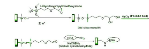
[image:3.595.309.550.382.462.2]The preparation of a silica monolith for the immobilization of HSA was also examined, using the epoxy immobilization method for attaching proteins to silica monoliths [36]. However, it is known that the epoxy method tends to give lower activities and lower protein coverage for HSA than other amine-based coupling methods [37]. Work in Rangan's study[38] used the Schiff base, an immobilization method which gives better results for HSA when used with other support materials [39]. NaBH4 phosphate solution (pH 8.2) is used to reduce double bond C=N to single bond C–N, which may pose great danger to researchers’ life. A mild reducing agent(sodium cyanoborohydride) was also present during this reaction to reduce the Schiff base to a more stable secondary amine linkage[38], which is illustrated in Figure 1.
Fig. 1. Reactions for the preparation of an HSA silica monolith. Abbreviation: HSA, human serum albumin.
Through another way, HSA is immobilized via its sulfhydryl groups [40]. Amino-silica is activated by succinimidyl
4-(N-maleimidomethyl)cyclohexane-1-carboxylate(SMCC) followed by reaction with a sulfhydryl group of the protein. Similarly, amino-silica is activated by succinimidyl iodoacetate (SIC) for reaction with a sulfhydryl group. Maleimide-activated silica(the SMCC method) or iodoacetyl-activated silica (the SIA method) is used for these methods. It is found that the SMCC and SIA methods gave HSA-based monolith with comparable or improved activity and stability, compared to those made by the Schiff base method.
Often, the preparation of a silica monolith for the AGP selector has been immobilized onto epoxy-activated supports [41],[42] or through the hydrazide immobilization method, a technique that has been shown to give site-selective coupling for glycoproteins like AGP, as demonstrated in previous work with silica particles [43],[44]. Prior to immobilization, the carbohydrate residues on AGP were oxidized under mild conditions to generate aldehyde groups, approximately, five reactive aldehyde groups per AGP molecule[45].
preparation of monolith, is also evolving. Liu et al. [46] reported fabrication of silica-based monolithic capillaries with physically adsorbed avidin as chiral selector. Enantiomeric separations of amino acid derivatives, several organic acids, menadione sodium bisulfite, warfarin and N-methylpseudoephedrine were achieved in both nano-HPLC and CEC mode. Theoretical plate numbers of 122,000 per meter for nano-HPLC and 242,000 per meter for CEC were observed.
There have been several attempts to prepare particle-based monolith, using silica particles bearing a chiral selector without the need for end frit preparation. In all cases, the immobilization of the packing material inside the capillary requires one additional preparation step. Such capillaries have been obtained, for example, by packing silica material into a tube followed by subsequent sintering of the whole packing material [47], by passing a sol solution of silicate [48] or alkoxysilanes through a pre-packed column [49], or by pumping a methacrylate solution through the column prior to polymerization[50]. Another attempt to prepare particle-based capillaries without end frits was published by Kato et al [51]. They prepared particle-loaded phases using a sol-gel technique by suspending silica particles containing a chemically bonded chiral selector in a solution of tetraethyl orthosilicate, which was forced into a piece of tubing.
Prepared particle-loaded monoliths were characterized by means of electron microscopy. An almost homogeneous distribution of silica particles could be achieved by various optimizations of the polymerization mixture used. Interestingly, silica particles were only slightly crosslinked by the polymeric backbone, leaving the majority of the silica particle surface unaffected. As chiral separation takes place only at the surface of silica particles bearing the chiral selector, this should lead to good separation performance.
D. Molecularly Imprinted Polymer Monoliths
Molecularly imprinted polymers (MIPs), another popular technique for enantioselective binding-sites creation, have a potential in the separation of chiral compounds, predicting not only the recognition ability but also the elution order [52]-[54]. Applications of MIP as separation media in LC, CE and CEC for chiral separation have been extensively investigated[55],[56]. The MIP monolith of CEC has the advantage of minimal chemical consumptions, especially the imprinted molecule.
To date, the imprinting of small molecules has been well-established and considered almost routine. However, the imprinting of biomacromolecules, such as proteins and peptides, continues to be a significant challenge due to difficulties with large molecular sizes, structural complexity, environmental sensitivity of the templates, and significantly reduced non-covalent template monomer interactions in aqueous media [57]-[59]. Despite these difficulties, there is still a strong incentive to synthesis MIPs of biomacromolecules and quite a few attempts have been made with more or less success.
For example, an approach using systematic optimization for the formation of an albumin MIP has been prepared by imprinting albumin using a copolymer comprising 3-dimethylaminopropylmethacrylate and tetraethylene
glycol dimethacrylate in a mole ratio of 1 to 8. Cytochrome c, lysozyme and myoglobin were used in competitive rebinding experiments to compete with the polymer's native template with all protein species present at 0.0004 g/mL. It has been investigated that 0.125 mole ratio of monomer to crosslinker, 6.04 wt.% water content with respect to the mass of the monomer complex, 60h polymerization time at 38 °C, and with 0.47% albumin in the prepolymerization monomer complex can obtain the optimum condition[60].
Three major challenges can be encountered for MIP taking proteins as template. First challenge is diffusional limitations in large molecular weight for molecularly imprinted polymers, which is attributed to the high crosslinking required to achieve recognition. Another problem is the difficulty of protein dissolution in the monomer solution and the low solubility of these structures in non-aqueous solvents. Most of the protein-imprinted polymers demonstrated to date show very low template rebinding, which forms the third challenge. Few exhibit rebinding capacity high enough for some applications such as bioseparation. Among these, Guo’s group[61]-[63]fabricated bovine hemoglobin (Hb)-imprinted polymers by the combined use of chitosan, presenting a high Hb rebinding capacity of above 20 mg/g wet gels, while the non-imprinted polymers(NIPs) bound very little Hb. However, Guo found that these high binding results are difficult to explain reasonably. Through X-ray diffraction and scanning electronic microscope investigations, a remarkable property changes of the NIPs after the washing process is confirmed. These findings[64] indicate that the non-specific adsorption resulting from the template removing process rather than the imprinted sites generated on the MIPs themselves may account for the high template rebinding capacity of the reported protein-imprinted polymers.
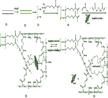
representatively selected for imprinted templates(Fig. 2). Under the optimum conditions, the obtained hybrid silica-based MIP monolith showed higher binding affinity for template than its corresponding non-imprinted (NIP) monolith. The imprinted factor (IF) for BSA and Lyz reached 9.07 and 6.52, respectively. Moreover, the hybrid silica-based MIP monolith displayed favorable binding characteristics for template over competitive protein. Compared with the imprinted silica beads and in situ organic polymer-based hydrogel MIP monolith, the hybrid silica MIP monolith exhibit better chromatographic performances[71].
Fig.2. Schematic representation of preparation procedures of hybrid silica-based MIP monolithic column. (1) Bare stainless steel column; (2) naked silica skeleton prepared by sol–gel process; (3) silanols on the surface of silica monolith; (4) vinylation on the surface of silica monolith; (5) formation of MIP coating on the surface of silica monolith;(6) removal of template protein.
The combination of MIP with chiral separation through macromolecules, at early times, only came from papers comparing the prepared MIP with commercially available chiral stationary phases. For example, the baseline separation of nilvadipine enantiomers thorough MIP was comapared with columns based on the protein (ovomucoid or α-acid glycoprotein) [72].
However, apart from applications of bioseparation and biosensors, no imprinting of protein has been applied for chiral separation. This may due to that macromolecular MIP is so difficult to fabricate and even if MIP was successfully prepared, the predetermined specificity would lose its halo. Another reason for not being reported in the field of enatiomer separation is probably that MIP with small molecules like mimic enatiomers have already met the demand of chiral analysis. Despite of above reasons, application of protein MIP for enatiomer separation still needs to be discussed and studied, for it can efficiently avoid requirement of highly-purified one enatiomer as temaplate and can provide more active sites for chiral separation.
E. Evaluation of monoliths
After fabrication of monolith, often a crucial step need to be undertaken, that is the evaluation process through various parameters from different perspective. However, up to now, in contrast with traditional chromatographic columns, no
strict or universal assessment system has been established. Paper between paper and column between column, even if the same type, would appear totally different types of measurements.
Several methods have been proposed to compare the performance of monolithic and packed columns, through the Hans Poppe plots [73], through various kinetic plots [74], and through column impedance[75], and of a few other approaches. The column impedance is a performance index that relates the hold-up time of a column and its efficiency [75],to the mobile phase viscosity and the inlet pressure available. The initial success of these various methods owes much to the initial ignorance of the scientific community regarding the properties of monolithic columns and their advantages and inconvenient compared to those of packed columns. Their use has much contributed to clarify the situation.
Pore size distribution of monolithic columns has always been one of the most challenging issues and often characterized by a bimodal pore size distribution[76], throughpores controlling the column permeability, and mesopore structure controlling the mass transfer kinetics and the column efficiency[77]. Porogenic distributions are often measured by Mercury Intrusion (MI), Scanning Electron Microscopy(SEM), and Nitrogen Adsorption method, which have turned a routine test[78]-[82].
Protein content is of great importance in monoliths with protein as chiral selectors. Through infrared spectroscopy of bare monolithic material and monolith with immobilized chiral selectors, their comparison and adsorption strength can evaluate whether the successful immobilization is achieved or not. Another method to estimate protein content is to add some indicator. For example, an estimate of the total protein content in the monolith is obtained by injecting a 0.1% (w/v) solution of copper sulfate onto the AGP columns. This method uses copper sulfate as a probe for the overall ion-exchange capability of the immobilized proteins [83]. Using carbamazepine, an analyte that has a single primary site on AGP, to make frontal analysis can also estimate the binding capacity and total amount of protein in the AGP silica monolith.
IV. CONCLUSIONS
REFERENCES
[1] C. Millot, J. Chromatogr. B 797 (2003) 131.
[2] Y.Tanaka, S.Terabe, J. Biochem. Biophys. Meth. 48 (2001) 103.
[3] F. Svec, T.B. Tennikova, Z. Deyl (Eds.), J. Chromatogr. Libr. 67,
Elsevier, Amsterdam,2003.
[4] T. Kimura, A. Shibukawa, K. Matsuzaki, Pharm. Res. 23 (2006) 1038.
[5] J. Ghuman, P.A. Zunszain, I. Petitpas, A.A. Bhattacharya, M. Otagiri,
S. Curry, J.Mol. Biol. 353 (2005) 38.
[6] E.J. Franco, H. Hofstetter, O. Hofstetter, J. Sep. Sci. 29 (2006) 1458.
[7] M, Lämmerhofer. J. Chromatogr. A 1217(2010) 814.
[8] H. Xuan, D.S. Hage, Anal. Biochem. 346 (2005) 300.
[9] R. Mallik, H. Xuan, D.S. Hage, J. Chromatogr. A 1149 (2007) 294.
[10] E. Hazai, J. Visy, I. Fitos, Z. Bik´ adi, M. Simonyi, Bioorg. Med. Chem.
14 (2006)1959.
[11] J. Haginaka, C. Seyama, N. Kanasugi, Anal. Chem. 67 (1995) 2539.
[12] F. Zsila, Y. Iwao, Biochem. Biophys. Acta 1770 (2007) 797.
[13] R. Mallik, J. Tao, D.S. Hage, Anal. Chem. 76 (2004) 7013–7022.
[14] J.J. Mart´ ınez-Pla, Y. Mart´ ın-Biosca, S. Sagrado, R.M.
VillanuevaCama˜ nas, M.J. Medina-Hern´ andez, J. Chromatogr. A 1048 (2004) 111.
[15] J.J. Mart´ ınez-Pla, Y. Mart´ ın-Biosca, S. Sagrado, R.M.
VillanuevaCama˜ nas, M.J. Medina-Hern´ andez, Anal. Chim. Acta 507 (2004) 175.
[16] A. Lavecchia, S. Cosconati, E. Novellino, E. Calleri, C. Temporini, G.
Massolini,G., et al. J. Mol. Graphics Modell. 25 (2007)773.
[17] I. Petitpas, A.B. Bhattacharya, S. Twine, M. East, S. Curry, J. Biol.
Chem. 276(2001) 22804.
[18] J. Stahlberg, H. Henriksson, C. Divne, R. Isaksson, G. Pettersson, G.
Johansson, et al. J. Mol. Biol. 305 (2001) 79.
[19] M. Ye, H. Zou, Z. Liu, R. Wu, Z. Lei, J. Ni. J. Pharmaceut. Biomed.
Anal. 27(2002) 651.
[20] H. Hofstetter, O. Hofstetter,V. Schurig, J. Microcol. Sep. 10(1998)
287.
[21] C. Fujimoto. Anal. Sci. 18 (2002) 19.
[22] F. Qin, C. Xie, Z. Yu, L. Kong, M. Ye, H. Zou. J. Sep. Sci. 29
(2006)1332.
[23] D. Wistuba, V. Schurig. J. Sep. Sci.29 (2006) 1344.
[24] B. Preinerstorfer, M. Laemmerhofer. Electrophoresis 28 (2007) 2527.
[25] I. Tanret, D. Mangelings, Y.V. Heyden. J. Chromatogr.Sci.
47(2009)407.
[26] S. Birnbaum, S. Nilsson, Anal. Chem. 64(1992) 2872.
[27] T. Koide, K. Ueno.Anal. Sci. 14 (1998) 1021.
[28] C. Gatschelhofer, et al. J. Biochem. Biophys. Methods 69(2006 ) 67.
[29] C. Yao, et al. Talanta 82(2010) 1332.
[30] J. Krenkova, Z. Bilkova, F. Foret, J. Sep. Sci. 28 (2005) 1675.
[31] D. Wistuba. J. Chromatogra. A 1217(2010)941
[32] M. Kato, N. Matsumoto, K. Sakai-Kato, T. Toyo’oka , J. Pharmaceut.
Biomed.Anal. 30 (2003) 1845.
[33] K. S. Kato, M. Kato, H. Nakakuki, T. Toyo’oka, J. Pharmaceut.
Biomed. Anal. 31 (2003) 299.
[34] M. Kato, K. Sakai-Kato, N. Matsumoto, T. Toyo’oka, Anal. Chem. 74
(2002) 1915.
[35] M. Kato, H. Saruwatari, K. Sakai-Kato, T. Toyo’oka, J. Chromatogr. A
1044 (2004) 267.
[36] E. Calleri, C. Temporini, E. Perani, A. D. Palma, D. Lubda, G.
Mellerio, et al. J. Proteome Res. 4(2005) 481.
[37] R. Mallik, T. Jiang, D.S. Hage, Anal. Chem. 76 (2004) 7013.
[38] R. Mallik, D. S. Hage, J. Pharmaceut. Biomed. Anal. 46(2008) 820.
[39] R. Mallik, D.S. Hage, J. Sep. Sci. 29 (2006) 1686.
[40] R. Mallik, C.Wa, D.S. Hage, Anal. Chem. 79 (2007) 1411.
[41] E. Calleri, G. Massolini, D. Lubda, C. Temporini, F. Loiodice, G.
Caccialanza, J. Chromatogr. A 1031 (2004) 93.
[42] E. Calleri, C. Temporini, E. Perani, C. Stella, S. Rudaz, D. Lubda, et al. J. Chromatogr. A 1045 (2004) 99.
[43] H. Xuan, D.S. Hage, Anal. Biochem. 346 (2005) 300.
[44] H. Xuan, D.S. Hage, J. Sep. Sci. 29 (2006) 1412.
[45] M.G. Schmid, J. Koidl, C. Freigassner, S. Tahedl, L. Wojcik, T.
Beesley, et al. Electrophoresis 25(2004) 3195.
[46] Z. Liu, K. Otsuka, S. Terabe, M. Motokawa, N. Tanaka,
Electrophoresis 23 (2002) 2973.
[47] D. Wistuba, V. Schurig. Electrophoresis 21(2000)3152.
[48] G. Chirica, V.T. Remcho. Electrophoresis 20(1999)50.
[49] Q. Tang, M.L. Lee. J Chromatogr A. 887(2000)265.
[50] G. Chirica, V.T. Remcho. Anal Chem. 72(2000)3605.
[51] M. Kato, M.T. Dulay, B. Bennett, J.R. Chen, R.N. Zare.
Electrophoresis 21(2000)3145.
[52] X. Huang, F. Qin, X. Chen, Y. Liu, H. Zou, J. Chromatogr. B 804
(2004) 13.
[53] J. Yin, G. Yang, Y. Chen, J. Chromatogr. A 1090 (2005) 68.
[54] P. Spegel, L. Schweitz, S. Nilsson, Anal. Chem. 75 (2003) 6608.
[55] J. Courtois, G. Fisher, B. Sellergren, K. Irgum, J. Chromatogr. A 1109
(2006) 92.
[56] H. Kim, G. Guiochon, Anal. Chem. 77 (2005) 93.
[57] T. Shiomi, M. Matsui, F. Mizukami, K. Sakaguchi. Biomaterials
26(2005)5564.
[58] Y. Li, H.H. Yang, Q.H. You, Z.X. Zhuang, X.R. Wang. Anal Chem
78(2005)317.
[59] D.M. Hawkin, D. Stevenson, S.M. Reddy. Anal Chim Acta 542(2005)
61.
[60] C.H. Hu, T.C. Chou, Microchem. J. 91 (2009) 53.
[61] T.Y. Guo, Y.Q. Xia, G.J. Hao, M.D. Song, B.H. Zhang. Biomaterials.
25(2004) 5905.
[62] T.Y. Guo, Y.Q. Xia, J. Wang, M.D. Song, B.H. Zhang. Biomaterials
26(2005) 5737.
[63] Y.Q. Xia, T.Y. Guo, G.J. Hao, M.D. Song, B.H. Zhang, B.L. Zhang.
Biomacromolecules 6(2005)2601.
[64] G.Q. Fu, H. Yu, J.Zhu. Biomaterials 29(2008) 2138.
[65] L. Schweitz, P. Spegel, S. Nilsson, Electrophoresis 22 (2001) 4053.
[66] Y.L. Xu, Z.S. Liu, H.F.Wang, C. Yan, R.Y. Gao, Electrophoresis 26
(2005) 804.
[67] Q.L.Deng, Z.H. Lun, R.Y.Gao, L.H. Zhang,W.B. Zhang, Y.K. Zhang,
Electrophoresis 27 (2006) 4351.
[68] J. Matsui, T. Kato, T. Takeuchi, M. Suzuki, K. Yokoyama, E. Tamiya,
I. Karube,Anal. Chem. 65 (1993) 2223.
[69] J.J. Ou, X. Li, S. Feng, J. Dong, X.L. Dong, L. Kong, M.L. Ye, H.F.
Zou, Anal. Chem.79 (2007) 639.
[70] H.F.Wang, Y.Z. Zhu, X.P. Yan, R.Y. Gao, J.Y. Zheng, Adv. Mater. 18
(2006) 3266.
[71] [Z. Lina, F. Yang, X. He, X. Zhao, Yukui Zhang. Journal of
Chromatography A, 1216 (2009) 8612.
[72] Q. Fu, H. Sanbe, C. Kagawa, K.-K. Kunimoto, J. Haginaka, Anal.
Chem.75 (2003) 191.
[73] H. Poppe, J. Chromatogr. A 778 (1997) 3.
[74] S. Eeltink, P. Gzil, W.T. Kok, P.J. Schoenmakers, G. Desmet, J.
Chromatogr. A 1130 (2006) 108.
[75] P.A. Bristow, J.H. Knox, Chromatographia 10 (1977) 279.
[76] G.S. Chirica, V.T. Remcho, J. Chromatogr. A 924 (2001) 223.
[77] G. Guiochon, J. Chromatogr. A 1168(2007)101.
[78] K. Nakanishi, N. Soga, J. Am. Ceram. Soc. 74 (1991) 2518.
[79] K. Nakanishi, N. Soga, J. Non-Cryst. Solids 139 (1992) 1.
[80] K. Nakanishi, N. Soga, J. Non-Cryst. Solids 139 (1992) 14.
[81] N. Ishizuka, H. Kobayashi, H. Minakushi, K. Nakanishi, K. Hirao, K
Hosoya, et al. J. Chromatogr. A 960 (2002) 85.
[82] N. Tanaka, H. Kobayashi, N. Ishizuka, H. Minakushi, K. Nakanishi, K.
Hosoya, et al. J. Chromatogr. A 965 (2002) 35.