Abstract — In recent years, biometrics eye detection methods have become popular tools of significance. The exact eye localization technique is important for analysis, and has direct effect in the subsequent steps of normalization, feature extraction and matching. In this paper, after considering merits and demerits of many advanced reported techniques, while keeping in mind, the required fast processing response time of detection to prevent Posterior Capsular Rupture, amalgamates technique has been proposed to obtain a precise, fast and robust method in localizing different segments of the eye, in particular separation of the pupil, iris and sclera. On the contrary to the majority of the current available procedures which make assumptions about circularity or elliptically shape of the iris and pupil, this proposed method aims to capture exact boundaries of these segments while considering minimal detection time. The technique has been intended to be used as part of a complication prevention procedure during eye surgeries. The results of the above studies indicate the effectiveness of the suggested procedure in real time operation.
Keywords — Cataract surgery, PCR, Localization, Pupil detection, Iris boundary detection
I. INTRODUCTION
RECENT advances in biometrics technology have shown great success for verification and identification purposes. This technology has aimed to capture the biological features or behavioral characteristics of the examined subject [1]. Amongst the diverse and widely used biometrics techniques, those detecting the biological features of iris are most promising and the success has been recognized by all in this field [1-4].
The first reliable iris recognition system was introduced by J. Daugman in 1993 [5]. Since then many new approaches have been proposed, aiming to improve the accuracy level and the response time. Despite the variations in the suggested methodologies, in majority of cases the procedural steps included segmentation, normalization, feature extraction and matching [6]. From this, it can be suggested that the first and most important step in the iris recognition is to identify and segment different features of the eye, in particular the iris. Therefore, it is only in the case of high Manuscript received March 21,2012; revised April 12, 2012
A. Ektesabi, PhD candidate, Faculty of Engineering and Industrial Sciences, Swinburne University of Technology, John Street, Hawthorn, Australia, 3122 (e-mail: [email protected] ).
Professor A. Kapoor, Associate Dean Research, Faculty of Engineering and Industrial Sciences, Swinburne University of Technology, John Street,
precision in the localization step that reliable and accurate results can be obtained in the subsequent steps.
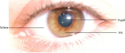
For the purpose of this study, image processing concepts, similar to those used in biometrics iris recognition systems are proposed to be used on video recordings captured during eye surgeries to extract important features including the iris, pupil and sclera, as shown in Figure 1. The identified features are then analyzed. This real time analysis provides feedback to the surgeon and in the case of a complication an alarm system would inform the operator to stop and revise the following steps in the surgical procedure. This is the first time this approach has been proposed in the field of ophthalmology. So far there have been significant improvements on instrumentations as well as surgical procedural methodologies but none have been able to assist the surgeon directly intra-operatively. This study aims to provide real time assistance to both experienced and less experience surgeons and residents during the surgery.
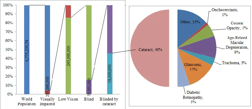
It is possible to apply this concept to different ophthalmological surgical procedures. In this case, the studies condition is cataract. Cataract is a condition, known as opacification of the lens, where the protein structures of the lens are denatured, causing blurred vision in patients [7]. Studies performed by the World Health Organization (WHO) indicate that 48% of the blindness in the world is caused by cataract, with majority living in the developing countries. The global causes of blindness and the affected population in the world is illustrated in Figure 2 [8].
Moreover, cataract is an age related condition [9] and so with the continuous increase in an overall life expectancy of the world population, the need to prevent and treat this has become of great significance.
Preventing the formation of cataract has proven to be difficult with today’s everyday life style. The studies indicate that nearly everyone would require the need for surgery at
Complication Prevention of Posterior Capsular
Rupture using Image Processing Techniques
A.
Ektesabi,
Member IAENG,IEEE,
A. Kapoor
[image:1.612.332.551.219.313.2]R
some stage of their lives [9]. However, certain factors have proven to be successful in reducing the probability of contracting cataract.
These include a healthy life style and diet, well economic and financial status, short term exposure to UV lights and regular visits to the optometrists [10-12].
The most common treatment for cataract is surgery. Cataract Surgeries, have been performed over centuries, originating back to 800 BC [12], where they used to implement a method known as “couching” to dislodge the lens into the posterior segment of the eye without any lens replacing the natural lens of the eye. It was not until the 20th century when Harold Ridley introduced the use of Intra-Ocular Lenses (IOL). Since then the common used techniques were the Extra-Capsular Cataract Extraction (ECCE), Intra-Capsular Cataract Extraction (ICCE) and most recently the Phacoemulsification was used for the surgery. For the purpose of this study, the data is obtained from the phacoemulsification procedure.
Despite the significant improvements in surgical instrumentations and procedures, certain complications might occur intra-operatively, including Posterior Capsular Rupture (PCR), Cystoid Macular Edema, Endophtalmitis and Retinal detachment [13-16].
The most common intra-operative complication is PCR with the average rate of 4.8% [17]. A male patient at the age of 75, who cannot lie flat and has conditions such as diabetic retinopathy, vitreous opacities, white cataract, glaucoma, pseudoexfolation, small pupil size, axial length greater than or equal to 26.0 mm, the use of the α-blocker doxazosin is most likely at risk [18].
Some of the post-operative effect of PCR occurrence is an increase in the recovery time of time of the patient as well as side effects on the overall visual outcome. This is due to the higher chance of induced astigmatism [12, 13].
Despite the low percentage of occurrence of PCR, due to the large number of surgeries performed yearly, the number of patients affected by this complication is significant. Hence an improvement in the surgical procedure would have great
impact on lives of patients, and also the associated costs and resources for the surgeries would be greatly reduced.
In section two, some of the most commonly used biometric techniques and procedures are examined.
This is then followed by the proposed technique and procedure for PCR prevention in section three.
Lastly the results are discussed and the importance and advantages of this technique is illustrated in the conclusion.
II. CURRENT DETECTION TECHNIQUES AND PROCEDURES Significant improvements in the methodology of iris localization have been performed in the field of biometrics. Several of these techniques have been examined and two of the most prominent works in this field are presented.
A. Daugman
Daugman [5] use of integral differential operator (3), to detect circular edges by use of maximum gray level contrast between the two neighboring sections, has proven to be successful in many cases.
The proposed operator (1) consists of the integral operator (3) and a smoothing Gaussian filter (2).
z y x z
y x
ds r
y x I r r G
, , ,
, 2
,
max
(1)
22
2
1 r
e r
G
(2)
x
yzds r
y x I
, , 2
,
(3)
Where, the integral operator has the radius r in the
x,y coordinates.B. Wildes
[image:2.612.90.518.59.245.2]Circular edge detector followed by the Hough Transform was the proposed technique by Wildes [3]. This method is based on the optimization technique (4) and has high computational costs.
n
r n
r r r
g g
g g
n 1
,
1
, , ,
8 1
(4)
Where, the gradient of the normalized image at
,
r
isg
,r.III. PROPOSED TECHNIQUE
To detect PCR, minute variations in the eye are to be detected. The key to detect these changes during the surgery is precise detection and analysis of the pupil and iris.
Figure 3, indicates the steps involved in the image processing steps, as well as the overall aim of the project.
A. Factors to consider
Prior to any implementation, certain factors should be kept under consideration.
Firstly, before to the surgery procedure, the patient must undergo health checks. Certain information obtained at this point are crucial and to be noted by the surgeon. Additionally, some of the obtained readings can be helpful in the interpretation of the results and so the detection of the complication.
Furthermore, for the purpose of this study, the accuracy of readings is very important. Therefore, the exact, non-elliptical boundaries of both pupil and iris were to be determined. As a result, methodologies involving circular or elliptical approximations were ignored.
Moreover, this identification should occur in real time and the feedback should inform the surgeon of the possibility of complication before it occurs. As a result, the computational time for the detection should have been kept to be minimal.
This can be done in two ways, one is to apply the image processing techniques which are less computationally costly and secondly by reducing the factors to be taken under consideration while doing the analysis. One of which includes, ignoring of eyelashes and eyelids as claps are used during the surgery to keep the eyes open. Additionally, the head is kept steady during the surgery and so the possible noise caused by head movement can be ignored.
However, since in the majority of cases the patients are under local anesthetic, extra movements of the eye are expected to be seen during the surgery and should be taken under consideration while interpreting the data.
B. Image Acquisition
The foremost step in any image processing analysis is to capture high quality images with good resolution. The obtained images in this case, should be captured from the video camera recording the surgery. Hardware filters are placed, to reduce the noise caused by minimal motion. The use of illumination compensations is also suggested to reduce the effect of lighting variations during the surgery.
C. Image Manipulation
To reduce the processing times, the RGB images are to be transformed to gray level images. The unwanted regions in the images are removed by software filtering.
D. Iris and pupil Localization
There are several techniques which can be implemented to localize the iris and pupil. However, the accuracy of these techniques and their processing times should be considered. To improve the accuracy of the detection, the two different procedures are chosen and run simultaneously. Since they are running concurrently, the procedural time will not vary significantly to those of a single technique being run by individually.
E. Normalization
In this stage filters are implemented to remove the noises in the image. Likewise, the unwanted reigns, such as the sclera can be removed as well, reducing the processing steps for the following stages.
F. Feature Extraction
At this stage the required special features of the iris and pupil are extracted and are taken for further analysis.
G. Masks
Masks are created, capturing the areas of interest. The two separate implemented procedures each create their own mask. These two masks are then combined, forming a single mask, which can then be taken to the next stage of the procedure.
H. Matching and Interpretation
Matching the results occurs by comparing each individual masked image with those of previously complicated situations.
If during this comparison the results match those of the complicated cases, the alarm would sound off, and then the results are displayed. It is then that the surgeon is required to stop and revise the surgical procedures being performed.
In the case where no complication is observed, the results are displayed directly and the surgery continues as planned.
IV. RESULTS AND DISCUSSIONS
To obtain usable data and to maximize the detection accuracy, hardware filtering is used in this paper. This reduced the effect of motion and light reflection on the surface of the eye.
The images have been pre-processed before segmentation. This included sampling and software filtering. The images were sampled in order to reduce the computational time. Furthermore, to reduce disturbances caused by motion and laminations software filtering were adapted.
In the segmentation stage, binary and gray scaled images were used to improve the computational time. In this study, total eye detection was not of interest, only the pupil and iris have been considered.
Failure in early detection of PCR could lead to catastrophic results in real time processing of the data during the surgery. This study shows that the proposed method can be used for exact detection of deviations leading to PCR, by prompt recognition of any variation in shape of the segments.
As mentioned earlier, different localization techniques can be implemented. Since the detection time has a significant role in real time capturing of features, simpler but precise techniques are of interest. In this case, for the separation of iris and pupil boundaries, the two methods of thresholding [21] and active contour procedure [22] were used. Each of the above two techniques, would provide good estimation for the interested boundaries. In the proposed system by merging the two techniques, enhancement of the final accuracy of the detection was observed. Hence, the results obtained showed similar outcomes of each technique, but the two masks were slightly different and so the combinations of the two masks were used to minimize the final error in the matching and interpretation step.
Simultaneous processing and implementation of the two methods, reduced the overall detection time, almost equal to time taken by each method. Though, the overall detection time was similar to each, but by combing them, the precision of detection was improved significantly. The measured time of this procedure was less than one second, which is similar to reported techniques so far but with higher accuracy in detection. This suggests the possibility of detecting an error and responding to it within an appropriate time, which in turn is a novel application of the high speed image processing technique in PCR detection during operation.
V. CONCLUSION
The study is aimed to predict the possibility of detecting PCR, using image processing techniques. The use of currently reported techniques in the field of biometrics, in particular iris localization has been suggested for capturing the required features of the eye, specifically the iris and the pupil. To improve the accuracy of the localization, integration of two or more techniques is suggested to be run simultaneously. The combined detected features are then compared with the previous cases of PCR, in order to help surgeons to predict the occurrence of such complication during the surgery. Currently the detection occurs in less than one second, with the aim to improve on this, in the near future.
REFERENCES
[1] W. T. Lin, et al., "Fast and robust iris recognition," Imaging Science
Journal, The, vol. 57, pp. 128-139, 2009.
[2] C. H. Daouk, et al., "Iris Recognition," presented at the IEEE Symposium on Signal Processing and Information Technology, Marrakesh, 2002.
[3] R. P. Wildes, "Iris Recognition: An Emerging Biometric Technology,"
Proceeding of the IEEE, vol. 85, pp. 1348-1363, 1997.
[4] P. W. Richard, "Iris recognition: an engineering biometric,"
Proceeding of the IEEE, pp. 1354-1356, 1997.
[5] J. Daugman, "High confidence visual recognition od persons by a test of statistical independence," IEEE Transactions. PAMI, vol. 15, pp. 1148-1161, 1993.
[6] H. Mehrabian and P. Hashemi-Tari, "Pupil Boundry Detection for Iris Recognition Using Graph Cuts," in Proceedings of Image and Vision
Computing New Zealand 2007, Hamilton, New Zealand, 2007, pp.
77-82.
[7] E. L. Lamoureux, et al., "The impact of cataract surgery on quality of life," Current Opinion in Ophthalmology, vol. 22, pp. 19-27, 2011. [8] World Health Organisation. (2004, June 2011). Global pattern of
blindness changes with success in tackling infectious disease and as
population ages. Available:
http://www.who.int/mediacentre/news/notes/2004/np27/en/
[9] R. Khanna, et al., "Cataract surgery in developing countries," Current
Opinion in Ophthalmology, vol. 22, pp. 10-14, 2011.
[10] M. Zare, et al., "Risk factors for posterior capsule rupture and vitreous loss during phacoemulsification," Journal of Ophthalmic and
Vision Research, vol. 4, pp. 208-212, 2009.
[11] J. C. Javitt, et al., "Geographic-Variation in Utilization of Cataract-Surgery," Medical Care, vol. 33, pp. 90-105, 1995.
[12] P. T. Ashwin, et al., "Advances in cataract surgery," Clinical and
Experimental Optometry, vol. 92, pp. 333-342, 2009.
[13] S. Pershing and A. Kumar, "Phacoemulsification versus extracapsular cataract extraction: where do we stand?," Current Opinion in
Ophthalmology, vol. 22, pp. 37-42, 2011.
[14] M. C. Wu and A. Bhandari, "Managing the broken capsule," Current
Opinion in Ophthalmology, vol. 19, pp. 36-40, 2008.
[15] E. Chan, et al., "Complications of cataract surgery," Clinical and
Experimental Optometry, vol. 93, pp. 379-389, 2010.
[16] A. Coombes and D. Gartry. (2003). Cataract surgery.
[17] Jackson B. et.al., "Cataract suregry performed by residents: risk analysis," Revista Brasileira de Oftalmologia, vol. 69, pp. 301-305, 2010.
[18] N. Narendran, et al., "The Cataract National Dataset electronic multicentre audit of 55 567 operations: risk stratification for posterior capsule rupture and vitreous loss," Eye, vol. 23, pp. 31-37, Jan 2009. [19] Y.-z. Shen, et al., "A New Iris Locating Algorithm," Proceedings of
the 16th International Conference on Artificial Reality and
Telexistence - Workshops, pp. 438-441, 2006.
[21] D. A. Chernyak, "Iris-based cyclotorsional image alignment method for wavefront registration," IEEE Transactions on Biomedical
Engineering, vol. 52, pp. 2032-2040, 2005.
[22] J. Jomier, et al., "Automatic quantification of pupil dilation under stress," International Symposium on Biomedical Imaging: Nano to