Original Article
Neuroleukin regulates the metastasis of non-small cell
lung cancer cells through the Wnt/β-catenin
signaling pathway
Changqing Shao1, Hongfei Ma1, Lili Jiang2, Hong Liu1
1Department of Thoracic Surgery, The Affiliated Central Hospital of Qingdao University, 127 Siliu South Road,
Shibei District, Qingdao City 266000, Shandong Province, China; 2Department of Traditional Chinese Medicine,
The Affiliated Central Hospital of Qingdao University, 127 Siliu South Road, Shibei District, Qingdao City 266000, Shandong Province, China
Received January 21, 2019; Accepted April 11, 2019; Epub July 15, 2019; Published July 30, 2019
Abstract: Purpose: We investigate the mechanisms of neuroleukin (NLK) in the metastasis of non-small cell lung cancer (NSCLC). Methods: A549 cells were separated into an si-NLK group, an NC group, and a control group. The expression of NLK was measured by qRT-PCR. The proliferation of A549 cells was measured using cell cloning and an MTT assay, and the migration ability of the A549 cells was measured using a scratch test, and the invasiveness of A549 cells was tested isomg a Transwell invasion assay. Additionally, immunofluorescence was used to observe the distribution of β-catenin in the cells, and western blot was applied in measuring the expressions of β-catenin, cyclin D1, and c-myc. Results: After transfection with NLK-siRNA, the expression of NLK was distinctly decreased compared to the control and NC group (P < 0.05). The proliferation in the si-NLK group was prominently slower than it was in the other two groups (P < 0.05). The cell cloning, migration, and invasiveness abilities in the si-NLK group was markedly decreased compared to the control and NC groups (P < 0.05). A high positive expression of β-catenin in the nuclei was detected in the control and NC groups. Moreover, in the si-NLK group, it was mainly expressed in the membranes, and the expression showed a downward trend. By comparison with the control and NC groups, the expressions of c-myc, β-catenin, and cyclin D1 in the si-NLK group were markedly down-regulated (P < 0.05). Conclusions: NLK significantly promotes the metastasis of NSCLC cells by adjusting the Wnt/β-catenin signaling pathway, which could provide a new target for controlling the metastasis of NSCLC at the gene level.
Keywords: NLK, non-small cell lung cancer, cell proliferation, metastasis, Wnt/β-catenin pathway
Introduction
Non-small cell lung cancer (NSCLC), including squamous cell carcinoma, adenocarcinoma, and large-cell carcinoma, makes up more than 85% of all lung cancer cases [1]. NSCLC is con-sidered the main subtype of lung cancer, with a high incidence and poor prognosis [2]. The common causes of NSCLC include smoking, occupational and environmental exposure, ion-izing radiation, chronic lung infections, genetic factors, and air pollution. At present, chemo-therapy, radiochemo-therapy, and surgical resection are the main treatments for NSCLC [3, 4]. However, the current treatment techniques are involve a long treatment cycle, and the cancer is prone to recurrence. In order to alleviate the
suffering of patients, a new method is urgently needed to diagnose and treat NSCLC quickly and effectively.
NSCLC [7]. The inhibition of NLK expression can significantly promote the proliferation of NSCLC cells [8]. Along with extensive research on the treatment of NSCLC targets, a number of studies have been done on the relevant ways for the treatment of cancer. The Wnt path-way is an important signal transduction system, and mutations in the Wnt/β-catenin pathway-associated protein expression lead to a variety of growth-related pathologies and cancers [9]. Vilchez’s research has shown that the up-regu-lation of the Wnt/β-catenin pathway takes part in maintaining tumor initiation cells, drug resis-tance, tumor progression, and metastasis [10]. Importantly, NSCLC cell proliferation can be promoted by regulating the Wnt/β-catenin pathway [11, 12].
Specifically, NLK is recognized as a key regula-tor of many cancers. However, the mechanism of NLK and the related signaling pathways in NSCLC are still unclear. In this study, we exam-ined the effects of NLK on NSCLC metastasis through the Wnt/β-catenin pathway from cell multiplication, migration, and invasion in A549 cells, which may provide new ideas for the clini-cal treatment of NSCLC.
Methods
Cell culture
A549 cells (cell Bank of Shanghai Institute of Cell Biology, Chinese Academy of Sciences) were cultured in an RPMI 1640 medium con-taining 100 IU/mL penicillin/streptomycin and 10% fetal calf serum at 37°C and 5% CO2. Single cell suspension was obtained by trypsin digestion, then the cells were washed with 0.01 mol/L PBS and suspended at about 1 × 10⁶ cells/mL. The cells were passaged once every other day and in the rapid growth phase were used for subsequent experiments.
Cell transfection
The A549 cells were divided into a control group (conventional culture), an NC group (transfected by siRNA-NC), and an si-NLK group (transfected by siRNA-NLK). After digestion, centrifugation, and rinsing with sterile saline, the cells were added to a six-well plate. After cell fusion reached 60%, DMEM-f12 complete medium (Hyclone Company) was replaced by serum-free DMEM-f12 medium for 1 h. Then
250 μL Opti-MEM with 5 μL (100 pmol) siRNA reagents (Shanghai Jima Pharmaceutical Te- chnology Company, Shanghai, China) and 250 μL Opti-MEM with 5 μL lipo2000 reagent were mixed and incubated at 25°C for 20 min. Finally, the mixed droplets were added to the cells in the six-well plate and transfected in a 37°C incubator for 6-8 h. Then the culture medium was converted to a DMEM-f12 complete medi-um with 10% FBS.
QRT-PCR assay
Trizol reagent was used to extract total RNA. The ratio of OD260/OD280 was determined by nucleic acid quantitative proteinometer to iden-tify the purity of RNA. The reverse transcription reaction was used to synthesize the template of cDNA using a PCR amplifier, and the qRT-PCR was performed using an ABI 7500 quantitative PCR (Thermo Fisher, Singapore). QRT-PCR was processed based on the following conditions: pre-treatment at 95°C for 10 min, following by 40 cycles of 95°C for 5 s, 60°C for 34 s and 60°C for 30 s. The 2-ΔΔCt method was used to analyze the data. β-actin was used as the inter-nal reference. The special primers used were as follows: NLK-F, 5’-CAGATTTTGCGAGGTTTG- 3’; NLK-R, 5’-AGGAGATTCCCTGGCTTA-3’; β-ac- tin-F, 5’-GCAAGGTCATCCCTGAGCTGA-3’; β-ac- tin-R, 5’-ACGCCTGCTTCACCACCTTC-3’.
MTT assay
After transfection for 48 h, the A549 cells of each experimental group were suspended in complete medium by trypsinization and inocu-lated at 2,000 cells per well on a 96-well plate. Then the cells were cultured in an incubator at 37°C and 5% CO2. A total of 10 μL of 5 mg/mL MTT (Roche, Shanghai, China) was added to each well for 4 h, and then 100 μL of DMSO (TEDIA, USA) was added to terminate the reac-tion. The absorbance (A) at 490 nm was mea-sured with a microplate reader (SpectraMax® M5/M5e, California, USA), and the growth curve was plotted.
Cell cloning assay
30 min, stained with Giemsa for 20 min, and the number of cloned cells was finally counted. Each group was repeated 3 times.
Scratching test
About 5 × 10⁵ cells were added to each well of the 6-well plate, and serum medium was added for culture. When cell fusion reached 80%, a 10 μL tip was used to draw a trace perpendicular to the horizontal line on the cell surface. Then, a serum-free medium was added into the delin-eated cells. Subsequently, the cells were ob- served and photographed with an optical micro-scope at 0 h and 24 h after scratching, and then the cell migration distance was calculat-ed. Finally, the scratch healing rate was reck-oned using Image Tool (Bechtel Nevada, Ca- lifornia, USA).
Transwell invasive assay
After transfection for 48 h, approximately 1 × 10⁵ A549 cells were added to the Transwell upper chamber with the inner membrane coat-ed with Matrigel, and 200 μL of FBS-free cell culture medium was added. A total of 500 μL of complete medium was added to the lower chamber, the upper chamber was placed, and the entire Transwell chamber was cultured in an incubator for 24 h. Then, the cells and the medium inside the upper chamber were wiped off with a cotton ball, and then the upper cham-ber was immersed in 4% formaldehyde at 4°C for 8 min. Subsequently, the Transwell upper chamber was inverted, and the outer mem-brane was stained with a drop of crystal violet solution for 10 min. Next, the crystal violet
solu-tion was washed away by PBS solusolu-tion, and the membrane was dried. The number of invasive cells was calculated using an inverted mi- croscope.
Immunofluorescence
After transfection for 24 h, the cells were digested with 0.25% trypsin, and incubated in a 37°C incubator for 48 h. Then, the cells were rinsed thrice in PBS, fixed at 25°C for 20 min with 4% paraformaldehyde and permeabilized with 0.1% Triton followed by blocking with 1% BSA for 30 min at 25°C. The cells were incu-bated overnight with a 1:100 diluted monoclo-nal antibody against β-catenin. Next, the cells were incubated for 1 h at 37°C with an FITC-labeled secondary antibody. Subsequently, DAPI was used to stain the nucleus for 10 min under light-proof conditions. The staining re- sults were observed using a confocal micro-scope (LeiCa microsystem Heidelberg GmbH, Germany).
Western blotting
After transfection, the cells were treated with a RIPA cell lysate (Beijing Beyotime Biotechnolo- gy Co., Ltd., China), and the total protein was extracted. The equivalent protein was subject-ed to SDS-PAGE and transferrsubject-ed onto the mem-branes. Subsequently, it was blocked with 5% skim milk powder for 1 h. These membranes were separately incubated overnight at 4°C with the following primary antibodies: 5% BSA-containing β-catenin antibody (Cell Signaling Technology, Massachusetts, USA), cyclin D1 antibody (Santa Cruz, California, USA), or c-myc antibody (Santa Cruz, California, USA). The me- mbranes were incubated with an HRP-labeled anti rabbit secondary antibody (Zhongshan, Beijing, China) for 1 h at 25°C. This experiment used β-actin as an internal reference, and the protein was quantified using luminescence image analyzer software.
Statistical analysis
[image:3.612.90.287.68.200.2]All data were represented by mean values ± SD. SPSS 21.0 (SPSS Inc., Chicago, USA) was used for inter-group comparisons with one-way analysis of variance and post-hoc comparisons. Statistical significance was accepted at P < 0.05.
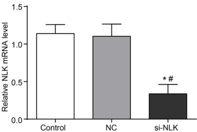
Figure 1. The effect of NLK knockdown was verified by qRT-PCR. *P < 0.05 (versus control group), #P <
Results
Effects of NLK knock down
After the transfection of NLK-siRNA, the expres-sion of NLK was observably less than the expressions of the control and NC groups (P < 0.05) (Figure 1). It was confirmed that knocking down NLK was successful.
suggesting that the knockdown of NLK inhibit-ed the proliferation of A549 cells.
Knockdown NLK inhibited migration and inva-sion of A549 cell lines
The fusion rate of scratch cells was observed 24 h after transfection. Compared with the other two groups, the cell fusion rate of the
si-Figure 2. The knockdown of NLK inhibited the pro-liferation of A549 cells. Note: (A) MTT assay, (B) Cell cloning assay. *P < 0.05 (versus control group), #P <
0.05 (versus NC group).
Figure 3. A scratch test de-tected the effect of knocking down NLK on cell migration. Note: (A) Migration of A549 cells under a lighted micros-copy (× 400), (B) Scratch re-pair rate. *P < 0.05 (versus
control group), #P < 0.05
(versus NC group).
Knockdown of NLK inhibited the proliferation of A549 cells
To further study the potential effects of NLK on A549 cell proliferation, we knocked-do- wn NLK by siRNA. As shown in Figure 2A, with the prolon-gation of culture time, the pro-liferation of A549 cells in the si-NLK group was observably slower when it was compared to the control and NC groups. In the first 5 days, the A490 values of the si-NLK group were remarkably lower than those in the other two groups (P < 0.05).
[image:4.612.89.526.61.648.2]tion and invasion of NSCLC cells by mediating the Wnt/β-catenin pathway. Remarkably, NLK is involved in the devel-opment and progression of tumors by phosphorylating multiple transcription factors, which is extremely important in a variety of signal transduc-tion pathways [13]. Silencing NLK could significantly inhibit cell proliferation and tumori-genicity in vitro and in vivo [14]. In this study, the knock-down of NLK inhibited the pro-liferation, migration, and in- vasion of A549 cells. Similarly, Li et al. [15] have demonstrat-NLK group was prominently slower. After the
si-NLK transfection, the scratch repair rate in the si-NLK group was clearly lower than it was in the control and NC groups (P < 0.05, Figure 3). The above experimental findings showed that the knockdown of NLK expression inhibit-ed the migration of A549 cells.
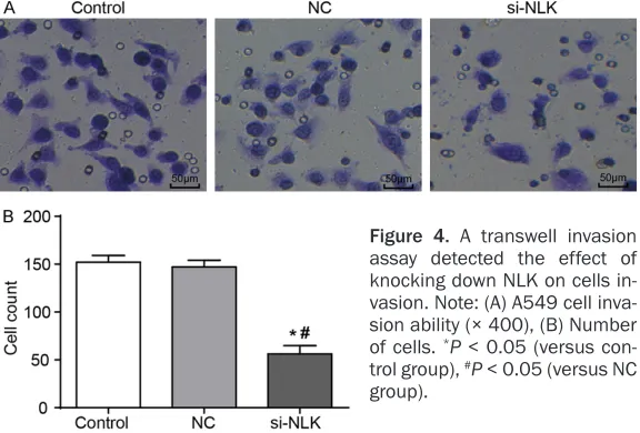
After knocking down NLK, the invasive ability of the A549 cells in the si-NLK group was distinct-ly lower than it was in the control and NC groups. Moreover, the number of cells pass- ing through the polycarbonate membrane de- scended markedly (P < 0.05, Figure 4), which suggested that the knockdown of NLK expres-sion inhibited the invaexpres-sion of A549 cells.
The knockdown of NLK inhibited the Wnt/β-catenin signaling pathway
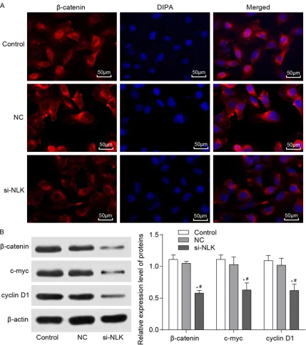
An immunofluorescence staining analysis indi-cated that a high level of β-catenin in the nuclei was found in the control and NC groups. Conversely, β-catenin in the si-NLK group was mainly expressed in the cytoplasms, and the level of β-catenin was reduced after transfec-tion (Figure 5A). A Western blot analysis show- ed that the expressions of c-myc, β-catenin and cyclin D1 were down-regulated in the si-NLK group more so than in the control and NC groups (P < 0.05) (Figure 5B). These results su- ggested that the knockdown of NLK expression inhibited the Wnt/β-catenin pathway activity.
Discussion
In this research, we verified that the knock-down of NLK inhibited the proliferation,
migra-ed that the absence of NLK impmigra-edes the prolif-eration and development of colorectal tumors. The knockdown of NLK could also inhibit SCLC cell growth and metastasis [16]. Moreover, xenograft tumor mice show that knocking out NLK reduces the ability of cells to form tumors [12]. These findings indicate that the knock-down of NLK is beneficial to inhibiting the devel-opment of NSCLC.
In particular, the sensitization of the Wnt/β-catenin pathway is associated with tumorigen-esis, development, and metastasis. For in- stance, the activation of the Wnt/β-catenin sig-naling leads to an increase in glioma cells [17]. The negative regulation of β-catenin-associated complexes could reduce transcriptional activa-tion of downstream target genes and inhibit tumorigenesis [18]. Additionally, β-catenin pla- ys a key role in epithelial cell migration. It has been proved that Hsp27 promotes β-catenin nuclear transport induced prostate cancer metastasis [19]. Masoumi and Sumihito et al.
[20, 21] state that NLK can negatively regulate the Wnt signaling pathway.
[image:5.612.90.378.73.268.2]Remarkably, c-myc is a common proto-onco-gene which encodes proteins involved in nor-mal cell differentiation and proliferation. In addition, the expression of c-myc is induced by the Wnt/β-catenin signaling pathway [22]. Un- der certain conditions, the overexpression of c-myc could induce cell transformation and tumor formation [23]. Moreover, the upregula-tion and targeting of c-myc in NSCLC results in increased cell activity and invasiveness [24]. A recent study also has verified that the content of c-myc in NSCLC tissues is distinctly higher
Figure 4. A transwell invasion assay detected the effect of knocking down NLK on cells in-vasion. Note: (A) A549 cell inva-sion ability (× 400), (B) Number of cells. *P < 0.05 (versus
con-trol group), #P < 0.05 (versus NC
than it is in healthy tissues, and the positive expression rate of c-myc proteins is clearly cor-related with the grade of tumor differentiation [25]. It was further shown that reducing the level of c-myc is beneficial to inhibit the devel-opment of NSCLC.
Moreover, cyclin D1 gene is a highly conserved member of the cell cycle family, and its abnor-mal expression could be used as an indicator
[image:6.612.91.520.70.554.2]for cancer diagnosis. Cyclin D1 strengthens the invasion and metastasis of cancer cells through cytoplasmic mechanisms [26]. Malusecka et al. [27] found that in 57% patients with cyclin D1-positive tumors, the tumors were associat-ed with an overexpression of cyclin D1. Rese- archers also illustrate that the expression of cyclin D1 in NSCLC tissues is obviously higher than it is in healthy tissues [28]. The progres-sion of the NSCLC cell cycle is inhibited by
down-regulating cyclin D1 [29]. In this work, the expressions of c-myc and cyclin D1 were down-regulated more in the si-NLK group than they were in the control and NC groups, suggesting that the knockdown of NLK expression inhibits Wnt/β-catenin pathway activity in NSCLC cells.
In conclusion, the current study provides new insights into the role of NLK in A549 cells. It reveals that the knockdown of NLK can inhibit A549 cell proliferation, migration and invasion through the Wnt/β-catenin pathway, which co- uld provide a new target for controlling the metastasis of NSCLC at the gene level.
Disclosure of conflict of interest
None.
Address correspondence to: Hong Liu, Department of Thoracic Surgery, The Affiliated Central Hospital of Qingdao University, 127 Siliu South Road, Shibei District, Qingdao City 266000, Shandong Province, China. Tel: 84961779; Fax: +86-0532-84961779; E-mail: liuhong267@163.com
References
[1] Listed N. Chemotherapy in non-small cell lung cancer: a meta-analysis using updated data on individual patients from 52 randomised clini-cal trials. Non-small cell lung cancer collabora-tive group. BMJ 1995; 311: 899-909.
[2] Zhan S, Wang C and Yin F. MicroRNA-29c inhib-its proliferation and promotes apoptosis in non-small cell lung cancer cells by targeting VEGFA. Mol Med Rep 2018; 17: 6705-6710. [3] Kratz JR, He J, Van Den Eeden SK, Zhu ZH, Gao
W, Pham PT, Mulvihill MS, Ziaei F, Zhang H, Su B, Zhi X, Quesenberry CP, Habel LA, Deng Q, Wang Z, Zhou J, Li H, Huang MC, Yeh CC, Segal MR, Ray MR, Jones KD, Raz DJ, Xu Z, Jahan TM, Berryman D, He B, Mann MJ, Jablons DM. A practical molecular assay to predict survival in resected non-squamous, non-small-cell lung cancer: development and international valida-tion studies. Lancet 2012; 379: 823-832. [4] Garon EB, Rizvi NA, Hui R, Leighl N, Bal-
manoukian AS, Eder JP, Patnaik A, Aggarwal C, Gubens M and Horn L. Pembrolizumab for the treatment of non-small-cell lung cancer. New Engl J Med 2015; 372: 2018-2028.
[5] Wu D, Liu J, Chen J, He H, Ma H and Lv X. MiR-449a suppresses tumor growth, migration and invasion in non-small cell lung cancer by tar-geting HMGB1-mediated NF-κB signaling way. Oncol Res 2019; 27: 227-235.
[6] Zhang Y, Wang Y and Wang J. MicroRNA-584 inhibits cell proliferation and invasion in
non-small cell lung cancer by directly targeting MTDH. Exp Ther Med 2018; 15: 2203-2211. [7] Chen J, Han Y, Zhao X, Yang M, Liu B, Xi X, Xu X,
Liang T and Xia L. Nemo-like kinase expression predicts poor survival in colorectal cancer. Mol Med Rep 2015; 11: 1181-1187.
[8] Liting L, Chunhua W, Buyou C, Mei L, Yifei L, Tingting N, Yi Y, Yanhua L, Xia C and Guoxin M. Nemo-like kinase (NLK) inhibits the progres-sion of NSCLC via negatively modulating WNT signaling pathway. J Cell Biochem 2013; 115: 81-92.
[9] Nusse R and Clevers H. Wnt/β-catenin signal -ing, disease, and emerging therapeutic mo-dalities. Cell 2017; 169: 985-999.
[10] Vilchez V, Turcios L, Marti F and Gedaly R. Tar- geting Wnt/β-catenin pathway in hepatocellu -lar carcinoma treatment. World J Gastroenterol 2016; 22: 823-832.
[11] Ding L, Yao W, Lu J, Gong J and Zhang X. Up- regulation of circ_001569 predicts poor prog-nosis and promotes cell proliferation in non-small cell lung cancer by regulating the Wnt/β-catenin pathway. Oncol Lett 2018; 16: 453-458.
[12] Yang CT, Li JM, Li LF, Ko YS and Chen JT. Sto- matin-like protein 2 regulates survivin expres-sion in non-small cell lung cancer cells through β-catenin signaling pathway. Cell Death Dis 2018; 9: 425.
[13] Li M, Zhang S, Wang Z, Zhang B, Wu X, Weng H, Ding Q, Tan Z, Zhang N, Mu J, Yang J, Shu Y, Bao R, Ding Q, Wu W, Cao Y, Liu Y. Prognostic significance of nemo-like kinase (NLK) expres -sion in patients with gallbladder cancer. Tumor Biol 2013; 34: 3995-4000.
[14] Suwei D, Liang Z, Zhimin L, Ruilei L, Yingying Z, Zhen L, Chunlei G, Zhangchao L, Yuanbo X, Jinyan Y, Gaofeng L, Xin S. Erratum to: NLK functions to maintain proliferation and stem-ness of NSCLC and is a target of metformin. J Hematol Oncol 2015; 8: 11.
[15] Li SZ, Zeng F, Li J, Shu QP, Zhang HH, Xu J, Ren JW, Zhang XD, Song XM and Du RL. Nemo-like kinase (NLK) primes colorectal cancer progres-sion by releasing the E2F1 complex from HDAC1. Cancer Lett 2018; 431: 43-53. [16] Lv M, Li Y, Tian X, Dai S, Sun J, Jin G and Jiang
S. Lentivirus-mediated knockdown of NLK in-hibits small-cell lung cancer growth and metas-tasis. Drug Des Dev Ther 2016; 10: 3737-3746.
[17] Yan Z, Wang J, Wang C, Jiao Y, Qi W and Che S. miR-96/HBP1/Wnt/β-catenin regulatory cir -cuitry promotes glioma growth. FEBS Lett 2016; 588: 3038-3046.
Nan Da Xue Xue Bao Yi Xue Ban 2007; 32: 985-991.
[19] Cordonnier T, Bishop JL, Shiota M, Nip KM, Thaper D, Vahid S, Heroux D, Gleave M and Zoubeidi A. Hsp27 regulates EGF/β-catenin mediated epithelial to mesenchymal transition in prostate cancer. Int J Cancer 2015; 136: E496-E507.
[20] Masoumi KC, Daams R, Sime W, Siino V, Ke H, Levander F and Massoumi R. NLK-mediated phosphorylation of HDAC1 negatively regu-lates Wnt signaling. Mol Biol Cell 2017; 28: 346-355.
[21] Sumihito T, Osamu I, Shinya K, Misa N, Yuichi S, Ryuta M, Asuka N, Kenji O, Taro K and Shizuo A. Zipper-interacting protein kinase (ZIPK) modulates canonical Wnt/beta-catenin signaling through interaction with Nemo-like kinase and T-cell factor 4 (NLK/TCF4). J Biol Chem 2011; 286: 19170-19177.
[22] Zhang S1 Li Y, Wu Y, Shi K, Bing L, Hao J. Wnt/ β-catenin signaling pathway upregulates c-Myc expression to promote cell proliferation of P19 teratocarcinoma cells. Anat Rec (Hoboken) 2012; 295: 2104-2113.
[23] Pennanen M, Hagström J, Heiskanen I, Sane T, Mustonen H, Arola J and Haglund C. C-myc ex-pression in adrenocortical tumours. J Clin Pathol 2018; 71: 129-134.
[24] Hu T and Lu YR. BCYRN1, a c-MYC-activated long non-coding RNA, regulates cell metasta-sis of non-small-cell lung cancer. Cancer Cell Int 2015; 15: 1-8.
[25] Li HS, Lv RQ and Liu L. Correlation of CT indica-tors of NSCLC and pathological features and the expression level of p53 and c-myc. Eur Rev Med Pharmaco 2018; 22: 135-141.
[26] Fusté NP, Ferrezuelo F and Garí E. Cyclin D1 promotes tumor cell invasion and metastasis by cytoplasmic mechanisms. Mol Cell Oncol 2016; 3: e1203471.
[27] Malusecka E, Zborek A and Krzyzowska-Gruca S. Changes in expression of pRb, p16 and cy-clin D1 in non-small cell lung cancer: an im-munohistochemical study. Folia Histochem Cyto 1999; 37: 19-24.
[28] Du B, Wang Z, Zhang X, Feng S, Wang G, He J and Zhang B. MicroRNA-545 suppresses cell proliferation by targeting cyclin D1 and CDK4 in lung cancer cells. PLoS One 2014; 9: e88022.