Original Article
Establishment of a three-dimensional digital model
database of inguinal compound tissue flaps
with multiple perforating artery branches
and its pilot application
Yiheng Chen, Zhijie Li, Peng Luo, Shi Li, Tinggang Chu, Weiyang Gao
Department of Hand and Plastic Surgery, The Second Affiliated Hospital and Yuying Children’s Hospital of Wen-zhou Medical University, WenWen-zhou, Zhejiang Province, China
Received January 13, 2019; Accepted March 11, 2019; Epub June 15, 2019; Published June 30, 2019
Abstract: Objective: The aim of the current study was to investigate the efficacy of three-dimensional digital model-assisted inguinal compound tissue flaps with multiple perforating artery branch (TDMA-ICTFMP) transplantation on traumatic wound plastic surgery in the upper limbs of patients. Methods: Sixty-eight patients with upper limb trauma were divided into the control group and observation group, according to a random number table, with 34 patients in each group. Patients in the control group were treated with inguinal compound tissue flap (ICTF) trans-plantation, while patients in the observation group received TDMA-ICTFMP transplantation. Results: There were 33 patients with effective treatment and 29 patients with excellent and good cosmetic results in the observation group, significantly higher than those in the control group. Perforating vessel-related data collected in the observa-tion group was basically consistent with relevant data found during the operaobserva-tion. Levels of accuracy of collected data and degrees of damage in the donor site were better than those in the control group. Operation times and flap healing times in the observation group were significantly less than those in the control group. Eleven patients had complications in the observation group, significantly lower than that in the control group. Conclusion: TDMA-ICTFMP transplantation improves treatment effects for patients with upper limb trauma, providing better upper limb function and improved cosmetic results.
Keywords: Inguinal compound tissues flaps, three-dimensional digital model-assisted transplantation, upper limb trauma, treatment efficacy
Introduction
Upper limbs are featured by strong mobility.
Due to the requirements of life and work, some
people are exposed to dangerous environ-ments, such as high-temperature solids, as
well as liquids and gases, adding risk of trauma
to the upper limbs [1]. Trauma in the upper limbs has a great impact on the health and lives of patients. If untreated, it may cause se- vere infections, leading to amputation [2]. In-
guinal flap transplantation is a common tech
-nique for clinical treatment of bone and soft tis -sue injuries [3]. Some studies have found that, in the treatment of trauma in the upper limbs, it is necessary not only to repair the wounds, but also to repair defective blood vessels. Current
inguinal flap transplantation practices have cer-tain deficiencies, leading to treatment failure
for upper limb trauma [4]. A recent study showed that establishment of a three-dimen-sional model database of targeted tissues is helpful for surgical design and improvement of therapeutic effects of burn tissues [5]. How- ever, the establishment of a three-dimensional model database is complicated. It has not been
clinically applied on a large scale. Design pa-rameters of some databases require further improvements to meet the requirements of clin -ical surgery.
In the current study, the practicability and oper-ability of a three-dimensional model database were further improved by optimizing databases,
with an aim of enhancing treatment efficacy. Through patient image data of skin soft tissue
defects, a three-dimensional digital model of
perforating artery branches was constructed. Based on this three-dimensional digital model, a system dedicated to three-dimensional mo- del management and data extraction, analyses and comparisons of each patient were estab-lished. Results provide strong support for guid-ance of personalized surgical design of inguinal
tissue flaps in the future.
Materials and methods
General data
Sixty-eight patients with trauma in the upper
limbs, treated in The Second Affiliated Hospital
and Yuying Children’s Hospital of Wenzhou
Medical University, from February 2017 to
Fe-bruary 2018, were selected. They were divided into two groups according to a random number table. Thirty-four patients in the control group, including 19 males and 15 females, aged 27-41
years, were treated with inguinal flap transplan -tation. Thirty-four patients in the observation group, including 16 males and 18 females, aged 23-48 years, were treated with three-di-
mensional digital model-assisted inguinal flap
transplantation.
Inclusion criteria: Patients without a history of inguinal surgery; Patients with complete clinical data. Exclusion criteria: Patients with liver and
kidney failure; Patients receiving treatment for the first time in other hospitals; Patients with
poor compliance. This study was conducted in
compliance with the Declaration of Helsinki
and approved by the Ethics Committee of The
Second Affiliated Hospital and Yuying
Chil-dren’s Hospital of Wenzhou Medical University. Informed consent was obtained from all enrolled patients and their families.
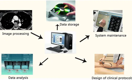
Establishment of a three-dimensional digital model database of inguinal compound tissue flaps with multiple perforating artery branches
Methods for establishing a three-dimensional digital model database of inguinal compound
tissue flaps with multiple perforating artery branches were as follows [6]. First, continuous
enhanced computed tomography (CT) images of the upper limbs were imported into Amira software (Visage Imaging CO.). The threshold was adjusted through the threshold slider on an isosurface and inguinal compound tissue
flaps were reconstructed on an isosurface. Directions and locations of perforating vessels of the compound tissue flaps were observed
and recorded. Next, pixel allocation, noise reduction, and smoothness levels of the
mate-rial image were sequentially performed. Image
color was then adjusted. Image data was
parti-tioned and connected with existing data. Data
sets were input into the software for compound
tissue flap reconstruction, observing the ana -tomic relationship between compound tissue
flaps and perforating vessels in multi-direction -al and stereoscopic ways. Relevant data was applied to surface or volume reconstruction of
hyalinized skin and subcutaneous tissues.
Cut-ting was performed according to the direction of the perforating vessels and the general shape of soft tissue defects. At the same time, the software was written in C++ language.
Database establishment adopted the embed
-ded database Vista DB 2.1. SQL 92 compatible v-SQL was used to develop the custom search engine, meeting the requirements of expansion to T-SQL in the future. After the database was
established, each module was debugged. Ap- plication testing included the stability of the running database, whether the items of
data-base design meet the requirements of clinical
data of surgical treatment of inguinal
com-pound tissue flaps with multiple perforating artery branches, and the flexibility of custom fields. According to application testing, each module and the overall database worked
sm-oothly, indicating that database establishment was completed (Figure 1).
Therapeutic methods
Patients in the control group were treated
with inguinal flap transplantation [6]. Primary
debridement of damaged tissues was carried out after local anesthesia. According to patient
tissue wounds, the wound area of skin soft tis -sue was assessed. Incisions were performed
from the inguinal skin to deep fascia. The flap
was cut off in the midline of the rectus
abdomi-nis. The flap area was slightly larger than the
wound area. Attention was paid to reduce
ner-vous system injuries. Postoperative inflamma -tion and infec-tions were controlled. Patients in the observation group were treated with
three-dimensional digital model-assisted inguinal
jected with 80 mL of iohexol, as as low-osmolar contrast agent, CT scans were performed. CT scan data was imported into the database management system. Vascular routes and mu-
tual relationships were confirmed through CT film analysis. After the three-dimensional model database of inguinal compound tissue flaps was debugged and verified, the most relevant
data points and characteristics of a three-dimensional model in the database were ex- tracted automatically with the combination of
patient CT image features. This was done to fit
out a most suitable perforating vessel model for patients. The model optimized surgical pl- ans to improve success rates of
transplanta-tion of tissue flaps. Suitable room tempera -tures were maintained in the operating room to prevent patients from spasming during surgery.
Dezocine or propofol was administered to con -trol pain and maintain sedation. Patients were prohibited from exercising to avoid injury at the
scar hyperplasia in local skin, and a little pig -mentation; 3) General: Patients had scar reduc-tion by about a half and mild pigmentareduc-tion; 4) Poor: Patients had many scars and obvious pig-mentation [7]. The excellent and good rate of cosmetic results = (the number of excellent rate + the number of good rate)/total number of patients * 100%.
Statistical analysis
SPSS 21.0 was used to analyze data. Me- asurement data are expressed as mean ± standard deviation. Student’s t-test was appli- ed to detect between-group differences. Enu- meration data are shown as percentages.
Chi-squared test was also applied. Comparison of
enumeration data for less than 40 patients
was performed using Fisher’s exact probabi-lity. P<0.05 indicates statistically significant
[image:3.612.90.371.71.242.2]differences.
Figure 1. Establishment of a three-dimensional digital model of inguinal compound tissue flaps with multiple perforating artery branches and data-base development.
Table 1. Analysis of general clinical data
Group Control (n=34) Observation (n=34) t/χ2 P
Gender (male/female) 19/15 16/18 2.146 0.085 Age (years) 35.3±6.9 36.8±7.4 1.592 0.097
Weight (kg) 62.6±5.3 64.1±4.7 3.146 0.072
Flap area (cm2) 78.4±5.8 85.6±6.2 2.459 0.086
Long-term smoking (Yes/No) 15/19 13/21 2.715 0.081
Long-term drinking (Yes/No) 11/23 10/24 1.739 0.092 Average follow-up time (months) 15.1 14.8 3.267 0.069
surgical site. If the patient felt intense pain after surgery, an- algesics were administered. Outcome measurements and therapeutic effect evaluation
Main outcome measurements
were flap area and flap healing
times. Treatment effects were
classified into three degrees,
including excellence, improve-ment, and failure. Criteria of
excellence: Transplanted flaps survived and limb flexion and
extension function were nor-mal [6]; Criteria of improve-ment: Part of the transplanted
flaps survived and limb flexion
and extension function recov-ered, to some degree; Criteria of failure: Treatment failed. Total effective rate = (the ber of excellence + the num-ber of improvement)/total nu- mber of patients * 100%. Ev- aluation of cosmetic results was as follows: 1) Excellent:
Patients had smooth skin
[image:3.612.93.374.318.435.2]Results
General data
There were no significant differences in gender, age, weight, flap area, term smoking, long-term drinking, and average follow-up times
between the two groups (all P>0.05, Table 1). Treatment effects
In the observation group, there were 27 pa-
tients (79.5%) with excellent effects, signifi -cantly more than those in the control group (P<0.05). There were 6 patients (17.6%) with improved effects and 1 patient (2.9%) with
failed treatment, significantly less than those
in the control group (both P<0.05). In the obser-vation group, there were 33 patients (97.1%)
metic results, significantly more than those in
the control group (P<0.05). There were 16 patients (47.1%) with good cosmetic results,
showing no significant differences between the
two groups (P>0.05). There were 3 patients (8.8%) with general cosmetic results and 2 patients (5.9%) with poor cosmetic results,
sig-nificantly less than those in the control group
(both P<0.05). In the observation group, there were 29 patients (85.3%) with excellent and
good cosmetic results, significantly more than
those in the control group (P<0.05, Table 3). Complications
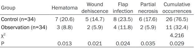
There were 3 (8.8%) patients with hematomas, 2 (5.9%) patients with wound dehiscence, 4
(11.8%) patients with flap infections, and 2
[image:4.612.89.384.92.342.2](5.9%) patients with partial necrosis in the Table 2. Comparison of clinical efficacy between the two groups (n,
%)
Group Excellence Improvement Failure Total effective rate
Control (n=34) 12 (35.3) 15 (44.1) 7 (20.6) 27 (79.4) Observation (n=34) 27 (79.5) 6 (17.6) 1 (2.9) 33 (97.1)
χ2 6.237 6.428
P 0.034 0.017 0.027 0.031
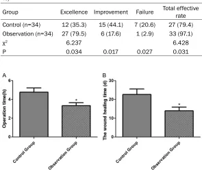
[image:4.612.90.385.411.486.2]Figure 2. Operation times and wound healing times. Compared with the control group, *P<0.05.
Table 3. Cosmetic results of wounds (n, %)
Group Excellent Good General Poor Excellent and good rate Control (n=34) 4 (11.8) 14 (41.2) 10 (29.4) 6 (17.6) 18 (53.0) Observation (n=34) 13 (38.2) 16 (47.1) 3 (8.8) 2 (5.9) 29 (85.3)
χ2 5.647
P 0.012 0.404 0.031 0.042 0.026
with effective treatment, si-
gnificantly more than tho- se in the control group (P< 0.05, Table 2).
Surgery related indicators
Perforating vessel-related information collected in the observation group (includ-ing the number of perforat-ing vessels, diameter, and starting location and direc-tion of the vessel) was basi-cally consistent with rele-vant data found during the operation. Accuracy levels of the collected data were
significantly better than tho-se in the control group.
De-grees of damage in the do- nor site in the observation group were lower than tho- se in the control group. Operation times (3.4±0.2
h) and flap healing times
(14.7±2.6 days) in the ob- servation group were signi-
ficantly less than those in
the control group (all P< 0.05, Figure 2).
Cosmetic results of wounds
cos-observation group, significantly less than
tho-se in the control group (all P<0.05). A total of
11 patients (32.4%) had complications, signifi -cantly less than those in the control group (P<0.05, Table 4).
Discussion
Compound tissue flaps with multiple perforat -ing artery branch transplantation has been a focus of plastic surgery [6]. Anatomy-related problems have also been proposed. Perforating
flaps and compound tissue flaps are new devel -opments in microsurgery. They are in confor-mity with the principle of tissue transplanta-tion, “effective repair and reconstruction on the recipient area leads to little damage on the donor site” [7, 8]. Clinical studies have found that the distribution of blood vessels in differ-ent humans may be extremely differdiffer-ent. Al- though distribution has a certain regularity, it
is not suitable for all patients. During the oper -ation, it is necessary to change the operation plan or abandon the operation for some pa- tients due to different blood vessel distribu-tion. Thus, many operations will fail due to
insufficient preparation [9, 10]. Therefore, it
is of great value to explore the application of three-dimensional digital model-assisted
ingui-nal flap transplantation for upper limb trauma.
In this study, a database of three-dimensional
digital model of inguinal compound tissue flaps
with multiple perforating artery branches was established with the combination of modern medical imaging, computer graphics and image processing, computational medicine, modern clinical anatomy, and osteology. Some studies have suggested that the introduction of digital medical technology into treatment of upper limb traumas can enhance the reliability, repro-ducibility, and traceability of surgery, improving clinical treatment effects [11, 12]. Some stud-ies have found that database technology can realize systematic and standardized
manage-needs of clinical work and research [13-15]. In
the development process, programmers were assisted by application personnel in
complet-ing a series work of design, development,
de-bugging, and improvement. Component-based software development was adopted, improving
the speed and quality of database develop
-ment, due to the large work quantity and short
construction period for the database [16, 17]. In the observation group, there were a total of 33 patients (97.1%) with effective treatment.
Operation time was 3.4±0.2 h and flap healing
time was 14.7±2.6 days. These values are
sig-nificantly better than those in the control group,
indicating that three-dimensional digital
model-assisted inguinal flap transplantation has good therapeutic effects and significantly improves postoperative upper limb function. Due to the
complex structure of muscles, blood vessels, and nerves of the human body, some patients
with severe skin and muscle tissue damage are difficult to treat. Three-dimensional digital mod
-el-assisted inguinal flap transplantation can
effectively assist physicians in assessing trau-matic wound features. This will help in deve- loping individualized surgical plans for patients,
significantly improving clinical treatment effects
[18]. One study, examining 52 upper limb
trau-ma patients with inguinal flap transplantation, found that patients had large flap areas, long
wound healing times, and poor treatment ef- fects of upper limb trauma. Results suggested
that using inguinal flap transplantation, alone,
could not accurately grasp the characteristics of damaged tissue, affecting patient treatment [19, 20]. Other studies have found that inguinal
[image:5.612.91.404.85.163.2]compound tissue flaps with multiple perforat -ing artery branches after three-dimensional reconstruction can be displayed transparently or in any combination. The three-dimensional tissue structure shows a clear overall structure with strong perception of solidity from diffe- rent angles. The relationship between tissues Table 4. Complications (n, %)
Group Hematoma dehiscenceWound infectionFlap necrosisPartial occurrencesCumulative
Control (n=34) 7 (20.6) 5 (14.7) 8 (23.5) 6 (17.6) 26 (76.5)
Observation (n=34) 3 (8.8) 2 (5.9) 4 (11.8) 2 (5.9) 11 (32.4)
χ2 4.216
P 0.013 0.021 0.024 0.035 0.029
ment of the three-di- mensional digital model of inguinal compound
is clear, at a glance, which is conducive to clini-cal treatment [21, 22].
In this study, there were 29 patients (85.3%) with excellent and good cosmetic results in the
observation group. This number was signifi -cantly larger than that in the control group, in- dicating that three-dimensional digital
model-assisted inguinal flap transplantation could
im-prove cosmetic results of patients and have
great effects on quality of life. A study found
that 58 patients with upper limb trauma, after three-dimensional digital model-assisted
ingui-nal flap transplantation, had up to 81.4% of
excellent and good rate of cosmetic results.
This was significantly higher than that after inguinal flap transplantation [23]. This is con
-sistent with present findings. In the current
study, a total of 11 patients (32.4%) had
com-plications in the observation group, significant -ly less than those in the control group. This indicates that three-dimensional digital
model-assisted inguinal flap transplantation could
effectively improve treatment and reduce inci-dence of complications. However, the three-dimensional digital model database of inguinal
compound tissue flaps with multiple perforat -ing artery branches established in this study has a short clinical application time. It is neces-sary to further improve and standardize opera-tion standards and procedures of the data-
base to meet the requirements for clinical
treatment.
In summary, three-dimensional digital
model-assisted inguinal flap transplantation shows a
three-dimensional dynamic anatomic structure
of normal anterolateral thigh flaps, vividly, pro -viding a visualized digital anatomic basis for
preoperative flap design. It effectively improves
treatment effects for patients with upper limb trauma. It also improves cosmetic results, sh- owing good clinical application value.
Acknowledgements
This work was supported by the Medical
He-alth Science and Technology Project of Zhe- jiang Provincial Health Commission (No. 2015- KYA160).
Disclosure of conflict of interest
None.
Address correspondence to: Yiheng Chen, De- partment of Hand and Plastic Surgery, The Second Affiliated Hospital and Yuying Children’s Hospital of Wenzhou Medical University, No. 109 Xueyuan West Road, Wenzhou 325027, Zhejiang Province, China. Tel: +86-0577-88879012; Fax: +86-0577-88879- 012; E-mail: [email protected]
References
[1] Kotick JD, Sandelin RS and Klein RD. Deep in-ferior epigastric perforator free flaps for use in complicated groin wound repair: a case report of severe groin scar contracture and review of pedicled and free flaps in groin wound repair. J Hand Microsurg 2017; 9: 101-106.
[2] Seo BF, Choi JY, Han HH, Oh DH, Rhie JW, Ahn ST and Moon SH. Perforators as recipients for free flap reconstruction of the inguinal and perineal region. Microsurgery 2015; 35: 627-633.
[3] Ray MD, Garg PK, Jakhetiya A, Kumar S and Pandey D. Modified skin bridge technique for ilio-inguinal lymph node dissection: a forgotten technique revisited. World J Methodol 2016; 6: 187-189.
[4] Westerman ME, Tausch TJ, Zhao LC, Siegel JA, Starke N, Klein AK and Morey AF. Ventral slit scrotal flap: a new outpatient surgical option for reconstruction of adult buried penis syn-drome. Urology 2015; 85: 1501-1504. [5] Zeltzer AA, Anzarut A, Braeckmans D,
Seiden-stuecker K, Hendrickx B, Van Hedent E and Hamdi M. The vascularized groin lymph node flap (VGLN): anatomical study and flap plan-ning using multi-detector CT scanner. The gold-en triangle for flap harvesting. J Surg Oncol 2017; 116: 378-383.
[6] Feng S, Xi W, Zhang Z, Tremp M, Schaefer DJ, Sadigh PL, Zhang W and Zhang YX. A reap-praisal of the surgical planning of the superfi-cial circumflex iliac artery perforator flap. J Plast Reconstr Aesthet Surg 2017; 70: 469-477.
[7] Scaglioni MF, Enrique Carrillo Jimenez L, Kuo YR and Chen YC. Pedicled posteromedial thigh (PMT) flap: a new alternative for groin defect reconstruction. Microsurgery 2017; 37: 339-343.
[8] Chao WN, Wang PH, Chen BR and Chen SC. Chimeric groin free flaps: design and clinical application. Microsurgery 2016; 36: 206-215. [9] Scaglioni MF and Suami H. Lymphatic anatomy
of the inguinal region in aid of vascularized lymph node flap harvesting. J Plast Reconstr Aesthet Surg 2015; 68: 419-427.
per-forator (TDAP) flap versus split skin graft. J Plast Reconstr Aesthet Surg 2014; 67: 1118-1124.
[11] Ding J, Su Y, Liu S, Yang Y, Zhou B, Yu Z, Xiao B and Guo S. A mouse model of vascularized skin transplantation. Ann Plast Surg 2017; 78: 576-581.
[12] Miyamoto S, Fujiki M, Nakatani F, Kobayashi E, Sakisaka M and Sakuraba M. Reconstruction of complex groin defects after sarcoma resec-tion. Ann Plast Surg 2017; 78: 443-447. [13] Hummelink S, Schultze Kool LJ and Ulrich DJ.
Displaying inguinal lymph nodes before trans-plantation in a deep inferior epigastric perfora-tor flap breast reconstruction using an innova-tive projection method. J Plast Reconstr Aesthet Surg 2016; 69: 376-380.
[14] Jia Y, Xu J, Kang Q, Zhang C and Chai Y. Re-verse-flow lateral tarsal island flap for covering the great toe donor site of wraparound flap. Ann Plast Surg 2016; 77: 445-449.
[15] Liu HL, Pang SY, Lee CC, Wong MM, Chung HP and Chan YW. Orthotopic transfer of vascular-ized groin lymph node flap in the treatment of breast cancer-related lymphedema: clinical re-sults, lymphoscintigraphy findings, and pro-posed mechanism. J Plast Reconstr Aesthet Surg 2018; 71: 1033-1040.
[16] Gupta MK, Patel AP and Master VA. Technical considerations to minimize complications of inguinal lymph node dissection. Transl Androl Urol 2017; 6: 820-825.
[17] Mukai Y, Watanabe T, Sugimoto M, Kimata Y and Namba Y. Vaginoplasty with a pudendal-groin flap in male-to-female transsexuals. Acta Med Okayama 2017; 71: 399-405.
[18] Chatterjee A, Kosowski T, Pyfer B, Fisher CS, Tchou JC and Maddali S. A cost-utility analysis comparing the sartorius versus the rectus fem-oris flap in the treatment of the infected vascu-lar groin graft wound. Plast Reconstr Surg 2015; 135: 1707-1714.
[19] Al-Qattan MM and Al-Qattan AM. Defining the indications of pedicled groin and abdominal flaps in hand reconstruction in the current mi-crosurgery Era. J Hand Surg Am 2016; 41: 917-927.
[20] Tong D, Liu Y, Wu LW, Zhu S, Zhu J and Chen S. Free groin flap for aesthetic and functional do-nor-site closure of the anterolateral thigh flap. J Plast Reconstr Aesthet Surg 2016; 69: 1116-1120.
[21] Zelken JA, AlDeek NF, Hsu CC, Chang NJ, Lin CH and Lin CH. Algorithmic approach to lower abdominal, perineal, and groin reconstruction using anterolateral thigh flaps. Microsurgery 2016; 36: 104-114.
[22] Sato S, Nakamura Y, Teramoto Y, Yeh YW, Maruyama H, Nakamura Y, Fujisawa Y, Fujimo-to M and YamamoFujimo-to A. A novel approach for inguinal lymph node dissection without ingui-nal skin incision for invasive extramammary paget disease. Dermatol Ther 2015; 28: 351-354.