Original Article
Long non-coding RNA growth arrest-specific transcript 5
(GAS5) protects ovarian cancer cells from apoptosis
Jiayin Gao1, Beidi Wang1, Min Mao2, Song Zhang3, Meiling Sun4, Peiling Li1
1Department of Obstetrics and Gynecology, The Second Affiliated Hospital of Harbin Medical University, Harbin, China; 2Department of Biopharmaceutical Sciences, College of Pharmacy, Harbin Medical University (Daqing), Daqing, China; 3Department of Medical Service, The First Affiliated Hospital of Harbin Medical University, Harbin, China; 4Department of Nursing, The Second Affiliated Hospital of Harbin Medical University, Harbin, China Received March 16, 2016; Accepted July 12, 2016; Epub September 1, 2016; Published September 15, 2016
Abstract: Epithelial ovarian cancer (EOC) is a main cause of death in malignant tumor of women genital system. This study aims to investigate the underlying role of growth arrest-specific transcript 5 (GAS5) in EOC. In vivo expression of GAS5 in 60 EOC specimens was evaluated by quantitative reverse transcription QRT-PCR, which used to study the differences of GAS5 expression between EOC tissues and normal ovarian epithelium. There were no significant differences of GAS5 expression between normal ovarian epithelium and benign epithelial lesions; however, GAS5 expression was lower in EOC tissues compared with normal ovarian epithelial tissues (6.44-fold), which was closely related to lymph node metastasis (P=0.025) and tumor node metastasis stage (P=0.035). Moreover, exogenous GAS5-inhibited proliferation promoted apoptosis and decreased migration and invasion in ovarian cancer cells. Finally, through Western blot analysis, overexpression of GAS5 protein could decrease the expression of Cyclin A, Cyclin D, Cyclin E, and PCNA. Conclusions: This study revealed that GAS5 is down-regulated in EOC specimens, and GAS5 inhibits EOC cell proliferation, migration, and invasion, and promotes the cell apoptosis. GAS5 can serve as a novel therapeutic target in patients with EOC.
Keywords: Long non-coding RNA, epithelial ovarian cancer, GAS5, apoptosis, mitochondrion
Introduction
Ovarian cancer is the main cause of death in malignant tumor of women genital system, which owns a high morbidity in developing countries. Although, rapid progress in diagno-sis and treatment of ovarian cancer [1], it is still the fifth major cause of death in women cancer patients. Of all ovarian carcinoma cases, Epi- thelial ovarian cancer (EOC) accounts for 90% of morbidity [2]. Moreover, EOC can spread to peritoneal cavity via peritoneal fluid, leading to the inefficiency of surgery and chemotherapy treatment. Thus, a better understanding of the mechanisms involved in EOC and more effec-tive therapeutic approaches are urgently need-ed. It is also reported that the loss of function proteins such as, Kras, Brca1/2, Tp53, Rb, and PTEN will induce epithelial ovarian cancer [3-5]. Long non-coding RNA (lncRNA) is longer than 200 nucleotides but does not translate into
recep-tors and promotes cells apoptosis [18], which had been originally identified in leukemia cells and NIH3T3 cells [19]. It is also reported that the inhibition of mammalian target of rapamy-cin (mTOR) pathway via rapamyrapamy-cin depends on GAS5 [20]. GAS5 negatively regulates miR-21 possibly through the RNA-induced silencing complex [21]. Additionally, it had also shown that GAS5 is down-regulated in some cancer cell lines and tissues [22-25]. Some research showed GAS5 inhibited malignant pleural me- sothelioma (MPM) cell growth by inhibiting hedgehog and PI3K/mTOR signal pathway in MPM [24]. GAS5 as a tumor suppressor in non-small-cell lung cancer (NSCLC) was mediated by p53-independent and p53-dependent path-ways [26]. Furthermore, GAS5 could inhibit E2F1 and cyclin D1 and thus leads to decreased gastric cancer cell proliferation [25]. However, the role of GAS5 in ovarian cancer is not known up to now.
The fundamental mechanisms of GAS5 on tumorigenesis remain largely unknown, despite GAS5 has been indicated to take part in sup-pression on malignant tumor. The potential mechanisms can be related to the fact that GAS5 regulates nonsense-mediated RNA decay pathway [20, 27] or down-regulates c-Myc [28]. These findings provide strong evidence that GAS5 plays an important role in guiding the cell fate toward inhibiting proliferation.
Signaling pathways in cancer cell biology are increasingly being used to investigate the mechanisms underlying tumor relapse and dor-mant behavior in many tumors [29, 30]. some evidences suggest that the MAPK pathway plays an important role in several tumors [31]. The ERK signaling cascade is one of most extensively studied MAPK pathways, hyperacti-vation of which has been demonstrated to play major roles in tumor cell proliferation and metastasis. while clinical and experimental evi-dence is consistent with most negative regula-tors of the ERK pathway being tumor suppres-sors [32]. The potential role of ERK pathway- dependent proliferation pathway in the prolif-eration effect of GAS5 has not been explored. In our study, we demonstrated a significant decrease in the expression of GAS5 in EOC tis-sues, which was associated with clinicopatho-logical parameters. Moreover, the overexpres-sion of GAS5 obviously inhibited the proliferation
of ovarian cancer cell lines. Together, these results reveal an evidence for proliferation reg-ulation between lncRNA GAS5 and ovarian can-cer, indicating a potential target of diagnosis and gene therapy in the disease.
Materials and methods
Cell lines and tissue samples
The human ovarian cancer HO8910 (Bioleaf Biotech Co., Ltd, Shanghai, China) and A2780 cells (Chuanbo Biotechnology Co., Ltd, Nanjing, China) were grown in RPMI-1640 (Nyclone, Beijing, China) and DMEM medium (Nyclone, Beijing, China), respectively, supplemented with 10% fetal bovine serum (FBS) (Sijiqing, Zhejiang, China) in a humidified incubator (37°C, 5% CO2).
Specimens of EOC tissue (n=60, without radia-tion or chemotherapy), normal ovarian epitheli-al tissues (n=13) and benign ovarian epitheliepitheli-al lesions (n=10) were collected at the Second Affiliated Hospital of Harbin Medical University (China) between Mar 2012 and Apr 2014 (median age years 55, range 37-79). The sam-ples of normal ovarian tissue were collected from hysterosalpingo-oophorectomy following uterine myoma, endometriosis, or adenomyo-sis. Written informed consent was obtained from all participants. The study was approved by the Human Ethnics Committee of the Second Affiliated Hospital of Harbin Medical University (NO.: 2015-yan-167).
Real-time polymerase chain reaction
18S rRNA (GenePharma, Shanghai, China; for-ward primer: 5’-TTTGACTCAACACGGGAAACC-3’; reverse primer: 5’-CACGGAATCGAGAAAGAGC- TATC-3’; probe: CCGGACACGGACAGGATTGACA- GAT) served as the endogenous control. All of the real-time PCRs were performed in triplicate. The relative quantification of GAS5 expression was calculated using the 2-ΔΔCT method relative to 18s rRNA.
Transfection
To overexpress GAS5, plasmid pCDNA-GAS5 was constructed by Shanghai GenePharma Co., Ltd of China containing the whole genome sequence of GAS5 (NCBI Reference Sequence: NR_002578.2). The plasmid carrying pCDNA-GAS5 (1 μg/μl) was transfected into ovarian cancer cell lines by using Lipofectamine 2000 (Invitrogen, CA, USA). (Lipofectamine 2000: pCDNA-GAS5=1:1.4).
Terminal deoxynucleotidyl transferase dUTP nick-end labeling (TUNEL)
The HO8910 and A2780 cells were transfected with pCDNA-GAS5 or empty vector, and cul-tured in six-well plates for 48 h. They were then fixed with 4% paraformaldehyde solution, the apoptotic cells were labeled using the in situ cell death detection kit (Beyotime, Shanghai, China), the fluorescence was detected by using a Nikon ECLIPSE TE2000-S fluorescence micro -scope (Nikon, Tokyo, Japan) and counted by Image Pro-Plus 6.0. (Media Cybernetics, MD, USA) in three different experiments, the red fluorescence marked cells were served as apoptotic cells.
Cell apoptotic analysis
HO8910 and A2780 cells (1~2 × 10⁵) were treated with a pcDNA-GAS5 or an empty vector; then, these were placed in 6-well plates. After 24-h incubation, the cells were trypsinized and then fixed in 70% ethanol for 3 h at 4°C; 3 h later, the cells were incubated with propidium iodide (20 ×) and RNase-A (50 ×) for 30 min in the dark. Cells were collected and analyzed for apoptosis using a flow cytometer (BD Bio-sciences, NJ, USA) after propidium iodide stain -ing. The results were analyzed by BD Accuri C6 software. The experiments were repeated at least thrice.
Transwell assay
Transwell assays were performed using a Costar chamber (Corning Costar Corp, MA, USA). The bottom chambers were filled with a culture medium containing 10% FBS. Different samples of tansfected HO8910 or A2780 cells (5 × 10⁴) were suspended in a culture medium without FBS, and then the cells were seeded into the upper chambers. The transwell cham-ber containing an 8-μm pore size polycarbon -ate membrane filter was co-ated either with (for invasion) or without (for migration) matrigel. After 48 h of culture at 37°C, the upper layer of cells were removed before visualization, and the cells on the lower surface were fixed and stained with 0.5% crystal violet. The cells were counted by Image Pro-Plus 6.0. (Media Cybe- rnetics, MD, USA) in five random fields and pho -tographed. The experiments were performed three times.
Cell proliferation and viability
Cell proliferation and viability of HO8910 and A2780 were evaluated using 3-[4,5-dimethyl-thiazol-2-yl]-2,5-diphenyl-tetrazoliumromide (MTT) (Amresco, OH, USA) assay. Briefly, after 6 h transfection with pCDNA-GAS5, about 5 × 10³ cells per well were seeded in a 96-well plate at 37°C. Each well was repeated six times. After further incubation with different times (24 h, 48 h, and 72 h), 20 μl MTT (0.5 mg/ml) was added to each well and further incubated for 4 h. Then the medium was removed and dimethylsulfoxide was added to dissolve the MTT formazan crystals. The cell viability and proliferation were determined by OD450 value. The experiments were performed three times.
Colony formation assay
Western blot analysis
The pCDNA-GAS5- or empty vector-transfected HO8910 and A2780 cells were lysed using a lysis buffer (Beyotime, Shanghai, China) that contained the phenylmethanesulfonyl fluoride. The protein concentrations were determined by
Statistical analysis
All statistical analysis was performed with SPSS 17.0 (Chicago, IL, USA). The data were presented as means ± standard error of mean from at least three independent experiments. Statistical analysis was performed using
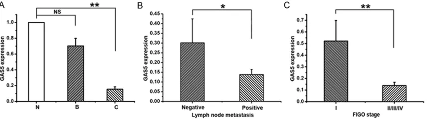
[image:4.612.94.522.73.194.2]chi-Figure 1. Relative lncRNA GAS5 expression in EOC tissues and the clinical significance. Relative expression of ln -cRNA GAS5 was analyzed by real-time PCR in ovarian normal epithelial tissues (N, n=13), ovarian benign epithelial lesions (B, n=10), and epithelial ovarian cancers (C, n=60). LncRNA GAS5 expression was normalized to 18s r-RNA expression. The data are presented as a fold-change in the tumor tissues relative to the normal tissues. B. The cor-relation between GAS5 and lymph node metastasis. Expression of lncRNA GAS5 was significantly lower in patients with lymph node metastasis than those without lymph node metastasis. C. The correlation between GAS5 expres-sion and clinical stage. Expresexpres-sion of lncRNA GAS5 was significantly lower in patients with an advanced clinical stage than those with an early stage. All values are denoted as means ± SEM. from at least three separate experi-ments. *P<0.05, **P<0.01. *P<0.05 represents negative lymph node metastasis compared with positive lymph node metastasis, **P<0.01 represents normal ovarian epithelium compared with EOC tissues and FIGO stage of I compared with II/III/IV.
Table 1. Correlation between GAS5 expression and clinico-pathological parameters of EOC
Relative GAS5 expression
Clinicopathological parameters No. of cases Low High P-valuea
Age (years) 0.381
≤50 22 13 9
>50 38 18 20
Differentiation 0.944
Well, moderate 26 14 12
Poor 34 18 16
Location 0.965
Unilateral 25 13 12
Bilateral 35 18 17
Lymph node metastasis 0.025b
Positive 40 28 12
Negative 20 8 12
FIGO stage 0.035b
I 15 5 10
II/III/IV 45 29 16
aChi-squared test; bP<0.05. GAS5, growth arrest-specific transcript 5; EOC, epithelial ovarian cancer.
square test, Student t test, or one-way analysis of variance followed by a post hoc test, where appropriate. Correlaction were estimated by Pearson’s correlation analysis. Differences were considered to be significant at P<0.05.
Results
GAS5 is down-regulated in EOC tissues
To investigate the expression of lncRNA GAS5 in normal and EOC patients, real-time PCR was used to examine the GAS5 expression in nor-mal ovarian epithelial tissues, benign ovarian epithelial lesions, and EOCs. The expression of GAS5 in normal ovarian epithelial tissues (N) and benign epithelial lesions (B), showed no statistical significance (Figure 1A). While, there appears to be a significant reduction of GAS5 expression in EOCs (C) compared with normal ovarian epithelial tissues (6.44-fold) (Figure 1A). Then, the clinicopathological parameters,
such as age, differentiation, location, lymph node metastasis, and FIGO stage, were ana-lyzed to assess the expression of GAS5 and clinical significance of EOC. As shown in Figure 1B, 1C, and Table 1, samples with lymph node metastasis and advanced FIGO stage owned the lower expression of GAS5. These data indi-cate that the decreased expression of GAS5 is related to EOC.
Effect of GAS5 on apoptosis in ovarian cancer cell lines
[image:5.612.92.522.70.337.2]Apoptosis-resistant phenotype is the major fea-ture of cancer cells [33]. To study the role of GAS5 in apoptosis, A2780 and HO8910 cells transfected with pCDNA-GAS5 or empty vector were monitored using flow cytometry. As shown in Figure 2A-C, ectogenic GAS5 obviously pro-moted the apoptosis of the cells. Similarly, TUNEL staining showed that the number of apoptotic cells was more in the GAS5-over-
expressed cells than in the controls (Figure 2D-F). The results mentioned above suggest that GAS5 displays a critical function in pro-apoptosis of ovarian cancer cells.
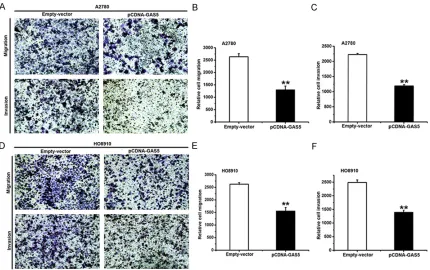
Effect of GAS5 on migration and invasion in ovarian cancer cell lines
Most ovarian cancer patients die of tumor metastasis, and there are a few research re- ports about the relationship between lncRNAs and neoplastic metastasis [34, 35]. To study the effect of GAS5 in vitro on migration and invasion of ovarian cancer cell lines, a Costar chamber without (for migration) or with (for invasion) matrigel was used. The treatment of A2780 and HO8910 cells is the same as before. Compared with the ones treated with empty vector, the ability of migration and invasion had been weakened in GAS5-overexpressed A2780
(Figure 3A-C) and HO8910 (Figure 3D-F) cells. Conclusively, GAS5 can inhibit migration and invasion in ovarian cancer cells.
Effect of GAS5 on proliferation in ovarian cancer cell lines
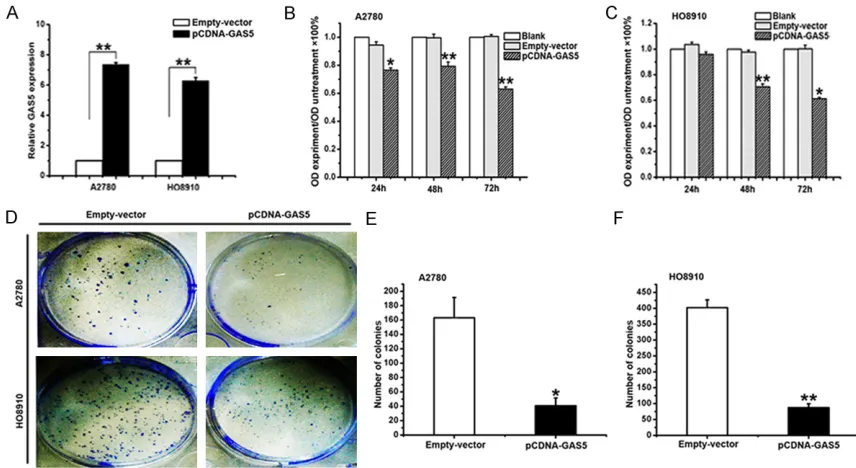
[image:6.612.93.523.71.341.2]With the aim of manipulating the GAS5 expres-sion in ovarian cancer cells, the pCDNA-GAS5 or an empty vector was transfected into A2780 and HO8910 cells. After 24 h of transfection, the level of GAS5 was well up-regulated in A2780 (74-fold) and HO8910 (63-fold) cells, respectively (Figure 4A). To ascertain the role of GAS5 in the proliferation of EOC, the overex-pression of A2780 and HO8910 cells of GAS5 were analyzed. MTT assay was used to assess the biological role of GAS5 in proliferation. Compared with the cells transfected with empty vector, the ones transfected with pCDNA-GAS5 demonstrated significantly decreased viability
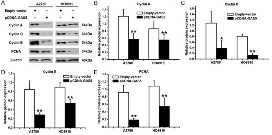
(Figure 4B and 4C). Besides, it was found that the overexpression of GAS5 greatly weakened the ability of colony forming using the colony formation assay (Figure 4D-F). It is well known that Cyclin A, Cyclin D, Cyclin E and PCNA are the symbols of proliferation, which were used to study the role in GAS5-inhibited proliferation of ovarian cancer cells. As shown in Figures 5A,
4B-E, there was an decrease in the expression of Cyclin A, Cyclin D, Cyclin E and PCNA in A2780 and HO8910 cells that were downex-pressed by the gene of GAS5. The results dem-onstrated that GAS5 could inhibit the prolifera-tion of ovarian cancer cells.
Discussion
Several important observations were demon-strated in this study. First, compared with thelial tissues of normal ovary and benign epi-thelial ovarian lesions, the depressed expre- ssion of GAS5 was detected in EOC. Meanwhile, GAS5 expression appeared to be significantly correlated to lymph node metastasis and FIGO
stage of EOC. Second, in vitro, the capability of apoptosis was strengthened, the ability of inva-sion and migration were weakened, and the ability of proliferation was decreased by GAS5 overexpression.
[image:7.612.89.517.72.306.2]GAS5 was found down-regulated in EOC tissues than in the normal ovarian epithelium, and lower GAS5 correlated with more transferred lymph nodes and advanced FIGO stage. Consistently, earlier studies showed that GAS5 was down-regulated in NSCLC compared with the adjacent normal lung tissues; more impor-tantly, lower GAS5 expression correlated with larger tumor size and advanced clinical stage [26]. Additionally, findings showed that GAS5 expression was markedly depressed in gastric cancer tissues, and the lower GAS5 mainly appeared in the larger tumor size-an advanced pathologic stage. Moreover, patients with lower GAS5 expression had poorer disease-free sur-vival and overall sursur-vival [25]. More recently, studies showed that the GAS5 expression was decreased in cervical cancer tissues than in
the adjacent normal tissues, and depressed GAS5 expression was correlated with the adv- anced FIGO stage, deeper invasion, and more lymph node metastasis. Patients with lower GAS5 expression had shown poorer overall sur-vival [36]. Taken together, GAS5 may serve as a tumor suppressor in human tumor.
Cell proliferation is a complex process that involves a variety of regulatory mechanisms and is closely related to tumorigenesis. The findings have indicated that GAS5 inhibits pro -liferation through significantly different intron or exon composition of these GAS5 transcripts [23]. Meanwhile, the current study found that GAS5 induced proliferation related proteins, Cyclin A, Cyclin D, Cyclin E and PCNA, (released more from the ovarian cancer cells) were expressed decreasingly by GAS5, indicating that the proliferation pathway is required for the effect of GAS5 in human ovarian cancer cells. Taken together, these findings prove that lncRNA GAS5 acts as a tumor suppressor in human EOC, However, there are some points that still need to be resolved: (1) more patients samples should be collected to verify the cred-ibility of GAS5 repression function in EOC, (2)
the interacting molecular mechanisms and signal pathways on how GAS5 changes the mitochondrion-independent apoptosis should be clarified, and (3) the mechanisms of decreased capabilities of migration and inva-sion by GAS5 in ovarian cancer cells should be explicated.
In conclusion, the findings of this study have shown that GAS5 overexpression can inhibit proliferation, promote apoptosis, and reduce migration and invasion in ovarian cancer cells. In addition, GAS5 expression is lower in EOC tis-sues than in the normal ovarian epithelium, and the lower expression of GAS5 is correlated to more lymph node metastasis and advanced FIGO stage of EOC, indicating that GAS5 may play a role as a suppressor of tumorigenesis for EOC patients. These findings have major impli -cations for devising a strategy to ovarian can-cer treatment target.
Acknowledgements
[image:8.612.90.524.73.288.2]This work was supported byHeilongjiang prov-ince science and technology research Found- ation (GA14C101-06).
Disclosure of conflict of interest
None.
Address correspondence to: Peiling Li, Department of Obstetrics and Gynecology, The Second Affiliated Hospital of Harbin Medical University, 148 Baojian Road, Harbin 150081, Heilongjiang Province, China. Tel: 86-451-86605343; Fax: 86-451-86605343; E-mail: [email protected]; Meiling Sun, Department of Nursing, The Second Affiliated Hospital of Harbin Medical University, 148 Baojian Road, Harbin 15-0081, Heilongjiang Province, China. Tel: 86-451-86605341; Fax: 86-451-86-451-86605341; E-mail: sm- [email protected]
References
[1] Siegel R, Naishadham D, Jemal A. Cancer sta-tistics. CA Cancer J Clin 2013; 63: 11-30. [2] Naora H. The heterogeneity of epithelial
ovari-an covari-ancers: reconciling old ovari-and new para-digms. Expert Rev Mol Med 2007; 9: 1-12. [3] Mullany LK, Fan HY, Liu Z, White LD, Marshall
A, Gunaratne P, Anderson ML, Creighton CJ, Xin L, Deavers M, Wong KK, Richards JS. Mo-lecular and functional characteristics of ovari-an surface epithelial cells trovari-ansformed by KrasG12D and loss of Pten in a mouse model in vivo. Oncogene 2011; 30: 3522-3536. [4] Quinn BA, Brake T, Hua X, Baxter-Jones K,
Lit-win S, Ellenson LH, Connolly DC. Induction of ovarian leiomyosarcomas in mice by condition-al inactivation of Brca1 and p53. PLoS One 2009; 4: e8404.
[5] Szabova L, Yin C, Bupp S, Guerin TM, Schlomer JJ, Householder DB, Baran ML, Yi M, Song Y, Sun W, McDunn JE, Martin PL, Van Dyke T, Difilippantonio S. Perturbation of Rb, p53, and Brca1 or Brca2 cooperate in inducing meta-static serous epithelial ovarian cancer. Cancer Res 2012; 72: 4141-4153.
[6] Gutschner T, Diederichs S. The hallmarks of cancer: a long non-coding RNA point of view. RNA Biol 2012; 9: 703-719.
[7] Clark MB, Johnston RL, Inostroza-Ponta M, Fox AH, Fortini E, Moscato P, Dinger ME, Mattick JS. Genome-wide analysis of long noncoding RNA stability. Genome Res 2012; 22: 885-898.
[8] Tee AE, Ling D, Nelson C, Atmadibrata B, Ding-er ME, Xu N, Mizukami T, Liu PY, Liu B, Cheung B, Pasquier E, Haber M, Norris MD, Suzuki T, Marshall GM, Liu T. The histone demethylase JMJD1A induces cell migration and invasion by up-regulating the expression of the long non-coding RNA MALAT1. Oncotarget 2014; 5: 1793-1804.
[9] Gutschner T, Hammerle M, Diederichs S. MALAT1 -- a paradigm for long noncoding RNA function in cancer. J Mol Med (Berl) 2013; 91: 791-801.
[10] Gutschner T, Hämmerle M, Eissmann M, Hsu J, Kim Y, Hung G, Revenko A, Arun G, Stentrup M, Gross M, Zörnig M, MacLeod AR, Spector DL, Diederichs S. The noncoding RNA MALAT1 is a critical regulator of the metastasis phenotype of lung cancer cells. Cancer Res 2013; 73: 1180-1189.
[11] Brown JA, Bulkley D, Wang J, Valenstein ML, Yario TA, Steitz TA, Steitz JA. Structural insights into the stabilization of MALAT1 noncoding RNA by a bipartite triple helix. Nat Struct Mol Biol 2014; 21: 633-640.
[12] Alaiyan B, Ilyayev N, Stojadinovic A, Izadjoo M, Roistacher M, Pavlov V, Tzivin V, Halle D, Pan H, Trink B, Gure AO, Nissan A. Differential expres-sion of colon cancer associated transcript1 (CCAT1) along the colonic adenoma-carcinoma sequence. BMC Cancer 2013; 13: 196. [13] Ling H, Spizzo R, Atlasi Y, Nicoloso M, Shimizu
M, Redis RS, Nishida N, Gafà R, Song J, Guo Z, Ivan C, Barbarotto E, De Vries I, Zhang X, Fer-racin M, Churchman M, van Galen JF, Beverloo BH, Shariati M, Haderk F, Estecio MR, Garcia-Manero G, Patijn GA, Gotley DC, Bhardwaj V, Shureiqi I, Sen S, Multani AS, Welsh J, Yama-moto K, Taniguchi I, Song MA, Gallinger S, Casey G, Thibodeau SN, Le Marchand L, Tiiri-kainen M, Mani SA, Zhang W, Davuluri RV, Mi-mori K, Mori M, Sieuwerts AM, Martens JW, Tomlinson I, Negrini M, Berindan-Neagoe I, Foekens JA, Hamilton SR, Lanza G, Kopetz S, Fodde R, Calin GA. CCAT2, a novel noncoding RNA mapping to 8q24, underlies metastatic progression and chromosomal instability in co-lon cancer. Genome Res 2013; 23: 1446-1461.
[14] Xiang JF, Yin QF, Chen T, Zhang Y, Zhang XO, Wu Z, Zhang S, Wang HB, Ge J, Lu X, Yang L, Chen LL. Human colorectal cancer-specific CCAT1-L lncRNA regulates long-range chromatin inter-actions at the MYC locus. Cell Res 2014; 24: 513-531.
[15] Pan W, Zhou L, Ge M, Zhang B, Yang X, Xiong X, Fu G, Zhang J, Nie X, Li H, Tang X, Wei J, Shao M, Zheng J, Yuan Q, Tan W, Wu C, Yang M, Lin D. Whole exome sequencing identifies lncRNA GAS8-AS1 and LPAR4 as novel papillary thy-roid carcinoma driver alternations. Hum Mol Genet 2016; 25: 1875-84.
[16] Raho G, Barone V, Rossi D, Philipson L, Sor-rentino V. The gas5 gene shows four alterna-tive splicing patterns without coding for a pro-tein. Gene 2000; 256: 13-17.
and a member of the 5’-terminal oligopyrimi-dine gene family reveals common features of snoRNA host genes. Mol Cell Biol 1998; 18: 6897-6909.
[18] Kino T, Hurt DE, Ichijo T, Nader N, Chrousos GP. Noncoding RNA gas5 is a growth arrest- and starvation-associated repressor of the gluco-corticoid receptor. Sci Signal 2010; 3: ra8. [19] Coccia EM, Cicala C, Charlesworth A, Ciccarelli
C, Rossi GB, Philipson L, Sorrentino V. Regula-tion and expression of a growth arrest-specific gene (gas5) during growth, differentiation, and development. Mol Cell Biol 1992; 12: 3514-3521.
[20] Fingar DC, Blenis J. Target of rapamycin (TOR): an integrator of nutrient and growth factor sig-nals and coordinator of cell growth and cell cy-cle progression. Oncogene 2004; 23: 3151-3171.
[21] Zhang Z, Zhu Z, Watabe K, Zhang X, Bai C, Xu M, Wu F, Mo YY. Negative regulation of lncRNA GAS5 by miR-21. Cell Death Differ 2013; 20: 1558-1568.
[22] Pickard MR, Mourtada-Maarabouni M, Wil-liams GT. Long non-coding RNA GAS5 regu-lates apoptosis in prostate cancer cell lines. Biochim Biophys Acta 2013; 1832: 1613-1623.
[23] Mourtada-Maarabouni M, Pickard MR, Hedge VL, Farzaneh F, Williams GT. GAS5, a non-pro-tein-coding RNA, controls apoptosis and is downregulated in breast cancer. Oncogene 2009; 28: 195-208.
[24] Renganathan A, Kresoja-Rakic J, Echeverry N, Ziltener G, Vrugt B, Opitz I, Stahel RA, Felley-Bosco E. GAS5 long non-coding RNA in malig-nant pleural mesothelioma. Mol Cancer 2014; 13: 119.
[25] Sun M, Jin FY, Xia R, Kong R, Li JH, Xu TP, Liu YW, Zhang EB, Liu XH1, De W. Decreased ex-pression of long noncoding RNA GAS5 indi-cates a poor prognosis and promotes cell pro-liferation in gastric cancer. BMC Cancer 2014; 14: 319.
[26] Shi X, Sun M, Liu H, Yao Y, Kong R, Chen F, Song Y. A Critical Role for the Long Non-Coding RNA GAS5 in Proliferation and Apoptosis in Non-Small-Cell Lung Cancer. Mol Carcinog 2015; Suppl 1: E1-E12.
[27] Hay N, Sonenberg N. Upstream and down -stream of mTOR. Genes Dev 2004; 18: 1926-1945.
[28] Huang FJ, Chan WH. Effects of ochratoxin a on mouse oocyte maturation and fertilization, and apoptosis during fetal development. Envi-ron Toxicol 2016; 31: 724-735.
[29] Oskarsson T, Batlle E, Massague J. Metastatic stem cells: sources, niches, and vital path-ways. Cell Stem Cell 2014; 14: 306-321. [30] Takebe N, Miele L, Harris PJ, Jeong W, Bando
H, Kahn M, Yang SX, Ivy SP. Targeting Notch, Hedgehog, and Wnt pathways in cancer stem cells: clinical update. Nat Rev Clin Oncol 2015; 12: 445-464.
[31] Dhillon AS, Hagan S, Rath O, Kolch W. MAP ki-nase signalling pathways in cancer. Oncogene 2007; 26: 3279-3290.
[32] Murphy T, Hori S, Sewell J, Gnanapragasam VJ. Expression and functional role of negative sig-nalling regulators in tumour development and progression. Int J Cancer 2010; 127: 2491-2499.
[33] Meier P, Finch A, Evan G. Apoptosis in develop-ment. Nature 2000; 407: 796-801.
[34] Guttman M, Rinn JL. Modular regulatory prin-ciples of large non-coding RNAs. Nature 2012; 482: 339-346.
[35] Gupta RA, Shah N, Wang KC, Kim J, Horlings HM, Wong DJ, Tsai MC, Hung T, Argani P, Rinn JL, Wang Y, Brzoska P, Kong B, Li R, West RB, van de Vijver MJ, Sukumar S, Chang HY. Long non-coding RNA HOTAIR reprograms chroma-tin state to promote cancer metastasis. Nature 2010; 464: 1071-1076.