Original Article
Low expression of Dapper1 induces malignancy via the
Wnt signalling pathway and is associated with poor
prognosis in gastric cancer
Yang Yang1*, Hui Qiu2*, Kewei Jiang1, Jizhun Zhang3, Hongqing Zhuo3, Zhidong Gao1, Hui Zhang1, Peng Guo1,
Zhanlong Shen1, Xiaodong Yang1, Yingjiang Ye1, Shan Wang1
1Department of Gastroenterological Surgery, Laboratory of Surgical Oncology, People’s Hospital of Peking University, Peking University, Beijing, China; 2Department of Surgery, Cancer Hospital of Peking University, Beijing Institute for Cancer Research, Beijing, China; 3Department of Gastroenterological Surgery, Shandong Provincial Hospital Affiliated to Shandong University, Jinan, Shandong, China. *Equal contributors.
Received November 5, 2015; Accepted January 1, 2016; Epub February 1, 2016; Published February 15, 2016
Abstract: Gastric cancer is the second leading cause of cancer death and remains a major clinical challenge due to poor prognosis and limited treatment options. The Wnt signalling pathway is abnormally activated in gastric cancer
as well as many other malignancies. The aim of this study was to explore the clinical significance and prognostic
value of Dapper1 expression and its regulation of the Wnt signalling pathway in gastric cancer. Real-time PCR and immunohistochemistry were respectively performed to investigate the expression of DAPPER1 at mRNA and pro-tein level in both gastric cancer tissues and adjacent normal mucosa tissues. We found that DAPPER1 mRNA and
protein was commonly downregulated is gastric cancer tissues. The lower expression of DAPPER1 was significantly
correlated with tumor invasion, lymph-node metastasis, and TNM stage (P < 0.05). Survival analysis revealed that DAPPER1 is a prognostic predictor of gastric cancer (P = 0.046). Overexpression of DAPPER1 in SGC7901 cells
significantly inhibited cell proliferation while increasing apoptosis. Overexpression of DAPPER1 led to reduced ex
-pression of Dvl-2, β-catenin, survivin, and bcl-xl. Similarly, there was reduced ex-pression of survivin, the canonical
Wnt-signalling target gene. Finally, overexpressed DAPPER1 inhibited tumour growth of SGC7901 cells transplanted into athymic nude mice. In conclusion, our results suggest that downregulation of Dapper1 gene expression in hu-man gastric carcinoma promotes tumour development through regulating the canonical Wnt-signalling pathway.
Keywords: Gastric neoplasms, Dapper1, Wnt signalling pathway, prognosis
Introduction
Gastric cancer is a leading cause of cancer-related mortality worldwide [1]. Like other malignancies, the development of gastric can-cer is a multi-step process involving aberrant signal transduction. In particular, the Wnt sig-nalling pathway is abnormally activated in gas-tric cancer as well as many other malignancies [2]. Dishevelled (Dvl) is a central mediator of Wnt signalling in both the canonical and nonca-nonical pathways. It inhibits glycogen synthase kinase 3β-induced degradation of β-catenin. Dapper1 (Dpr1) is a Dvl-interacting protein that negatively regulates canonical Wnt signalling and is required for notochord formation [3, 4]. The human DAPPER1 gene is located in the genome at nucleotide position
compo-nents of the Wnt/β-catenin signalling pathway [7]. Human Dpr1 can inhibit Wnt signalling by promoting Dvl degradation in colorectal cancer [8, 9]. Dpr1 may play an important role in inhib-iting the development of malignant tumours and may become a new target for cancer diag-nosis and treatment. However, the expression profile of Dpr1 and its functional mechanism in gastric cancer have never been reported.
Materials and methods
Patients and tissues
Frozen specimens of gastric cancer and adja-cent normal mucosa were obtained prospec-tively from 30 consecutive patients undergoing surgical resection for gastric cancer at Peking University People’s Hospital in 2010. The sam-ples were immediately used for mRNA extrac-tion to measure DAPPER1 expression.
An additional group of 84 gastric-cancer pa- tients were confirmed by pathology and they underwent radical surgery at Peking University People’s Hospital from 2004 to 2006. No pre-operative chemotherapy or radiotherapy was administered. From these patients, paraffin-embedded specimens were obtained and were analysed for Dpr1 protein expression by immu-nohistochemistry. All patients were followed by direct evaluation or phone interview until death or June 2012.
This study was approved by the Human Ethics Committee of Peking University People’s Hospital. All patients provided informed con-sent, and no children were enrolled.
Reverse transcription polymerase chain reac-tion (RT-PCR)
Total RNA was extracted from liquid-nitrogen-frozen human tissue using Trizol. After chloro-form extraction, RNA was precipitated with iso-propanol. DNase I was used to remove genomic DNA contamination. cDNA was synthesized from the extracted RNA using Superscript II reverse transcriptase (GIBCO) at 42°C for 60 min. PCR conditions were as follows: 32 cycles of 94°C for 45 sec, 60°C for 45 sec, and 72°C for 45 sec. After PCR products were electropho-resed at 100 V for 30 min, a gel image was taken and analysed semi-quantitatively to determine the grey value.
Western blot
Whole-cell extracts from gastric cancer cell lines, BGC823, MGC803, and SGC7901, were mixed with 2× sample buffer and heated at 100°C for 5 min. Proteins (50 μg/lane) were separated on a 10% SDS-PAGE separating gel and 4% stacking gel and were transferred onto a nitrocellulose membrane. Membranes were blocked for 2 h in 2.5% Blotto pre-hybridization solution. After blocking, membranes were incu-bated with the appropriate primary antibody (Dpr1, Dvl-2, β-catenin, Survivin, Bcl-2, or Bcl-xl rabbit anti-human antibody) in 2.5% Blotto pre-hybridization solution at 4°C overnight. After washing the membranes three times with 2.5% Blotto pre-hybridization solution for 15 min each, they were incubated with horseradish peroxidase-conjugated secondary antibody in 2.5% Blotto pre-hybridization solution for 45 min. Subsequently, membranes were washed twice with Tris-buffered saline with Tween (TBS-T) and once with TBS and were developed using the enhanced chemiluminescence (ECL) detec-tion system. Quantitadetec-tion of the signal for Dpr1, Dvl-2, β-catenin, Survivin, 2, and Bcl-xl was performed using a Density Scanner (ZEISS automatic image analyser, VIDAS data analysis).
Immunohistochemistry
Human tissues embedded in paraffin wax were sectioned to a thickness of 4 mm. Slides were stained with primary antibody (rabbit anti-human Dpr1) using 3% H202 solution as a blocking agent. Slides were incubated at 4°C overnight in a 1:100 dilution of the primary anti-body and were washed three times with phos-phate-buffered saline (PBS) for 5 min each. Slides were incubated with horseradish peroxi-dase-conjugated secondary antibody and were developed with 3,3’-diaminobenzidine (DAB) solution.
Flow cytometry
ware, and differences were considered signifi-cant when P < 0.05. The Chi-square test was used to analyse the relationship between Dpr1 expression variation in tumour tissue and the clinicopathological parameters of 84 gas-tric-cancer patients. The Kaplan-Meier method was used to draw the survival curve of 84 gas-tric-cancer patients, and the Log-rank method was used to analyse the relationship between Dpr1 expression variation in tumour tissue and patient outcomes.
Results
DAPPER1 expression in gastric cancer tissues
DAPPER1 mRNA expression was significantly lower in tumour tissue than in adjacent normal mucosa from 17 of 30 gastric-cancer patients (Supplementary Figure 1). Dpr1 protein expres-sion was lower in tumour tissue than in adja-cent normal mucosa from 59 of 84 (70.2%) gastric-cancer patients. Immunohistochemistry results showed that Dpr1 protein was mainly localized in the cytoplasm, while the nucleus was free of expression (Supplementary Figure 2).
ml) and detected by flow cytometry. Arsenic trisulphide-induced acute promyelocytic leu-kaemia NB-4 apoptotic cells were used as a positive control.
Plasmid construct
Plasmids pcDNA3.1 (+) and pcDNA3.1-Dpr1 were gifts from Professor Chen Yeguang, Tsinghua University. Plasmid pcDNA3.1-Dpr1 was 7.9 kb in length and was comprised of a fragment of human DAPPER1 cDNA inserted into vector pcDNA3.1 (+) polyclonal site. pcDNA3.1 (+) is 5.4 kb in length. Its multiple clone site (MCS) was located at bases 895-1,010, which contained the XhoI and EcoRI restriction sites into which the DAPPER1 cDNA fragment was inserted. Sequences were con-firmed by restriction-enzyme digestion.
In vivo tumour xenograft study
Male athymic nude mice (6-week-old BALB-c/ nu/nu) were randomly assigned to three groups, the control group, empty-vector group, and experimental group (6 animals per group). The gastric cancer cell line SGC7901 was transfect-ed with plasmid pcDNA3.1 (+) or
pcDNA3.1-Dpr1. The cells (106) were
implanted subcutaneously in- to the bilateral forelimb axilla with a 26-gauge needle and 1-ml syringe. Mice were assessed at 1, 2, 3, 4, and 5 weeks after implantation, and tumour nodules were mea-sured using a vernier calliper. The long diameter (dmax) and the short diameter (dmin) of the subcutaneous tumours were recorded. Tumour vol-ume was calculated accord-ing to the followaccord-ing formula: Volume (V) = 0.52× dmin2 ×
dmax. A tumour growth curve was drawn. The animal study protocol was approved by the Animal Ethics Committee of Peking University People’s Hospital.
Statistical analysis
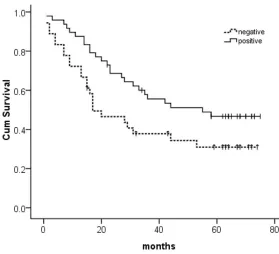
All data were analysed using SPSS 16.0 statistical soft-Figure 1. Relationship between Dpr1 expression and survival of 84 gastric
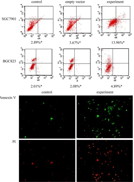
[image:3.612.91.370.71.325.2]apoptosis rate was significantly higher in the experimental group than in the control group and empty-vector group. The apoptosis rates were 4.89%, 2.01%, and 2.08%, respectively.
Effect of DAPPER1 overexpression on Wnt pathway proteins and their target genes
In SGC7901 cells, levels of Dvl-2 and β-catenin proteins were significantly lower in the experi-mental group than in the control group and empty-vector group. In SGC7901 cells, Survivin protein levels were significantly lower in the experimental group than in the control group and empty-vector group. Similarly, Survivin mRNA levels in SGC7901 cells were significant-ly lower in the experimental group than in the control group and empty-vector group (Figure 4). In BGC823 cells, there were no differences in Survivin protein or mRNA levels among the experimental group, control group, and empty-vector group 48 h after transfection (data not shown).
Effect of DAPPER1 gene overexpression on apoptosis-related protein Bcl-2 family mem-bers
There were no significant differences in Bcl-2 protein expression among the experimental group, control group, or empty-vector group in either cell line (data not show). Bcl-xl protein levels in both cell lines were significantly lower in the experimental group than in the control group and empty-vector group, while Bcl-xl mRNA levels did not significantly differ among the three groups (Supplementary Figure 5). Dpr1 protein expression was absent from
gas-tric tumour tissues of 36 patients (44%) and present in gastric tumour tissues of 48 patients (56%). The level of Dpr1 protein expression correlated with depth of tumour invasion (P = 0.046), lymph-node metastasis (P = 0.016), and TNM stage (P = 0.048) but not with the patient’s gender or age or degree of tumour dif-ferentiation (Borrmann type).
Kaplan-Meier survival curves showed that sur-vival of patients whose tumours were positive for Dpr1 protein expression was significantly higher than patients whose tumours were neg-ative for Dpr1 protein expression (Figure 1, P = 0.046).
Effect of DAPPER1 overexpression on gastric cancer cell lines
Dpr1 protein expression level was lower in SGC7901 cells than in MGC803 cells; β-catenin protein expression level was higher in SGC7901 than in BGC823 cells or MGC803 cells; Dvl-2 protein expression level was similar among the three gastric cancer cell lines (Supplementary Figure 3). Based on these results, SGC7901 and BGC823 cell lines were selected for further study.
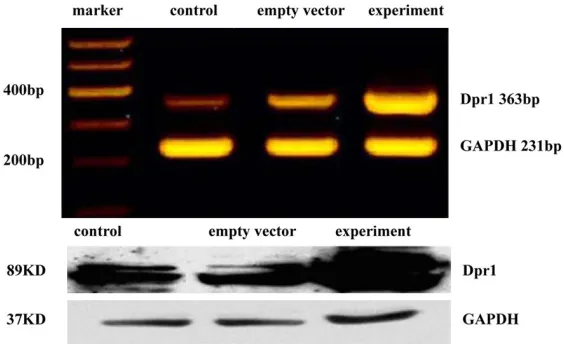
In SGC7901 cells, DAPPER1 mRNA expression was significantly higher in the experimental group than in the control group and empty-vec-tor group. Similarly, in SGC7901 cells, Dpr1 pro-tein level was significantly higher in the experi-mental group than in the control group and
empty-vector group (Figure 2). Proliferation of SGC7901 cells was reduced by 19% in the experimental group as com-pared to the control group and empty-vector group at both 48 and 72 h after trans-fection (P < 0.05) (Supple- mentary Figure 4).
[image:4.612.92.374.73.245.2]In SGC7901 cells, the apopto-sis rate was significantly higher in the experimental group than in the control group and empty-vector group (Figure 3). The apoptosis rates were 13.96%, 2.89%, and 3.67%, respectively. Simi- larly, in BGC823 cells, the Figure 2. DAPPER1 mRNA and Dpr1 protein levels in
Figure 3. Apoptosis in gastric cancer cells overexpressing DAPPER1 Upper: The lower right quadrant is early apop-totic cells; upper right quadrant is late apopapop-totic cells. *apopapop-totic rate. Lower: Apoptosis of SGC7901 cells assessed
by fluorescence microscopy at 100× magnification. Annexin V has green fluorescence and is located in the mem
In the present study, DAPPER1 mRNA levels were significantly lower in gastric tumour tissue than in the adjacent normal mucosa in 57% of cases (17/30), which is consistent with Yau’s finding from hepatocellular carcinoma [13]. Accordingly, Dpr1 protein levels were signifi-cantly lower in gastric tumour tissue than in the adjacent normal mucosa in 70% of cases
Effect of DAPPER1 overexpression on tumouri-genicity of SGC7901 cells in nude mice
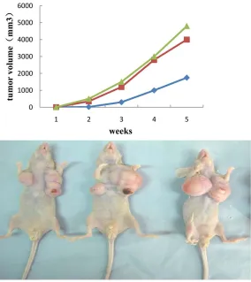
There was no swelling, ulceration, or other inflammatory response at the site of cell-cul-ture injection. Tumours of approximately 1-mm diameter could be seen at the injection site subcutaneously one week after the inoculation.
Xenograft growth rate was significantly lower in the experimental group than that in control group and empty-vector group. From the sec-ond week, tumour volume in the experimental group was significantly lower than that in the control group and empty-vector group. There was no significant difference in tu- mour size between the con-trol group and empty-vector group (Figure 5).
Discussion
[image:6.612.92.374.75.199.2]In 2002, Cheyette isolated a novel Dvl-binding protein from the Japanese toad body using two-dimensional elec-trophoresis technology [4]. Unlike previously character-ized Dvl-binding proteins, this protein inhibited the function of Dvl when bound to it; there-fore, Cheyette named it the Dapper (Dpr). Subsequently, Jushua divided Dpr proteins into Dpr1 and Dpr2 accord- ing to their functions [6]. Dpr1’s main function is in regulating the classic Wnt/β-catenin pathway, while Dpr2’s main function is in regulating non-classic Wnt pathways. Thereafter, Jushua, Barbara, Gloy, and Hikasa discovered that Dpr1 regulated cell prolif-eration and differentiation and played an important role in embryogenesis and organ-ogenesis [10-12]. Therefore, they speculated that Dpr1 might also influence tumour development.
Figure 4. Dvl-2, b-catenin, and surviving protein expressed in DAPPER1-over-expressing SGC7901 cells.
[image:6.612.94.378.251.570.2](59/84). Thus, we speculate that reduction of Dpr1 protein expression is associated with the development of gastric cancer. Furthermore, Dpr1 expression level was correlated with depth of tumour invasion, lymph-node metasta-sis, and TNM stage. Thus, we deduce that Dpr1 expression level is related to the development of gastric cancer.
Our immunohistochemistry staining results suggested that Dpr1 was mainly expressed in the cytoplasm, which is consistent with previ-ous reports [4]. In addition, high expression of Dpr1 was identified in the cytoplasm of normal stromal cells within the tumour tissue, and Dpr1 expression level was similar to that in the adjacent normal mucosa, suggesting that Dpr1 is generally expressed at a high level in normal human gastric tissue. Results from in-vitro experiments confirmed that DAPPER1 overex-pression inhibited cell proliferation, increased apoptosis, and reduced tumourigenicity of gas-tric cancer cells. Based on these results, we concluded that decreased DAPPER1 expres-sion is a feature of malignant tissue and is related to the development of gastric cancer. Dpr1’s functional mechanism in inducing gas-tric-cancer development remains unclear. Cheyette and others have suggested that Dpr1 inhibits the classic Wnt pathway [3, 4, 13]. Waxman has suggested that Dpr1 activates the classic Wnt pathway [6]. Cheyetteintroduced Dpr1 antisense oligonucleotides into HEK293 cells, which resulted in increased β-catenin expression [4]. Our study demonstrated that DAPPER1 overexpression in the gastric-cancer cell line SGC7901 inhibits β-catenin, the core protein of the classic Wnt pathway, as well as mRNA expression of Survivin, the classic Wnt-pathway target gene. These results are consistent with those of Cheyette’s study. In addition, we found relatively low β-catenin expression in the gastric-cancer cell line BGC823. DAPPER1 overexpression in those cells did not increase the expression of Survivin mRNA or protein, suggesting that Dpr1 did not activate the classic Wnt pathway. As such,we conclude that Dpr1 inhibits the classic Wnt pathway and thus promotes the development of gastric cancer.
Interestingly, we obtained a contradictory find-ing. Overexpression of DAPPER1 significantly reduced Survivin protein expression in the
gas-tric-cancer cell line SGC7901 but not in the gastric-cancer cell line BGC823. Nevertheless, overexpression of DAPPER1 significantly increased the apoptosis rate in both cell lines, indicating that Dpr1 may promote apoptosis through a Wnt-independent pathway. Therefore, we measured the expression of other apoptotic proteins (Bcl-2 and Bcl-xl) after overexpression of Dpr1. Overexpression of DAPPER1 decreased Bcl-xl protein expression in both SGC7901 and BGC823 cells. Catlett-Falcone reported that Stat3 bound to the Bcl-x gene at the APRE sequence of Stat3-binding sites, a fragment of about 600 bp, and initiated its transcription [14]. Jingand others have shown that the Wnt/ β-catenin signalling pathway upregulates Stat3 gene expression in mouse embryonic carcino-ma cells [15]. Based on these findings, we speculate that Dpr1’s effect on apoptosis is achieved through simultaneous interaction with the Wnt and Stat3 signal transduction pathways.
Most importantly, DAPPER1 expression was closely correlated with prognosis among gas-tric-cancer patients. Previous studies have indi-cated that lower levels of N-cadherin expres-sion are associated with reduced patient sur-vival. However, Dpr1 has never been used as a prognostic factor. Our results indicate that patients with Dpr1-positive tumours have bet-ter prognoses than those with Dpr1-negative tumours. Therefore, Dpr1 may be a valuable prognostic biomarker for resectable gastric cancer.
Conclusions
In conclusion, downregulation of DAPPER1 gene expression in human gastric carcinoma induced tumour development chiefly through regulating the Wnt signalling pathway. Dpr1 may be a valuable prognostic factor and a novel therapeutic target for human gastric cancer.
Acknowledgements
We are grateful to Mr. Chen Xiguang of Tsinghua University for his outstanding support. We wish to thank Professor Graeme J Poston for his assistance in the preparation of this manu-script. The study was supported by grant from The National Natural Science Fund (30801091).
Disclosure of conflict of interest
Address correspondence to: Drs. Shan Wang and Kewei Jiang, Department of Gastroenterological Surgery, Laboratory of Surgical Oncology, People’s Hospital of Peking University, 11 Xizhimen South Street, Xicheng District, Beijing, PR China. Tel: +86 10 8832 6600; Fax: +86 10 6831 8386; E-mail: [email protected] (SW); dr_jiangkewei@163. com (KWJ)
References
[1] Jemal A, Bray F, Center MM, Ferlay J, Ward E and Forman D. Global cancer statistics. CA Cancer J Clin 2011; 61: 69-90.
[2] Nusse R. Wnt signaling and stem cell control. Cell Res 2008; 18: 523-527.
[3] Zhang L, Gao X, Wen J, Ning Y and Chen YG. Dapper 1 antagonizes Wnt signaling by pro-moting dishevelled degradation. J Biol Chem 2006; 281: 8607-8612.
[4] Cheyette BN, Waxman JS, Miller JR, Takemaru K, Sheldahl LC, Khlebtsova N, Fox EP, Earnest T and Moon RT. Dapper, a Dishevelled-associated antagonist of beta-catenin and JNK signaling, is required for notochord formation. Dev Cell 2002; 2: 449-461.
[5] Katoh M and Katoh M. Identification and char -acterization of human DAPPER1 and DAPPER2 genes in silico. Int J Oncol 2003; 22: 907-913. [6] Waxman JS, Hocking AM, Stoick CL and Moon
RT. Zebrafish Dapper1 and Dapper2 play dis -tinct roles in Wnt-mediated developmental pro-cesses. Development 2004; 131: 5909-5921. [7] Lagathu C, Christodoulides C, Virtue S,
Cawthorn WP, Franzin C, Kimber WA, Nora ED, Campbell M, Medina-Gomez G, Cheyette BN, Vidal-Puig AJ and Sethi JK. Dact1, a nutrition-ally regulated preadipocyte gene, controls adi-pogenesis by coordinating the Wnt/beta-catenin signaling network. Diabetes 2009; 58: 609-619.
[8] Jiang X, Tan J, Li J, Kivimae S, Yang X, Zhuang L, Lee PL, Chan MT, Stanton LW, Liu ET, Cheyette BN and Yu Q. DACT3 is an epigenetic regulator of Wnt/beta-catenin signaling in colorectal cancer and is a therapeutic target of
histone modifications. Cancer Cell 2008; 13:
529-541.
[9] Su Y, Zhang L, Gao X, Meng F, Wen J, Zhou H, Meng A and Chen YG. The evolutionally con-served activity of Dapper2 in antagonizing TGF-beta signaling. FASEB J 2007; 21: 682-690.
[10] Brott BK and Sokol SY. Frodo proteins: modula-tors of Wnt signaling in vertebrate develop-ment. Differentiation 2005; 73: 323-329. [11] Gloy J, Hikasa H and Sokol SY. Frodo interacts
with Dishevelled to transduce Wnt signals. Nat Cell Biol 2002; 4: 351-357.
[12] Hikasa H and Sokol SY. The involvement of Frodo in TCF-dependent signaling and neural tissue development. Development 2004; 131: 4725-4734.
[13] Yau TO, Chan CY, Chan KL, Lee MF, Wong CM, Fan ST and Ng IO. HDPR1, a novel inhibitor of the WNT/beta-catenin signaling, is frequently downregulated in hepatocellular carcinoma: involvement of methylation-mediated gene si-lencing. Oncogene 2005; 24: 1607-1614. [14] Catlett-Falcone R, Landowski TH, Oshiro MM,
Turkson J, Levitzki A, Savino R, Ciliberto G, Moscinski L, Fernandez-Luna JL, Nunez G, Dalton WS and Jove R. Constitutive activation of Stat3 signaling confers resistance to apop-tosis in human U266 myeloma cells. Immunity 1999; 10: 105-115.
Supplementary Figure 1. DAPPER1 mRNA level in gastric carcinoma tissue and adjacent normal mucosa: Tx repre-sents tumor tissue, Nx reprerepre-sents adjacent normal mucosa.
Supplementary Figure 2. Dpr1 protein expression in gastric carcinoma tissue and adjacent normal mucosa: A:
Adja-cent normal mucosa. B: Tumor tissue (immunohistochemistry staining using the SP method at 100× magnification).
A: The cytoplasm of adjacent normal mucosal cells was stained dark brown, while the nucleus was not stained. B: The nuclei and cytoplasm of cancer cells were not stained. The cytoplasm of stromal cells was stained dark brown.
Supplementary Figure 3. Dpr1, b-catenin, and Dvl-2 protein expression in gastric cancer cell lines.