Original Article
Up-regulation of miR-324 suppresses cell apoptosis by
targeting DUSP1 in hepatocellular carcinoma
Wei-Liang Xiao1,2, Huan-Ling Zeng3, Yi-Zhuo Wang2, Qing Gou2, Ze-Jian Zhou2, Rong-De Xu2, Wei-Ke Li2,
Wen-Xing Zhuang2, Xiao-Ming Chen2
1Shantou University Medical College, Shantou, Guangdong, China; 2Department of Interventional Radiology, Can-cer Center, Guangdong General Hospital, Guangdong Academy of Medical Sciences, Guangzhou, China; 3Xiaolan Peoples’ Hospital of Zhongshan, Zhongshan, Guangdong, China
Received October 1, 2016; Accepted October 20, 2016; Epub February 1, 2017; Published February 15, 2017
Abstract: Backgrounds: Accumulating evidence suggested that microRNAs (miRNAs) are engaged in hepatocellular carcinoma (HCC). This study aimed to reveal the role of miR-324 in HCC and its potential mechanisms. Methods: The tissues from patients undergoing HCC resection were collected. Real time-PCR was used to test the expression of miR-324 and DUSP1 mRNA in tissues and HCC cell lines; western blot was used to detect the expression of DUSP1, AKT, MAP2K1 and p53 in the tissues and cell lines. In vitro experiment, wound healing assay was performed to test the migration ability of SK-HEP-1 cells and transwell assay was used to detect the invasion ability of SK-HEP-1 cells with the inhibitor of miR-324. Cell proliferation ability of HCC was detected by CCK-8 assay and apoptosis level was
measured by flow cytometry and TUNEL assay. Luciferase assay was used to confirm whether DUSP1-3’-UTR is the
target gene of miR-324. DUSP1 over expression plasmid was transfected to SK-HEP-1 cells to detected the rela-tionship between miR-324 and DUSP1. Results: The expression of miR-324 was dramatically up-regulated in HCC tissues and cells compared with matched normal carcinoma adjacent tissue and liver cells (P<0.05), meanwhile p53, MAP2K1, AKT and DUSP1 proteins showed different changes in HCC tissues. Furthermore, the migration and
invasion ability of SK-HEP-1 were decreased after transfected with miR-324 inhibitor. CCK-8 assay, flow cytometry and TUNEL assay showed miR-324 significantly promoted the proliferation and inhibited apoptosis of HCC cells. Luciferase reporter assay identified the 3’-UTR of DUSP1 mRNA contained a complementary sequence for miR-324.
DUSP1 could reverse anti-apoptosis role of miR-324. Conclusions: Our study provided a better understanding of miR-324 in HCC developing process. And our results may contribute to the development of miRNA-directed diagnos-tic and therapeudiagnos-tic against HCC.
Keywords: Hepatocellular carcinoma, MicroRNAs, DUSP1, tumor progression
Introduction
Primary liver cancer was the fifth most fre
-quently diagnosed cancer globally and the sec -ond leading cause of cancer death [1, 2], with hepatocellular carcinoma (HCC) now being the third reason of cancer-related mortality
world-wide [3]. And surgical resection, radiofrequency
ablation and liver transplantation are the effec-tive methods for clinical treatment of patients with liver cancer for the moment. However, the prognosis of liver cancer has not been ideal, and patients who received surgery within 2 years reappeared exceeds 50% and the 5-year survival rate for patients was less than 5% due to the late detection of the tumors and high
rate of recurrence and metastasis [4]. Hence, to further clarify the molecular mechanism of HCC invasion and metastasis is to develop new therapeutic strategies for the treatment of HCC. MiRNAs are small endogenous, noncoding RNAs that were 21-24 nucleotides in length and directed the posttranscriptional regulation of
gene expression by binding to sequences in a 3’-untranslated region (3’-UTR) of the target
cently, some studies found that miRNAs could play important roles in liver cancer initiation and progression. For example, upregulation of miR-144 led to inhibition of cell proliferation, cell cycle progression, chemoresistance, and other malignant biological behaviors in HCC [8].
Li et al. provided an unequivocal evidence for
critical oncogenic roles of the miR-675 in hepa-ocellular carcinoma and supported the notion
that miR-675 may be an alternative bona fide
promoting factor of hepatocellular carcinoma [9]. In the meantime, more and more miRNA related to liver cancer were gradually being dis-covered and studied.
Previous study showed that to identify plasma levels of miR-324 as a potential diagnostic bio-markers for early stage lung cancer, and detec-tion of plasma miR-324 levels may also serve as a prognostic marker for lung cancer patients [10]. MiR-324 could also be used as one of the biomarker in the diagnosis of HCC [11], but the
mechanism remains unclear. Dual-specificity
phosphatase-1 (DUSP1, also called MKP-1,
ERP) was initially identified in cultured murine
cells [12]. DUSP1 is one member of
dual-speci-ficity phosphatases which were recognized as
key players for inactivating different mitogen-activated protein kinase (MAPK) isoforms [13, 14]. DUSP1 played a role in cell proliferation, differentiation and transformation, cycle arrest, and apoptosis mainly by regulation of MAPK signaling [15, 16]. Previous study showed that the miRNA could target to DUSP1, which regu-lated the development of tumor cells through DUSP1 and its pathway [17]. Hence, we
hypoth-esized that miR-324 suppressed HCC cell apo- ptosis and induced the cell proliferation and migration through directly regulating DUSP1 in this study.
Materials and methods
Patients and samples
Hepatocellular carcinoma and their corre-sponding non-tumor tissues were collected at the time of surgical resection from 100 patients with liver cancer from 2014 to 2015 at Guang- dong General Hospital. All specimens were
con-firmed pathologically. Human tissues were immediately frozen in liquid nitrogen and stored
at -80°C refrigerator. Informed consent was signed by all patients and the study was ap- proved by the Ethics Committee of Guangdong General Hospital. All of the included patients met the following criteria: pathologically and
histologically confirmed HCC, no history of any
other malignant tumors, and no neoadjuvant therapy prior to the surgery.
Cell culture and transfection
The following human HCC cell lines were ob- tained from American Type Culture Collection (ATCC, Rockville, Maryland, USA): HepG2, SK- HEP-1, Huh-7, and Hep3B. The normal human liver LO2 cell line was also employed as normal control. All the cells were grown in RMPI 1640 medium with 10% fetal bovine serum (FBS) (Gibco, CA, USA), 1% of 100 U/ml penicillin and 1% of 100 mg/ml streptomycin sulfates. The
cells were incubated in humidified incubators
with 5% CO2 at 37°C.
MiR-324 inhibitor and the inhibitor control
(Negative control, NC) were synthesized chemi
-cally in Suzhou GenePharma Co., Ltd. (Suzhou,
China). Human DUSP1 gene was constructed into pcDNA3.1+ vector by Life Technologies (Invitrogen, CA, USA), and the empty vector was served as the negative control. MiR-324 inhibi-tor and pcDNA3.1+HA-DUSP1 or pcDNA3.1+HA empty vector were transfected after the cells
were cultured to 70-80% confluence by using
Lipofectamine 2000 (Invitrogen, CA, USA) acc-
ording to the manufacturer’s instructions.
Quantitative real-time PCR
Total RNA was extracted from the cell lines
and frozen tissue specimens with TRIzol rea-gent (Thermo Fisher Scientific). Complementary
[image:2.612.90.290.82.206.2]DNA was generated using a miScript Reverse Transcription Kit (Qiagen NV, Venlo, Netherla- nds). Primers for miR-324, U6 small nuclear
Table 1. Primers for qRT-PCR
Name Sequence
miR-324 F: 5’-GCCCCAGGTGCTGGGGGT R: 5’-GATGCGGGGAGGCATAGTCAG
U6 F: 5’-CTCGCTTCGGCAGCACA
R: 5’-AACGCTTCACGAATTTGCGT
DUSP1 F: 5’-GCGAAGAAGCCGAGGAGCCCG R: 5’-CGGGCTCCTCGGCTTCTTCGC
RNA (snRNA) (internal control), DUSP1 and
GAPDH were purchased from Suzhou
Gene-Pharma. The expression level of miRNA was
defined based on the threshold cycle (Ct), and
relative expression levels were calculated using the 2-ΔΔCt method, using the expression level of
the U6 snRNA as a reference gene. Each poly-merase chain reaction (PCR) was performed in triplicate. The primers for the examined genes were presented in Table 1.
Cell proliferation assay
The Cell Counting Kit-8 (CCK-8, Dojindo, Japan) assay was used for cell proliferation analysis
following the manufacturer’s instruction.
SK-HEP-1 and HepG2 cells with established stable expression after transfected with NC, miR-324 inhibitor, miR-324 inhibitor+pcDNA3.1 empty vector or miR-324 inhibitor+pcDNA3.1-DUSP1 were seeded at a density of 5×10³ cells per well
in 96-well plates and incubated for various peri-ods of time (0 h, 24 h, 48 h, 72 h). The absor-bance at 450 nm was measured using a elec-troluminescence immunosorbent assay reader
(Thermo Fisher Scientific, Waltham, MA).
Flow cytometry and TUNEL analysis of cell apoptosis
Cells were collected and washed twice with cold phosphate-buffered saline solution (PBS)
to remove floating cells before analysis by the
Annexin V-APC Apoptosis Detection Kit (KeyGEN Biotech, Nanjing, China). Apoptosis was
evalu-ated with a flow cytometry analyzer (BD
Biosci-ences, CA). Terminal deoxynucleotidyl transfer-ase-mediated deoxyuridine triphosphate in situ nick end labeling (TUNEL) detection kit (Roche, China) was used to demonstrate cell apoptosis
followed the manufacturer’s instructions. After
DAPI counterstain, the tissue section was exa-
mined and photographed with a fluorescence
microscope.
Wound-healing assay
Wound-healing assay was performed using
SK-HEP-1 cells. Cells were trypsinized and seeded in equal numbers into 6-well culture plates, and allowed to grow until confluent. When serum starvation for 24 hours, an artifi -cial homogenous wound (scratch) was created
onto the cell monolayer with a sterile 100 μL
tip. After scratching, the cells were washed with serum-free medium, complete media was
added, and microscopic images (20× magnifi -cation) of the cells were collected at 0 and 48 hours.
Cell invasion assay
Transwell chamber was used to examine cell invasion capability. SK-HEP-1 cells were trans-fected with miR-324 inhibitor and pcDNA3.1-DUSP1 or pcDNA3.1 empty vector following to
the manufacture’s information. Transfected cells were trypsinized and resuspended, 2.0×
104 cells in 200 μL RPMI 1640 medium were
placed into the upper chambers (8-mm pore
size; Corning, MA) after 6 h transfection. The lower chambers were filled with 500 μL com -plete medium with 10% FBS. The cells on the upper side of the inserts were softly scraped off after incubation for 12 h at 37°C. Cells that migrated to the lower side of the inserts were
fixed with 4% paraformaldehyde and stained
with crystal violet (1 μg/ml), and then the cells
from five independent, randomly chosen visual fields were counted under an immunofluores
-cence microscope (×100 magnification) for quantification of cells.
Luciferase reporter assay
Luciferase reporter assay was performed
according to the manufacturer’s instructions. Briefly, cells (3.0×10⁴) were seeded in triplicate
in 24-well plates overnight. Next, 100 ng of
pcDNA3.1-DUSP1-3’-UTR (wild type/mutant) or
control-luciferase plasmid plus 1 ng of pcDNA- 3.1 empty vector renilla plasmid (#E2810; Pr- omega, USA) were transfected into the cells using Lipofectamine® 2000 (Thermo Fisher
Scientific). Three independent experiments
were performed and the data was presented as the mean ± standard deviation (SD).
Statistical analyses
Data were presented as the mean value ± SD
and analyzed with SPSS 17.0 software (SPSS Inc., Chicago, IL, USA). Statistical significance
was determined by an analysis of one-way ANOVA analysis or two-tailed Student t-test. P-value of less than 0.05 was considered to be
statistically significant. All experiments were
Results
Expression of miR-324 was frequently upregu-lated in HCC tissues and cell lines
To demonstrate whether miR-324 was corre-lated with the progression in HCC cell lines, tis-sues and matched with adjacent non-tumor
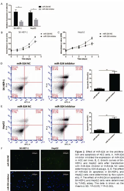
To investigate the effects of miR-324 on the proliferation of HCC cell lines, CCK-8 assay was performed. The OD=450 nm value in the CCK-8 assay revealed that miR-324 inhibited SK-HEP-1 and HepG2 cell proliferation compared with the miR-324 negative control group (Figure 2B and
2C). Annexin V staining showed that the per-Figure 1. Upregulation of miR-324 in HCC tissues and cell lines. A. The relative miR-324 expression in HCC lines was
much higher than that in the normal liver cell LO2 detected by qRT-PCR. B. The expression of miR-324 was increased
[image:4.612.95.522.75.226.2]in 100 HCC tissues than their matched adjacent non-tumor liver tissues. The data is shown as the means ± SD. *P<0.05; **P<0.001.
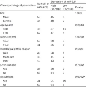
Table 2. Relationship between miR-324 and clinicopathological features of HCC patients
Clinicopathological parameters Number of cases (%)
Expression of miR-324 High
(15/100) Low (85/100) P-value
Sex 1.000
Male 53 45 8
Female 47 40 7
Age (years) 0.2843
≤60 48 37 11
>60 52 47 5
Diameter(cm) 1.0000
≤5.0 59 50 9
>5.0 41 35 6
Histological differentiation 0.1726
Well 33 28 5
Moderate 48 41 7
Poor 19 13 6
Liver cirrhosis 0.7832
Yes 37 30 7
No 63 54 9
Recurrence 0.0062*
Yes 31 21 10
No 69 64 5
Note: *Statistically significant (P<0.05).
liver tissues collected from
100 patients by qRT-PCR. The results confirmed that the
expression of miR-324 was
significant increased in cell
lines SK-HEP-1, Huh-7, Hep3B and HepG2 compared with that in normal HCC cell line LO2 (Figure 1A). As shown in
Figure 1B, the expression of miR-324 was found increased in HCC tissues with matched adjacent non-tumor liver tis-sues (P<0.05). However, no
statistically significant rela -tionships was found between miR-324 and any of the cli- nicopathological parameters except for recurrence (P= 0.0062) (Table 2).
Effect of miR-324 on the proliferation of HCC cells
[image:4.612.91.377.325.612.2]Figure 2. Effect of miR-324 on the prolifera-tion and apoptosis of HCC cells. A. miR-324 inhibitor inhibited the expression of miR-324 in HCC cell lines. B, C. Growth curves of SK-HEP-1 and HepG2 cells after transfection with miR-324 inhibitor or miR-324 NC were determined by CCK-8 assays. D, E. The effect of miR-324 on apoptosis in SK-HEP-1 and
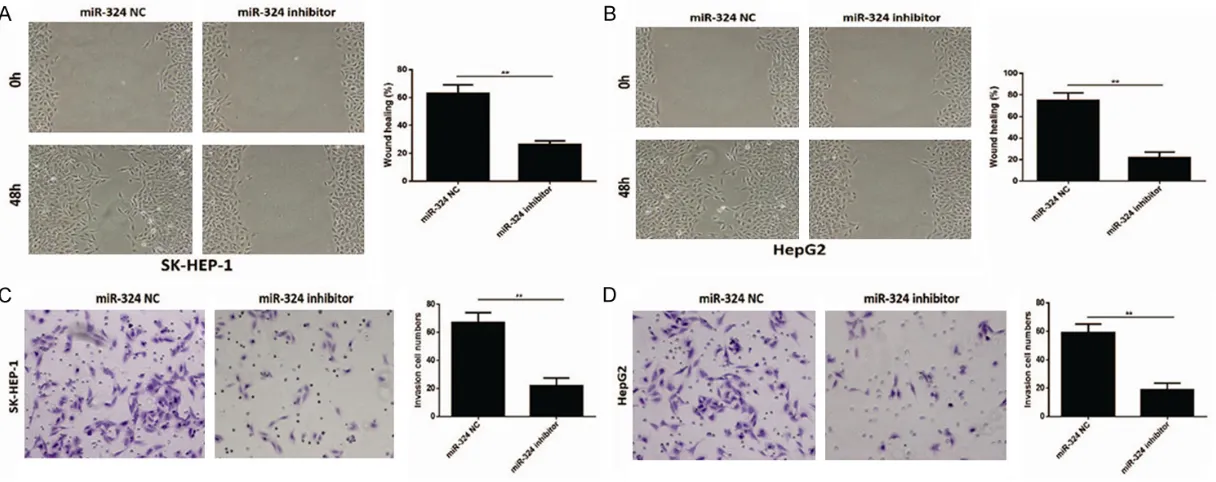
Figure 3. Effect of miR-324 on migration and invasion of HCC. A, B. The wound healing rate in SK-HEP-1 and HepG2 cells transfected with miR-324 inhibitor were
centage of apoptotic cells following miR-324 inhibitor was drastically increased relative to that in control groups (Figure 2D and 2E). TUNEL staining revealed that cell apoptosis of SK-HEP-1 and HepG2 cells transfected with
miR-324 inhibitor were significantly increased
compared with miR-324 NC group (Figure 2F). Effect of miR-324 on migration and invasion of HCC
To further investigate the biological significance
of miR-324 in HCC, wound healing assay and transwell assay were performed to detect the effect of miR-324 on migration and invasion of HCC cells. Wound-healing assay showed that the mobility of SK-HEP-1 and HepG2 cells trans-fected with miR-324 inhibitor evidently deceler-ated in rate in within 48 hours compared with negative controls showed in Figure 3A and 3B
(P<0.05). Transwell assay showed that SK- HEP-1 and HepG2 cells transfected with
miR-324 inhibitor resulted in a significant decrease
in invasive potential (P<0.05) (Figure 3C and
3D). Taken together, the expression of miR-324 suppressed the migration and invasion of HCC cells.
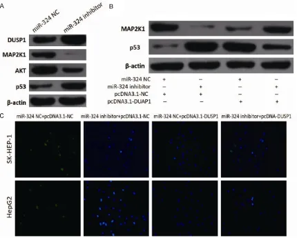
Function of miR-324 in HCC cells partially at-tributed to DUSP1
To investigate the underlying mechanism by which miR-324 regulates progression of HCC,
promoted the expression of DUSP1 mRNA (Figure 4C). Taken together, the results strongly
suggested that miR-324 could significantly sup -press the ex-pression of DUSP1 through
target-ing the 3’-UTR.
MiR-324 regulates apoptosis by affecting the expression of DUSP1
As showed in Figure 5A, we detected parts of protein level which were related to apoptotic pathway HCC cells. When the expression of miR-324 was downregulated by inhibitor, the protein level of DUSP1 and p53 were increased. Otherwise, the protein level of MAP2K1 and AKT were decreased. Furthermore, MAP2K1 and p53 showed different level changes
com-pared with β-actin consistent with apoptosis
change in SK-HEP-1 cells transfected with miR-324 NC, miR-miR-324 inhibitor, pcDNA3. 1-NC or pcDNA3.1-DUSP1 (Figure 5B), and the cells also showed different apoptosis after transfec-tion in SK-HEP-1 and HepG2 cell lines (Figure 5C).
Discussion
In the present study, we showed that the ex-
pression of miR-324 was significantly increased
in tissues of HCC patients compared with its carcinoma adjacent tissues. Over expression of miR-324 promoted migration, proliferation and Figure 4. Function of miR-324 in HCC cells partially attributed to DUSP1.
A. Prediction consequential pairing of target region between miR-324 and
DUSP1 by bioinformatics method and construction of reporter gene
plas-mids and point mutation plasplas-mids. B. miR-324 significantly suppressed the
luciferase activity that carried wild-type DUSP1 but not the mutant DUSP1.
C. Downregulated of miR-324 significantly increased the mRNA of DUSP1 in
SK-HEP-1 and HepG2 cells compared with negative control group. The data is shown as the means ± SD. *P<0.05; **P<0.001.
bioinformatic algorithms, in- cluding TargetScan and Star base, to predict the potential target of miR-324. Bioinfor- matics analysis of miRNA
rec-ognition sequences on
miR-324 revealed the presence of DUSP1 binding sites. Fur- thermore, we constructed the luciferase reports carrying the wild type and mutant type of
DUSP1 3’-UTR (Figure 4A). As shown in Figure 4B, luciferase assays indicated that the wild
type of 3’-UTR caused a sig
-nificant reduction in lucifrase activity, but mutant of 3’-UTR
inhibited apoptosis of HCC cells. Furthermore, we demonstrated DUSP1 as a target gene of miR-324 in SK-HEP-1 cells. Restoration of DUSP1 partly reverse miR-324 suppressed cell apoptosis in HCC cells.
Lots of evidence indicated that miRNAs are important regulators in various cellular pro-cesses and are recently extensively investigat-ed relating to cancer initiation, progression, diagnosis and treatment [6, 8, 18]. Dysregu- lation of miRNAs were often found in HCC, and
it was confirmed that most of them played an
important role in the progression and develop-ment of HCC [5]. For example, miR-384 was
significantly downregulated in HCC cells and tis -sues, and this resulted in HCC cell proliferation was suppressed [19]. Previous study also showed that miR-1299 inhibited cell
prolifera-tion and might be a target for HCC therapy [20]. MiR-324 was detected dysregulation expres-sion and as a potential prognostic markers in breast cancer [21]. Next, researchers showed that that miR-324 could be highly promising as diagnostic biomarkers for early stage LSCC with miR-1285 [10]. In the present study, we found that the expression of miR-324 was increased in different degrees both in tissues and cells of HCC. The proliferation, apoptosis and metastasis of HCC cells were changed when we inhibited or over-expressed miR-324. So we think that the up-regulation of miR-324 was one of the reasons for the development of liver cancer.
[image:8.612.92.522.72.417.2]early phase of cancer helped the tumor to evade JNK1-induced apoptosis, whereas down-regulation of DUSP1 allowed for proliferation and increased tumor mass in the more adva- nced stages of tumorigenesis [23]. DUSP1 was
one member of dual-specificity phosphatases which was recognized as key players for inacti -vating different mitogen-activated protein kin- ase (MAPK) isoforms [24]. Increased expres-sion of DUSP1 was also observed in other tu- mors, including colon, bladder, gastric, breast,
and lung cancer, which consequently inhibited
tumor cell apoptosis [16, 25, 26]. But on the other hand, previous studies found that the expression of DUSP1 is different in different tumors. For example, the expression of DUSP1 in hepatocellular carcinoma (HCC) decreased slightly compared with normal liver tissues as an ERK inhibitor, played a role in inhibiting the hepatocarcinogenesis [27]. The changes in the proliferation, apoptosis and metastasis of hepatocellular carcinoma cells in the present study may be due to the involvement of DUSP1 proteins.
The findings of the present study were in line
with those of previous evidence, which showed the increased expression of miR-324 in cancer cell lines and human tissues. The expression of miR-324 enhanced HCC cell proliferation and migration suggesting that increased expres-sion of miR-324 corrected with the malignant
potential of HCC. Furthermore, we identified
DUSP1 as a target of miR-324 in HCC cells. And DUSP1 was involved in p53 activation via the p38 MAPK/HSP27 pathway. Downregulating the DUSP1 may interrupt the positive regulato-ry loop between DUSP1 and p53, and then pro-mote HCC development and progression. Therefore, miR-324 was directly regulated the expression of DUSP1 protein, which was involved in the MAPK pathway, and ultimately affect the growth and metastasis of liver can-cer cells through affecting the expression of protein included in the pathway.
In conclusion, our study indicated that miR-324 was up-regulated in HCC patients and cells. In addition, miR-324 promoted proliferation and inhibited cell apoptosis of HCC by targeting DUSP1. Our study provided a better under-standing of miR-324 function in liver cancer
development, which may also be benefit for the
development of miRNA-directed diagnostic and therapeutic against HCC.
Acknowledgements
This study was supported by the National sicence foundation of China (80328532).
Disclosure of conflict of interest
None.
Address correspondence to: Xiao-Ming Chen, De- partment of Interventional Radiology, Cancer Center, Guangdong General Hospital, Guangdong Academy
of Medical Sciences, Guangzhou 510080, China.
E-mail: cxmdj@sina.com References
[1] El-Serag HB. Hepatocellular carcinoma. N Engl J Med 2011; 365: 1118-1127.
[2] Altekruse SF, McGlynn KA and Reichman ME. Hepatocellular carcinoma incidence, mortality, and survival trends in the United States from 1975 to 2005. J Clin Oncol 2009; 27: 1485-1491.
[3] Li D, Kang J, Golas BJ, Yeung VW and Madoff DC. Minimally invasive local therapies for liver cancer. Cancer Biol Med 2014; 11: 217-236. [4] Anwar SL and Lehmann U. MicroRNAs:
emerg-ing novel clinical biomarkers for hepatocellular carcinomas. J Clin Med 2015; 4: 1631-1650. [5] Yang J, Han S, Huang W, Chen T, Liu Y, Pan S
and Li S. A meta-analysis of microRNA expres-sion in liver cancer. PLoS One 2014; 9: e114533.
[6] Vosa U, Kolde R, Vilo J, Metspalu A and Annilo T. Comprehensive meta-analysis of microRNA expression using a robust rank aggregation ap-proach. Methods Mol Biol 2014; 1182: 361-373.
[7] Song JH and Meltzer SJ. MicroRNAs in patho -genesis, diagnosis, and treatment of gastro-esophageal cancers. Gastroenterology 2012; 143: 35-47, e32.
[8] Yu M, Lin Y, Zhou Y, Jin H, Hou B, Wu Z, Li Z, Jian Z and Sun J. MiR-144 suppresses cell pro-liferation, migration, and invasion in hepato-cellular carcinoma by targeting SMAD4. Onco Targets Ther 2016; 9: 4705-4714.
[9] Li H, Li J, Jia S, Wu M, An J, Zheng Q, Zhang W and Lu D. miR675 upregulates long noncoding RNA H19 through activating EGR1 in human liver cancer. Oncotarget 2015; 6: 31958-31984.
[10] Gao X, Wang Y, Zhao H, Wei F, Zhang X, Su Y, Wang C, Li H and Ren X. Plasma miR-324-3p and miR-1285 as diagnostic and prognostic
biomarkers for early stage lung squamous cell
[11] Wen Y, Han J, Chen J, Dong J, Xia Y, Liu J, Jiang Y, Dai J, Lu J, Jin G, Han J, Wei Q, Shen H, Sun B and Hu Z. Plasma miRNAs as early biomark-ers for detecting hepatocellular carcinoma. Int J Cancer 2015; 137: 1679-1690.
[12] Lau LF and Nathans D. Identification of a set of
genes expressed during the G0/G1 transition of cultured mouse cells. EMBO J 1985; 4: 3145-3151.
[13] Guan KL, Broyles SS and Dixon JE. A Tyr/Ser protein phosphatase encoded by vaccinia vi-rus. Nature 1991; 350: 359-362.
[14] Alessi DR, Smythe C and Keyse SM. The hu-man CL100 gene encodes a Tyr/Thr-protein
phosphatase which potently and specifically
inactivates MAP kinase and suppresses its ac-tivation by oncogenic ras in Xenopus oocyte extracts. Oncogene 1993; 8: 2015-2020. [15] Bang YJ, Kwon JH, Kang SH, Kim JW and Yang
YC. Increased MAPK activity and MKP-1 over-expression in human gastric adenocarcinoma. Biochem Biophys Res Commun 1998; 250: 43-47.
[16] Manzano RG, Montuenga LM, Dayton M, Dent
P, Kinoshita I, Vicent S, Gardner GJ, Nguyen P, Choi YH, Trepel J, Auersperg N and Birrer MJ. CL100 expression is down-regulated in ad-vanced epithelial ovarian cancer and its re-ex-pression decreases its malignant potential. Oncogene 2002; 21: 4435-4447.
[17] Wei X, Tang C, Lu X, Liu R, Zhou M, He D, Zheng D, Sun C and Wu Z. MiR-101 targets DUSP1 to regulate the TGF-beta secretion in sorafenib inhibits macrophage-induced growth of hepa-tocarcinoma. Oncotarget 2015; 6: 18389-18405.
[18] Hong TH and Park IY. MicroRNA expression
profiling of diagnostic needle aspirates from
surgical pancreatic cancer specimens. Ann Surg Treat Res 2014; 87: 290-297.
[19] Lai YY, Shen F, Cai WS, Chen JW, Feng JH, Cao J, Xiao HQ, Zhu GH and Xu B. MiR-384 regu-lated IRS1 expression and suppressed cell pro-liferation of human hepatocellular carcinoma. Tumour Biol 2016; 37: 14165-14171.
[20] Zhu H, Wang G, Zhou X, Song X, Gao H, Ma C, Chang H, Li H, Liu FF, Lu J and Ma J. miR-1299 suppresses cell proliferation of hepatocellular carcinoma (HCC) by targeting CDK6. Biomed Pharmacother 2016; 83: 792-797.
[21] Jayavelu ND and Bar N. Reconstruction of tem-poral activity of microRNAs from gene expres-sion data in breast cancer cell line. BMC Ge-nomics 2015; 16: 1077.
[22] Shen J, Zhang Y, Yu H, Shen B, Liang Y, Jin R, Liu X, Shi L and Cai X. Role of DUSP1/MKP1 in tumorigenesis, tumor progression and therapy. Cancer Med 2016; 5: 2061-2068.
[23] Gratton JP, Morales-Ruiz M, Kureishi Y, Fulton
D, Walsh K and Sessa WC. Akt down-regulation of p38 signaling provides a novel mechanism of vascular endothelial growth factor-mediated cytoprotection in endothelial cells. J Biol Chem 2001; 276: 30359-30365.
[24] Theodosiou A and Ashworth A. MAP kinase phosphatases. Genome Biol 2002; 3: RE-VIEWS3009.
[25] Wang HY, Cheng Z and Malbon CC. Overexpres-sion of mitogen-activated protein kinase phos-phatases MKP1, MKP2 in human breast can-cer. Cancer Lett 2003; 191: 229-237.
[26] Vicent S, Garayoa M, Lopez-Picazo JM, Lozano MD, Toledo G, Thunnissen FB, Manzano RG
and Montuenga LM. Mitogen-activated protein kinase phosphatase-1 is overexpressed in non-small cell lung cancer and is an indepen-dent predictor of outcome in patients. Clin Cancer Res 2004; 10: 3639-3649.