Original Article
Acute myocardial infarction and myocardial
ischemia-reperfusion injury: a comparison
Satwat Hashmi1, Suhail Al-Salam2
1Department of Biological and Biomedical Sciences, Agha Khan University, Stadium Road, Karachi-74800, Pakistan; 2Department of Pathology, College of Medicine and Health Sciences, United Arab Emirates University, ALAIN PO Box 17666, UAE
Received June 24, 2015; Accepted July 27, 2015; Epub August 1, 2015; Published August 15, 2015
Abstract: Myocardial infarction (MI) denotes the death of cardiac myocytes due to extended ischemia. Myocardial
reperfusion is the restoration of coronary blood flow after a period of coronary occlusion. Reperfusion has the poten -tial to salvage ischemic myocardium but paradoxically can cause injury, a phenomenon called as ‘reperfusion injury’ (IR). Standard histologic, immunohistochemical and Elisa techniques were used to study the histopathologic,
oxida-tive, apoptotic and inflammatory changes in MI and IR. The IL-6 levels in the LV of the MI group were significantly
raised as compared to the IR group (P=0.0008). Plasma IL-6 was also significantly increased in the MI group as com -pared to the IR group (P=0.031). MI model was also associated with increase in the neutrophil polymorphs number
in the infarction related myocardium as compared to the re-perfused myocardium. A significant increase in troponin
I level in the MI group as compared to the IR group is also seen (P=0.0001). Our IR model showed enhanced pro-apoptotic mediators like cleaved caspase-3 (P=0.005) and cytochrome c in the myocardium as compared to the MI
model. In conclusion, myocardial damage in MI is mainly due to ischemic necrosis and inflammatory mechanisms
while apoptosis is the main mechanism of cell death in IR in addition to limited ischemic necrosis.
Keywords: Heart, acute myocardial infarction, ischemia-reperfusion injury
Introduction
Coronary heart disease affects the heart due to the detrimental effects of acute myocardial infarction (MI) and Ischemia-reperfusion injury (IR). Understanding the mechanisms underlying these two processes is important as these two types of injuries are interrelated as well as different. Extending the period of acute myo-cardial ischemia for more than 20 minutes causes a “wave front” of cardiomyocyte death that begins in the subendocardium and extends
transmurally toward the pericardium [1]. This is
the reason that when a patient presents with an acute MI, the most effective therapeutic intervention is timely myocardial reperfusion using thrombolytic therapy or primary percuta-neous coronary intervention to salvage the
ischemic myocardium. The process of reperfu -sion, the very event critical for survival, can itself cause injury to the cardiomyocytes, a phenomenon called as the ‘reperfusion injury’ [2-4].
During acute myocardial ischemia, the lack of oxygen switches the cell metabolism to
anaero-bic respiration, with lactate accumulation, ATP
depletion, Na+ and Ca2+ overload and inhibition
of myocardial contractile function [5, 6]. Reperfusion results in reactivation of electron transport chain which generates reactive oxygen species (ROS). ROS induces opening of mitochondrial permeability transition pores, contributing to intracellular Ca2+ overload, lipid
peroxidation of cell membrane, and oxidative damage to DNA. In addition there is neutrophil accumulation in response to ROS, cytokines and complements. All these processes can independently induce cardiomyocyte death of the acutely ischemic myocardium [2-4].
Oxidative stress, apoptosis and inflammation
that can help reduce the myocardial infarct size. In order to investigate these processes, we compare the histopathologic, oxidative,
apoptotic and inflammatory changes in these
two models of MI and IR. Our MI model has
permanent left anterior descending (LAD)
artery ligation for 24 hours and our IR model
has LAD ligation for 30 min followed by reperfu -sion for 24 hours.
Material and methods
Murine model of myocardial infarction and ischemia reperfusion injury
Male C57B6/J mice (n=24) were divided into 24 hour MI group (n=8), IR group (n=8) and sham operated group (n=8). All experimental animal procedures are approved by the Animals Ethics Committee of the College of Medicine and Health Sciences, UAE University, protocol A12/10.
Male C57B6/J mice (male, age: 12-16 weeks; wt: 20-25 g) were anesthetized by an intraperi-toneal injection of a combination of ketamine
(100 mg/kg) and xylazine (10 mg/kg). The
mice were then intubated by transesophageal
illumination using a modified 22-gauge plastic cannula and fixed on the operating pad in the
supine position by taping all four extremities.
The mice were connected to a mouse ventilator
(Harvard apparatus Minivent Hugo Sachs Ele- ctronik) which supplied room air supplemented with 100% oxygen (tidal volume 0.2 ml/min., rate 120 strokes/min). Rectal temperature was continuously monitored and maintained within
36-37°C using a heat pad. The lead II ECG
(ADInstrument multi-channel recorder inter-faced with a computer running Power lab 4/30 data acquisition software) was recorded from needle electrodes inserted subcutaneously. MI was induced in the mice by permanently
occluding the LAD artery as described earlier
[7-10].
Briefly, the chest was opened with a lateral inci -sion at the 4th intercostal space on the left side
of the sternum. Next, the chest wall was retract-ed for better visualization of the heart. With minimal manipulation, the pericardial sac was
removed and the LAD was visualized with a ste
-reomicroscope (Zeiss STEMI SV8). An 8-0 silk suture was passed under the LAD and ligated 1
mm distal to left atrial appendage. Occlusion
was confirmed by observing immediate blanch -ing of the left ventricle at post ligation site. An
accompanying ECG recording showed charac
-teristic ST-Elevation which further confirmed
ischemia.
For our IR model, after the 8-0 silk suture was
passed under the LAD, a small 1 mm polyethyl
-ene tubing (PE) was placed on top of the LAD
and the suture was ligated on the top of the PE tubing without damaging the artery. Ischemia
was confirmed by observing immediate blanch
-ing of the left ventricle (LV) at post ligation site. An accompanying ECG also showed cor-responding ST-elevation. After 30 minutes of
ischemia the ligature is removed by cutting the knot on top of this PE tube. Reperfusion was
confirmed visually and by ECG changes. The chest wall was closed by approximating the
third and fourth ribs with one or two interrupted
sutures. The muscles returned back to their
original position and the skin closed with 4-0
prolene suture. The animal was gently discon -nected from the ventilator and spontaneous breathing was seen immediately. Post-operative analgesic (Butorphanol 2 mg/kg, s/c, 6 hourly) was given at the end of the procedure. Sham operated mice underwent exactly the same procedure described above, except that the
suture passed under the LAD is left open and
untied. According to the experimental protocol,
mice were sacrificed 24 hour after induction of myocardial infarction. The hearts were washed
in ice cold PBS, right ventricle and both atria dissected away and left ventricle immediately frozen in liquid nitrogen and later stored in
-80°C freezer. Blood was also collected in EDTA
vacutainers and centrifuged at 3000 RPM for
15 minutes. The plasma was collected, alliquot -ed and stor-ed at -80°C until further analysis. Heart samples from the same time point
follow-ing LAD ligation were fixed in 10% buffered
formal-saline for 24 hours. Histopathology
Hearts were excised, washed with ice-cold PBS and weighed. Each heart was sectioned into 4 equal transverse (coronal) sections, cassetted
and fixed directly in 10% buffered formalin.
Sections were dehydrated in increasing con-centrations of ethanol, cleared with xylene and
embedded in paraffin. Three-um sections were prepared from paraffin blocks and stained with
H&E stain was performed by de-waxing sections with xylene, rehydration with graded alcohol and washing in running tap water for 5
minutes. Tissue sections were then incubated
in haematoxylin for 5 minutes followed by
wash-ing in tap water. The slides were checked for
bluing of the nuclei. If the intensity of blue color is high, the slides were given a quick dip in 1% acid alcohol solution. Sections were then rinsed in running tap water until satisfactory blue color
of nuclei is achieved. The slides were then
stained with Eosin for 1 minute, washed in running tap water, dehydrated, cleared and mounted with DPX.
Immunohistochemistry
Three-micrometer sections were prepared and
mounted on aminopropyltriethoxysilane (APES) coated slides. After dewaxing with xylene and rehydration with graded alcohol, slides were placed in a 0.01 M citrate buffer solution (pH=6.0) and pre-treatment procedures to unmask the antigens was performed in a water bath at 90°C for 60 minutes. Sections were then treated with peroxidase block for 30 min-utes followed by protein block for 30 minmin-utes. Sections were later incubated overnight with anti-cleaved caspase-3 (Rabbit polyclonal, ASP
175, Cell Signaling Technology, USA), anti-Bcl2
(Mouse monoclonal, SP66, Cell Marque, USA ), anti-Myeloperoxidase (MPO) (Rabbit Polyclonal, 1:2000, Santa Cruz biotechnology, USA), and anti-Cytochrome c (Rabbit Polyclonal, 1:400, Santa Cruz biotechnology, USA) at 4°C. After conjugation with primary antibodies, sections were incubated with biotin-labeled secondary
antibody (Thermo Scientific, USA) for 20 min -utes at room temperature. Finally, sections were incubated with streptavidin-peroxidase complex for 20 minutes at room temperature
(Thermo Scientific, USA), DAB chromogen (Thermo Scientific, USA) was then added and
counter staining done with haematoxylin. Appropriate positive controls were used. For negative control, the primary antibody was not added to sections and the rest of the procedure was carried as above. Positive and negative controls were used in every batch of slides that were stained.
Morphometric analysis
Morphometric analysis of the number of neu-trophil polymorphs at 24-hour following MI and IR was done using ImageJ software (http:// rsbweb.nih.gov/ij/).
The number of neutrophil polymorphs was
determined by counting the number of
neutro-phil polymorphs in 10 randomly selected fields in the left ventricle in areas supplied by LAD
in all specimens followed by measuring the mean neutrophil polymorphs number in each specimen.
The apoptotic index was determined by
counting the number of cells expressing cleaved caspase-3 in 10 randomly selected
fields in the left ventricle in areas supplied by LAD in all specimens following by measuring
the mean apoptotic cells in each specimen. For cleaved-caspase labeling, cells were con-sidered positive when there was a nuclear and cytoplasmic staining pattern.
Enzyme linked immunosorbent assay
Left ventricular myocardial concentration of IL-6, IL-1β, cleaved caspase-3, Wnt-3, and total Akt-1 and plasma levels of IL-6 were deter -mined using DuoSet enzyme linked
immuno-sorbent assay (ELISA) Development kit (Mouse IL-6 (DY406), Mouse IL-1β/IL-1F2 (DY401), Mouse Akt-1 (DYC1775-5), Mouse Wnt-3 (DY1324), Mouse cleaved caspase-3 (Asp175) (DYC835-5), R&D Systems, Minneapolis, MN, USA) for sandwich ELISA, using standard proce -dure according to the manufacturer’s
instruc-tions. The levels were normalized to total pro
-tein concentrations. Briefly, 96-well plates
(Nunc-Immuno Plate MaxiSorp Surface (NUNC Brand Products, A/S, Roskilde, Denmark), were
coated with antibody specific for our proteins of
interest. Biotinylated detection antibody and streptavidin conjugated horseradish peroxida- se were used for detection of captured
anti-gens. The plates between steps were aspirated and washed 3 times using ELISA plate washer (BioTek ELx50). Captured proteins were visual
-ized using tetramethylbenzidine (TMB)/hydro -gen peroxide. Absorbance readings were made at 450 nm, using a 96-well plate
spectropho-tometer (BioTek ELx800). Concentrations in the
samples were determined by interpolation from a standard curve. Standards and samples were assayed in duplicate.
Troponin-I assay
Mouse cardiac troponin I levels in plasma were measured by using a high sensitivity mouse
cardiac troponin-I Elisa kit (2010-1-HSP, Life
Glutathione and superoxide dismutase (SOD) activity assay
Total glutathione level in the heart protein extract was measured by a Glutathione Assay kit (CS0260 Sigma-Aldrich). Glutathione
standard solutions were used to generate a
standard curve and GSH levels calculated
using Magellan 6 software. SOD activity was measured using the SOD determination kit (19160 Sigma-Aldrich). Appropriate blanks were set up and SOD activity was calculated according to the manufacturer’s instructions. Statistical analysis
Statistical analysis is done using IBM SPSS Statistics version 20. Data are presented in mean ± standard error (S.E). Statistically signi-
ficant differences (P<0.05) were calculated between groups by Student t test.
Results
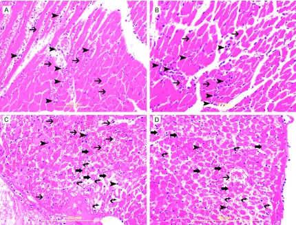
Histological changes in MI and IR models
The main histologic change in acute MI at
24-hour time point is coagulative necrosis of cardiomyocytes with heavy neutrophil polymor-
phs infiltration (Figure 1A, 1B). The ischemic
cardiomyocytes appear deeply eosinophilic with loss the cross striation and disappearance
of the nuclei. LV sections studied at 24-hour
following IR injury show focal necrosis of cardio-myocytes with prominent interstitial edema and accumulation of RBCs in the interstitial spaces with scattered neutrophil polymorphs
infiltration of the injured myocardium (Figure 1C, 1D).
Inflammatory mediators in MI and IR models
[image:4.612.91.521.71.398.2]IL-6 levels in the LV of the MI group were signi-ficantly raised as compared to the IR group
Figure 1. A & B. Represent high power views of MI heart sections showing ischemic cardiomyocytes appearing
deeply eosinophilic with loss of cross striations (thin arrow) and neutrophil polymorphs(arrow heads) flooding the
infarcted area. C & D. Represent high power views of IR heart sections showing injured cardiomyocytes (thick ar-rows) with prominent interstitial edema (curved arar-rows), RBCs in the interstitial spaces (thin arar-rows) and neutrophil
(130.9 ± 16.79 vs. 56.17 ± 4.63 pg/mg,
*P=0.0008) (Figure 2A). Plasma IL-6 was also
significantly increased in the MI group as com -pared to the IR group (479.3 ± 174.9 vs. 59.68 ± 9.61 pg/ml, *P=0.031) (Figure 2B). Heart LV
IL-1β concentrations were not significantly
different between the groups (Figure 2C).
Immunohistochemical staining of the heart
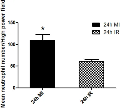
section with MPO showed significant differenc -es in the number as well as the intensity of staining of neutrophil polymorphs between the MI and IR groups (Figure 3). Morphometric analysis of the number of neutrophils was
significantly higher in the MI group (109.6 ±
13.73) as compared to the IR group (61.10 ± 3.990), *P=0.0032 (Figure 4).
Apoptotic markers in MI and IR models
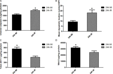
Heart LV cleaved caspase-3 levels were signifi -cantly increased in the IR group as compared to the MI group (2027 ± 93.47 vs. 1600.49 ± 89.44 pg/mg protein. *P=0.0053) (Figure 5A).
Morphometric analysis of apoptotic cells is
significantly higher in IR group than in MI group
(18.08 ± 2.930 vs. 9.364 ± 1.274 *P=0.0152)
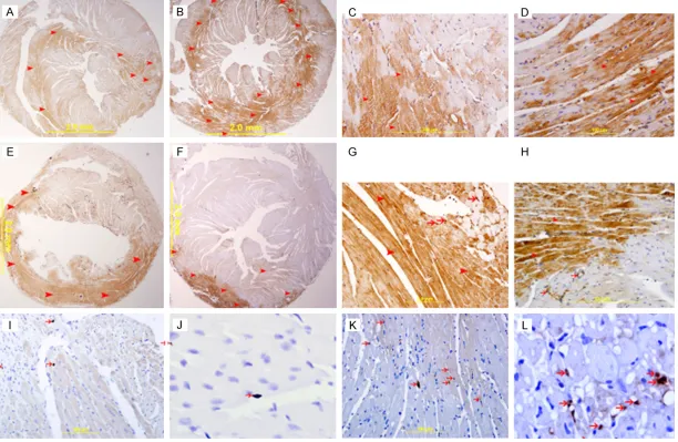
(Figure 5B). Immunohistochemical staining of the heart sections with cleaved caspase-3 also showed more apoptotic cells in the IR group (Figure 6K, 6L) as compared to the MI group (Figure 6I, 6J). Cytochrome c was also seen to be higher in the heart sections from the IR group (Figure 6B, 6D) as compared to the MI group (Figure 6A, 6C) by
immunohistochemis-try. The expression is cytoplasmic seen pre -dominantly in cardiomyocytes, but endothelial cells and neutrophil polymorphs also stained
positive for it. The anti- apoptotic protein Bcl-2
was also seen to be expressed by cardio-
myocytes and endothelial cells. The expression
was higher in the MI group (Figure 6E, 6G) as compared to the IR groups (Figure 6F, 6H). Total AKT-1 protein in MI and IR models
Heart LV AKT-1 levels were significantly higher
in the MI group as compared to the IR group (755.8 ± 70.58 vs. 417.4 ± 48.47 pg/mg protein. *P=0.0014) (Figure 5C).
Total Wnt-3 protein in MI and IR models
Heart LV Wnt-3 levels were significantly higher
in the MI group as compared to the IR group (32130 ± 1979 vs. 24420 ± 2704 pg/mg protein. *P=0.0402) (Figure 5D).
Total troponin I in MI and IR models
Plasma troponin I levels were significantly
increased in the MI group as compared to the Figure 2. The graphs represent (A) left ventricular
IL-6 concentrations (B) Plasma IL-6 levels and (C) left ventricular IL-1β concentrations in 24 hour MI and IR
IR group (12.58 ± 0.8158 vs. 3.727 ± 1.168 pg/mg protein. *P=0.0001) (Figure 7).
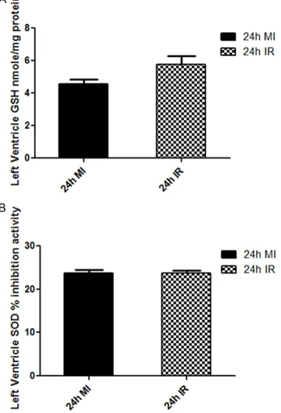
Antioxidant enzyme levels in MI and IR models
The levels of antioxidant enzymes are not sig
-nificantly different between the MI group and the IR group. Heart LV Total Glutathione levels
was 4.55 ± 0.31 nmol/mg in the MI group as compared to the IR group, which showed 5.76 ± 0.51 nmole/mg, P=0.06. Heart LV SOD inhibi -tion activity was also not different among the MI and IR group (23.71 ± 0.76% vs. 23.49 ± 0.49%, P=0.81) (Figure 8).
Discussion
For many years, it was thought that myocardial
reperfusion is only beneficial and that there was no cell death related to it [2, 11, 12]. Later
when cardiomyocytes death was seen in the re-perfused myocardium it was postulated that they are the already irreversibly damaged cardiomyocytes that were fated to die during
[image:6.612.90.524.71.397.2]ischemia [13]. The concept of ‘reperfusion
Figure 3. A & C. Representative section from LV in an area supplied by LAD at 24-hour following ligation of LAD showing many neutrophil polymorphs infiltrating the myocardium and expressing myeloperoxidase (arrow heads). B & D. Representative section from LV in an area supplied by LAD at 24-hour following ligation of LAD and reperfusion showing fewer number of neutrophil polymorphs infiltrating the myocardium and expressing myeloperoxidase (arrow heads). A & B. Immunoperoxidase streptavidin-biotin method. C & D. Alexa Fluor 488 immunoflyorescent technique.
Figure 4. Comparison of Mean number of neutrophil
[image:6.612.92.285.482.656.2]injury’ was presented when it was shown that reperfusion induced death in cardiomyocytes that were viable during ischemia [14]. Com- parisons between these two types of injuries is still continuing till today because of two main reasons: First, it is almost impossible to esti-mate the own effects of reperfusion [15] and second, despite advances in antithrombotic, anti-platelet and PCI technologies, there is still no effective way to prevent the myocardial reperfusion injury [5].
Our study attempts to show substantial differences in the local microenvironment of the myocardium between these two modes of injury and analyze these changes.
The MI group shows a significantly higher inflammatory activity associated with induction of IL-6 than IR group both in the LV and plasma. The MI group is also associated with a signifi
-cantly higher number of infiltrating neutrophil
polymorphs in the infarcted myocardium as compared to the re-perfused myocardium.
Neutrophil polymorphs that infiltrate the infarct
-ed myocardium at 24 hour following MI will secret their enzymes to digest necrotic tissue as well as will have undesired damaging effects on adjacent survived cardiomyocytes which
may potentiate the inflammatory response. We also show a significant higher level of troponin I
in the MI group when compared with the IR
group. Troponin I level is an indirect estimate of
the infarct size [16-18]; hence, a higher level of troponin I in the MI group when compared with IR group indicates more necrosis in the
myocar-dium in MI than in IR. The results comparing
the MI and IR groups show that in MI there is increased myocardial damage via ischemic
necrosis and inflammatory mechanisms. The IR group also shows inflammation but it is unclear whether the inflammatory response that accompanies an acute MI, during the first 30 minutes following ligation of LAD, contributes
to the pathogenesis of myocardial reperfusion injury or whether it is a microinvironmental reaction to the acute myocardial injury [19]. Our IR group shows higher cleaved caspase-3
[image:7.612.92.522.75.356.2]activities in the LV than in MI group. We also
Figure 5. The graphs represent (A) left ventricular cleaved caspase-3 concentrations (B) Left ventricular mean Apop
-totic bodies number/high power field (HPF), (C) Left ventricular total AKT-1 protein (D) Left ventricular Wnt-3 protein
8793 Int J Clin Exp Pathol 2015;8(8):8786-8796 Figure 6. (A & B) Represents low power view of heart sections showing cytochrome c expression in MI (A) and IR (B) groups. (C & D) Show high power view of MI (C)
and IR heart section expressing cytochrome c in the cytoplasm of cardiomyocytes. The intensity and number of positive staining in IR group is higher than the MI. Streptavidin-biotin immunoperoxidase method. (E & F) Represents low power view of heart sections expressing bcl2 in MI (E) and IR (F) groups. (G & H) Are the high power views of these sections showing increase in bcl2 immunostaining in cardiac myocytes (arrow heads) and endothelial cells (thin arrows) in the MI group (G)
compared to IR (H) group, Streptavidin-biotin immunoperoxidase method. (I & J) Represent high power views of heart sections from the MI group showing cleaved
[image:8.792.93.705.73.472.2]show a significantly higher AKT-1 in the LV in the MI group than IR group. AKT-1 activation has
anti-apoptotic activities on cardiac myocytes in MI and IR [20]. Many downstream targets of
AKT-1 have been shown to contribute to its
pro-survival effects such as phosphorylation of
BCL-2 family members [21]. Activation of AKT-1
has been shown to modulate pro-apoptotic
proteins through the phosphorylation of BCL-2
family members BAX and BAD leading to block-age of pro-apoptotic protein function and initia-tion of protective signaling cascades resulting
in anti-apoptotic effects [22, 23]. This will con -tribute to the higher cleaved caspase-3 activity in IR group than MI group.
We also show significantly higher Wnt-3 levels
in MI group than IR group. Activation of Wnt-3 has shown to be associated with an
anti-apop-totic effect [24, 25]. This will also contribute to
the higher cleaved caspase-3 activity in IR group than MI group.
The higher apoptotic activity in IR group than MI
group suggests that the main mode of cardio-myocyte death in IR is apoptosis in addition to
ischemic necrosis. It was shown by Lieberthalet al that the severity and duration of ATP deple -tion determines the mechanism of death: cells
with an intracellular ATP concentration below a
certain threshold become necrotic, whereas an
ATP value above that threshold induces apopto -sis [26, 27]. As MI model is associated with
more ATP depletion as compared to IR model, where reperfusion may replenish the ATP
stores, the main mechanism of cell death is caspase activated apoptosis in IR model. Perhaps the most interesting observation made in our study is the level of antioxidant enzymes measured in the MI and IR models.
We did not find significant differences between
the groups signifying that the level of anti- oxidant enzymes is approximately the same whether the myocardium is subjected to 24 hours of permanent ischemia or whether it is subjected to 30 min of ischemia followed by 24 hours of reperfusion. We think that this obser-vation should be looked at from a different
angle. There is ample evidence that the level of
oxidative stress generated in reperfusion inju-ries is considerably more than mere ischemic
injury. In the first few minutes of myocardial
reperfusion, a burst of oxidative stress [14, 28] is produced by a variety of sources. Organelles may begin to produce reactive oxygen species. Myocytes produce both hydrogen peroxide and
superoxide radicals [29]. The electron trans -port chain of mitochondria is also a potential source of free radicals in both the endothelial
cell and myocyte [30]. This detrimental oxida -tive stress mediates myocardial injury and
[image:9.612.92.286.75.220.2]cardiomyocyte death. The same oxidative
Figure 7. The graph represent plasma Troponin I con -centrations in MI and IR models.
[image:9.612.92.289.277.566.2]stress can lead to increase in the antioxidant capacity and so the level of antioxidant enzymes
capacity may reflect the oxidative stress [31]. The level of oxidative stress and subsequent
anti oxidant protection in IR is dependent on the time of ischemia before reperfusion is initiated and the reperfusion time [4, 32, 33]. However controversies do exist in this respect [34]. Our MI and IR model showing the same level of antioxidant capacity at 24-hour
follow-ing LAD ligation may be related to the particular
ischemia and reperfusion time in our models.
Our results are significant in terms of IR injuries
as the main goal in treatment of acute infarc-tion is early revascularizainfarc-tion with reperfusion. Reperfusion affects a larger portion of the left ventricle than infarction alone [35] so reperfu-sion injury may act as an independent determi-nate of cardiac remodeling in addition to infarct size.
In conclusion, the myocardial damage in MI is mainly due to ischemic necrosis with
accom-panying inflammation while apoptosis is the
main mechanism of cell death in IR in addition to limited ischemic necrosis.
The differences observed in these two models
as seen in our experiments show that these two processes of cardiomyocyte injury are indeed very distinct and the local microenvironment of the myocardium determines the particular roles of molecules and enzymes that are part of their pathogenesis.
Acknowledgements
The authors would like to thank The National
Research Foundation and the United Arab Emirates University for their support of this project, grant number 31MO76. In addition we would like to thank Ms Manjusha Sudhadevi
and Ms HibaTajEldin Naser from Department
of Pathology, College of Medicine & Health Sciences, United Arab Emirates University, for their technical support in tissue processing and staining.
Disclosure of conflict of interest
None.
Address correspondence to: Dr. Suhail Al-Salam, Department of Pathology, College of Medicine and Health Sciences, United Arab Emirates University,
ALAIN PO Box 17666, UAE. Tel: +97137137464; Fax:
+97137671966; E-mail: suhaila@uaeu.ac.ae
References
[1] Reimer KA, Lowe JE, Rasmussen MM, Jennings RB. The wavefront phenomenon of ischemic
cell death. Myocardial infarct size vs duration of coronary occlusion in dogs. Circulation 1977; 56: 786-794.
[2] Braunwald E, Kloner RA. Myocardial reperfu -sion: a double-edged sword? J Clin Invest 1985; 76: 1713-1719.
[3] Piper HM, García-Dorado D, Ovize M. A fresh
look at reperfusion injury. Cardiovasc Res 1998; 38: 291-300.
[4] Yellon DM, Hausenloy DJ. Myocardial reperfu -sion injury. N Engl J Med 2007; 357: 1121-1135.
[5] Hausenloy DJ, Yellon DM. Myocardial
ischemia-reperfusion injury: a neglected therapeutic tar-get. J Clin Invest 2013; 123: 92-100.
[6] Avkiran M, Marber MS. Na(+)/H(+) exchange inhibitors for cardioprotective therapy: prog-ress, problems and prospects. J Am Coll Cardiol 2002; 39: 747-53.
[7] Michael LH, Entman ML, Hartley CJ, Youker KA, Zhu J, Hall SR, Hawkins HK, Berens K,
Ballantyne CM. Myocardial ischemia and re-perfusion: a murine model. Am J Physiol 1995; 269: H2147-2154.
[8] Hashmi S, Al-Salam S. Loss of dystrophin stain -ing in cardiomyocytes: a novel method for de-tection early myocardial infarction. Int J Clin Exp Pathol 2013; 6: 249-257.
[9] Al-Salam S, Hashmi S. Galectin-1 in early acute myocardial infarction. PLoS One 2014; 9:
e86994.
[10] Hashmi S, Al-Salam S. Galectin-3 is expressed
in the myocardium very early post-myocardial infarction. Cardiovasc Pa-thol 2015; 24: 213-223.
[11] Kloner RA. Does reperfusion injury exist in hu -mans? J Am Coll Cardiol 1993; 21: 537-545. [12] Opie LH. Reperfusion injury and its pharmaco
-logic modification. Circulation 1989; 80:
1049-1062.
[13] Gottlieb RA, Burleson KO, Kloner RA, Babior BM, Engler RL. Reperfusion injury induces
apoptosis in rabbit cardiomyocytes. J Clin Invest 1994; 94: 1621-1628.
[14] Hearse DJ, Humphrey SM, Chain EB. Abrupt reoxygenation of the anoxic potassium-arrest-ed perfuspotassium-arrest-ed rat heart: a study of myocardial enzyme release. J Mol Cell Cardiol 1973; 5: 395-407.
[16] Hallén J, Buser P, Schwitter J, Petzelbauer P,
Geudelin B, Fagerland MW, Jaffe AS, Atar D.
Relation of cardiactroponin I measurements at 24 and 48 hours to magnetic resonance-deter-mined infarct size in patients with ST-elevation
myocardial infarction. Am J Cardiol 2009; 104: 1472-1477.
[17] Younger JF, Plein S, Barth J, Ridgway JP, Ball SG, Greenwood JP. Troponin-I concentration 72
h after myocardial infarction correlates with in-farct size and presence of microvascular ob-struction. Heart 2007; 93: 1547-1551. [18] Mair J, Wagner I, Morass B, Fridrich L,
Lechleitner P, Dienstl F, Calzolari C, Larue C,
Puschendorf B. Cardiac troponin I release cor-relates with myocardial infarction size. Eur J Clin Chem Clin Biochem 1995; 33: 869-872. [19] Vinten-Johansen J. Involvement of neutrophils
in the pathogenesis of lethal myocardial reper-fusion injury. Cardiovasc Res 2004; 61: 481-497.
[20] Armstrong SC. Protein kinase activation and myocardial ischemia/reperfusion injury. Cardiovasc Res 2004; 61: 427-436.
[21] Jamnicki-Abegg M, Weihrauch D, Pagel PS,
Kersten JR, Bosnjak ZJ, Warltier DC, Bienengraeber MW. Isoflurane inhibits cardiac myocyte apoptosis during oxidative and inflam -matory stress by activating Akt and enhancing Bcl-2 expression. Anesthesiology 2005; 103: 1006-1014.
[22] Aikawa R, Nawano M, Gu Y, Katagiri H, Asano T, Zhu W, Nagai R, Komuro I. Insulin prevents car -diomyocytes from oxidative stress-induced apoptosis through activation of PI3 kinase/ Akt. Circulation 2000; 102: 2873-2879. [23] Datta SR, Dudek H, Tao X, Masters S, Fu H,
Gotoh Y, Greenberg ME. Akt phosphorylation of
BAD couples survival signals to the cell-intrin-sic death machinery. Cell 1997; 91: 231-241. [24] Zhang X, Hu D, Zhang C, Zhong Q, Feng T,
Huang J. Overexpression of Wnt3 inhibits apoptosis of hepatic progenitor cells in vitro).
Nan Fang Yi Ke Da Xue Xue Bao 2014; 34:
46-50.
[25] Chera S, Ghila L, Dobretz K, Wenger Y, Bauer C, Buzgariu W, Martinou JC, Galliot B. Apoptotic
cells provide an unexpected source of Wnt3 signaling to drive hydra head regeneration. Dev Cell 2009; 17: 279-289.
[26] Lieberthal W, Menza SA, Levine JS. Graded ATP
depletion can cause necrosis or apoptosis of cultured mouse proximal tubular cells. Am J Physiol 1998; 274: F315-327.
[27] Shiraishi J, Tatsumi T, Keira N, Akashi K, Mano A, Yamanaka S, Matoba S, Asayama J, Yaoi T,
Fushiki S, Fliss H, Nakagawa M. Important role of energy-dependent mitochondrial pathways in cultured rat cardiac myocyte apoptosis. Am J Physiol Heart Circ Physiol 2001; 281: H1637-1647.
[28] Zweier JL, Flaherty JT, Weisfeldt ML. Direct
measurement of free radical generation fol-lowing reperfusion of ischemic myocardium. Proc Natl Acad Sci U S A 1987; 84: 1404-1407. [29] Rowe GT, Manson NH, Caplan M, Hess ML.
Hydrogen peroxide and hydroxyl radical media-tion of activated leukocyte depression of car-diac sarcoplasmic reticulum. Participation of the cyclooxygenase pathway. Circ Res 1983; 53: 584-591.
[30] Boveris A, Chance B. The mitochondrial gener
-ation of hydrogen peroxide. General properties
and effect of hyperbaric oxygen. Biochem J 1973; 134: 707-716.
[31] Bandeira SeM, Guedes GaS, da Fonseca LJ, Pires AS, Gelain DP, Moreira JC, Rabelo LA, Vasconcelos SM, Goulart MO. Characterization
of blood oxidative stress in type 2 diabetes mellitus patients: increase in lipid peroxidation
and SOD activity. Oxid Med Cell Longev 2012;
2012: 819310.
[32] Rochitte CE, Lima JA, Bluemke DA, Reeder SB, McVeigh ER, Furuta T, Becker LC, Melin JA.
Magnitude and time course of microvascular obstruction and tissue injury after acute myo-cardial infarction. Circulation 1998; 98: 1006-1014.
[33] Zhao ZQ, Nakamura M, Wang NP, Velez DA, Hewan-Lowe KO, Guyton RA, Vinten-Johansen
J. Dynamic progression of contractile and en-dothelial dysfunction and infarct extension in the late phase of reperfusion. J Surg Res 2000; 94: 133-144.
[34] Ytrehus K, Liu Y, Tsuchida A, Miura T, Liu GS, Yang XM, Herbert D, Cohen MV, Downey JM.
Rat and rabbit heart infarction: effects of anes-thesia, perfusate, risk zone, and method of in-farct sizing. Am J Physiol 1994; 267: H2383-2390.