Original Article
Programmed death-1 and PD-1 ligand-1 expression in
early onset gastric carcinoma and
correlation with
clinicopathological characteristics
Ping Wei1,2, Mulan Jin2, Xiang Zhou2, Xiumei Hu2, Ying Wang2
1Department of Pathology, Beijing Friendship Hospital, Capital Medical University, Beijing, China; 2Department of
Pathology, Beijing Chaoyang Hospital, Capital Medical University, Beijing, China
Received January 10, 2018; Accepted February 21, 2018; Epub April 1, 2018; Published April 15, 2018
Abstract:Purpose: Early onset gastric carcinoma (EOGC) is thought to be developed by a distinct molecular genetic profile of gastric carcinoma occurring at an older age. The aim of this study was to compare clinicopathological fea-tures and expression patterns of programmed death-1 (PD-1) and its ligand PD-L1 in young and older GC patients. Methods: Consecutive cases of GC presented to our hospital between 2007-2016 were collected. Clinicopathologi-cal features and overall survival data of EOGC patients (initially diagnosed at 40 years old or younger) were retro-spectively reviewed from hospital records and compared with data of GC in the elderly (GC-E) (age ≥60 years). We investigated expression of PD-1 and PD-L1 by immunohistochemistry in both GC groups. Staining results were cor-related with clinicopathological characteristics and overall survival. We then compared expression of 1 and PD-L1 with EOGC and GC-E. Results: Two thousands one hundred and forty five GC cases were collected. All 109 EOGC cases and 116 randomly selected GC-E cases were enrolled in the current study. Compared to GC-E patients, EOGC had a significantly higher proportion of female and gastric body location, a larger diameter, a higher proportion of low adhesive adenocarcinoma or diffuse gastric carcinoma, and a higher proportion of poorly differentiated tumors. PD-L1 was positively expressed in tumor cells in 43.6% (98/225) of GC cases and was weak to medium positive in most positive cases. The intensity of PD-L1 expression in tumor cells was significantly higher in EOGC (P=0.02). The positive rate of PD-L1 expression in tumor cells was significantly higher in EOGC than in GC-E (P=0.02). Conclusion: Clinicopathological characteristics of EOGC were distinctive from GC-E patients. EOGC had poorer prognosis com-pared to the older age group. Expression of PD-L1 in EOGC was significantly higher than in GC-E.
Keywords: Gastric carcinoma, early onset, PD-1, PD-L1, prognosis
Introduction
Gastric carcinoma (GC) is the second most common cause of cancer-related deaths world-wide [1]. Progressive GC has a very poor prog-nosis. Its 5-year survival rate is only 27.9%. GC mostly occurs in older adults, with an incidence peak among those older than 60 years [2, 3]. Over the past decade, GC incidence has declined every year, however, incidence of ear-ly-onset gastric carcinoma (EOGC), initially diag-nosed at 40 years old or younger, is increasing gradually [4] and accounts for 2.7%-15% of GC [5, 6].
Studies have shown that EOGC is different from GC in the elderly (GC-E-, initially diagnosed at >60 years old) in many clinicopathological
In recent years, much attention has been paid to the role of programmed death-1 (PD-1) and its ligand-1 (PD-L1) in tumor immune escape, as well as to immunosuppressive drugs against these molecules. PD-1 is a member of the CD28 superfamily, expressed on surfaces of immune cells such as activated T-cells and B-cells [17]. PD-L1, one of the two specific ligands of PD-1, is widely expressed on the surface of antigen-presenting cells, activated T-cells, and B-cells. The binding of PD-1 and PD-L1 can arrest CD8+ cytotoxic T-cells in the
G0/G1 phase and even induce apoptosis and functional depletion, thereby protecting cells from attacks by cytotoxic T-cells and achieving protective immunity in the body. This mecha-nism, however, may also be used by tumor cells. In many solid tumors, the tumor cells show normal expression of PD-L1 [18, 19], thereby triggering the PD-1/PD-L1 pathway to inhibit ability of T-cells to kill tumor cells, thus, reducing the body’s ability in tumor surveillance and facilitating immune escape by the tumor [20]. High expression of PD-L1 has been shown in 23.5%-63% of gastric cancer cells. Moreover, data from most researchers have associated high PD-L1 expression in GC cells with clinico-pathological features indicative of poor prog- nosis [21-23]. Younger people tend be more immunocompetent and immune escape of tumor cells likely helps promote rapid tumor progression. Does activating the PD-l/PD-L1 pathway lead to poor prognosis of EOGC? So far, no studies have investigated PD-l/PD-L1 expression in EOGC.
In our present study, we collected EOGC cases, summarizing their clinicopathological features and overall survival (OS). Moreover, we obser- ved PD-L1 and PD-1 expression in GC tumor tis-sue, surrounding interstitium, and tumor-infil-trating immune cells and comparatively ana-lyzed EOGC and GC-E in terms of clinicopa- thological features, survival, and PD-1 and PD-L1 expression.
Materials and methods
Patient data
We collected data and specimens of progres-sive gastric adenocarcinoma among patients that underwent radical gastrectomies from July 2007-June 2016 at Beijing Chaoyang Hospital, affiliated to the Capital Medical University of
China. All of the patients were Asian. Cases were divided into two groups by age: EOGC tially diagnosed at ≤40-year-old) and GC-E (ini-tially diagnosed at >60-year-old). We excluded cases that were outside of the age range, had incomplete data, insufficient tissue specimens for testing, or had distant metastases at the time of surgery. One hundred and nine cases of EOGC were collected and we also randomly selected 116 cases of GC-E as the control group.
We collected basic clinical information from patient hospital medical records and pathologi-cal information including age, sex, and distant metastases. All patients were followed up by telephone. OS was defined as time between ini-tial diagnosis and death due to any cause or the last date of follow up. The cut-off follow up time was December 31, 2016.
We also collected tumor clinicopathological features such as tumor site, size, Borrmann type [24], WHO histological classification [24], Lauren’s histological type [25], invasion depth, lymph node metastasis, vein invasion, lymphat-ic vessel invasion, perineural invasion, sur-rounding organ involvement, TNM stage, and number of infiltrated immune cells in tumors. We examined sites with the highest immune-cell densities between or around the tumors. Immune cells were counted in 3 high-power fields (HPF, ×400) selected at random and then averaged. Lymphoid follicles were exclud-ed from the count. Numbers of immune cells per HPF were defined as low (<50 cells/HPF); and medium (50-200 cells/HPF) or high: (>200 cells/HPF).
Immunohistochemical staining
Interpretational methods
Staining intensities and percentages were interpreted in tumor cells, stromal, and immune cells. For PD-L1 staining of tumor cells, mem-brane staining alone was taken as positive and further subjected to the following imm- unoreactivity scoring system (IRS) [27]. A: percentage of stained cells-0 (negative), 1 (≤1% positive), 2 (2%-10% positive), 3 (11%-50% positive), and 4 (>(11%-50% positive cells); and B: staining intensity-0 (no immunos- taining), 1 (weak staining/light yellow), 2 (mod-erate staining/brown), and 3 (strong stain- ing/dark brown). The addition of category A
average was taken as the positive percentage for each case.
Statistical analysis
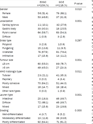
[image:3.612.94.412.97.517.2]Statistical analysis was mainly performed us- ing IBM SPSS Statistics for Windows, version 20.0 (IBM SPSS, Somers, NY, USA). PD-l and PD-L1 data were analyzed using Chi-squ- are test to compare groups and evaluate th- eir associations with various histological and clinicopathological factors. P<0.05 (two-tailed) was considered statistically signifi- cant. 5-year survival rate was calculated us- ing the Kaplan-Meier method. Survival curve Table 1. Comparison of clinicopathological features of gastric
carci-noma between early-onset (EOGC) and elderly (GC-E) groups EOGC
n=109 (%) n=116 (%)GC-E P value
Gender 0.010
Female 56 (51.4) 79 (68.1)
Male 53 (48.6) 37 (31.9)
Localization 0.001
Cardia/pylorus 11 (10.1) 32 (27.6)
Gastric body 33 (30.3) 18 (15.5)
Pyloric canal 64 (58.7) 63 (54.3)
Diffuse 1 (0.9) 3 (2.6)
Gross type 0.297
Polypoid 3 (2.8) 1(0.9)
Fungating 15 (13.8) 11 (9.5)
Ulcerated 74 (67.9) 91 (78.4)
Infiltrative 17 (15.6) 13 (11.2)
Tumour size 0.001
≤5 cm 60 (55.0) 89 (76.7)
>5 cm 49 (45.0) 27 (23.3)
WHO histologic type 0.011
Tubular 23 (21.1) 41 (35.3)
Papillary 0 (0.0) 4 (3.4)
Poorly cohesive 70 (64.2) 51 (44.0)
Mixed 16 (14.7) 19 (16.4)
Other rare types 0 (0.0) 1 (0.9)
Lauren type 0.001
Intestinal 20 (18.3) 46 (39.7)
Diffuse 72 (66.1) 46 (39.7)
Mixed 17 (15.6) 23 (19.8)
Grading 0.000
Well-differentiated 4 (3.7) 6 (5.2) Moderately differentiated 13 (11.9) 39 (33.6) Poorly differentiated 92 (84.4) 71 (61.2)
and B resulted in an IRS ranging from 0 and 7. A total score of ≤2 was considered negative and >2 was considered posi- tive.
test was conducted using the two-stage test method.
Results
Patients
Out of 2,145 GC patients that underwent radi-cal gastrectomy during the study time frame, 109 (5.0%) were EOGC patients (male: female was 1.06:1; median age: 36 years; range: 19-40 years). One hundred and sixteen cases of GC-E were selected at random for compari-son (male: female, 2.1:1; mean age: 69 years; median age: 68 years: range: 60-89 years).
Comparison of clinicopathological features between EOGC and GC-E
The EOGC group had a significantly higher per-centage of women than the GC-E group (31.9% vs 48.6%, P=0.010). Compared with GC-E, EOGC had a significantly higher percentage that occurred in the gastric body (P=0.003) and diameter of tumor >5 cm (P=0.001), wh- ereas GC-E had lower percentage that occurred in the gastric cardiac (P=0.003). There were significant differences between the two groups in terms of histological type (P=0.011) and grade (P=0.001). No significant differences were found between the groups in ratio of gross tumor type, invasion depth, lymphatic
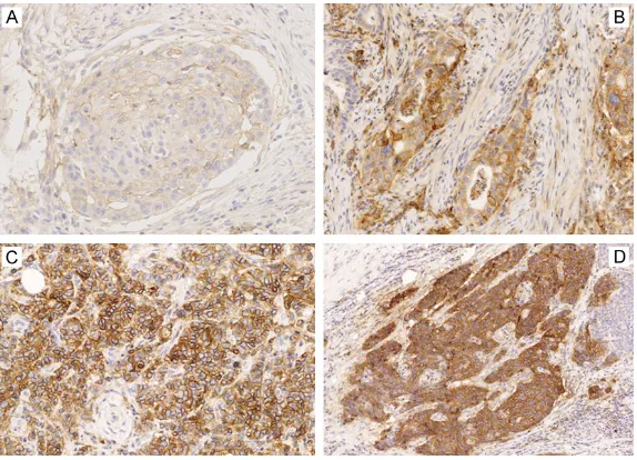
metasta-both groups, PD-L1 membrane staining was observed to varying degrees in tumor cells of 156 (69.3%) patients (Figures 2 and 3). Am- ong them, 96% of cases had ≤10% of PD-L1-positive tumor cells. According to the IRS sys-tem, PD-L1 positive cases accounted for 43.6% (98/225) of all cases. PD-L1 positive expres-sion in immune cells was seen in 49.8% cas- es (112/225) and positive expression in sur-rounding stroma was seen in more than half of the cases (120/225, 53%).
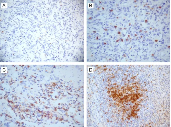
Unlike PD-L1, PD-1 was not expressed in tumor cells but was diffused or scattered expressed in TII and lymphoid follicles in all cases with dif-ferent degrees (median positive expression: 15%; range: 1%-80%) (Figure 4).
Comparison of PD-L1 and PD-1 expression in EOGC and GC-E
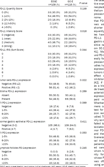
[image:4.612.91.376.71.293.2]Expression intensity of PD-L1 in the EOGC group was significantly higher than in GC-E group (P=0.016). IRS score, based on cell num-bers and staining intensity, also significantly differed between the two groups (P=0.044). Although EOGC group had a higher percentage of specimens with PD-L1-positive tumor cells (P=0.022), no significant difference was found between the two groups with regard to PD-L1 expression in the stroma, immune cells, or nor-mal gastric tissue (Table 2).
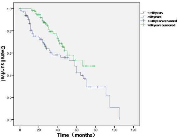
Figure 1. Comparison of overall survival of EOGC group and GC-E group. EOGC patients (blue) had poorer overall survival than GC-E patients (green) (P<0.05).
sis, veins, lymphatic vessel, neural invasion, tumour stage, or HER2 expression (P>0.05 for all). Detailed data is shown in Table 1.
All patients were followed up for 6-108 months (median: 32 months). 5-year OS rate was 42.7% in the EOGC group and 53.6% in GC-E group. The dif-ference in OS was statistically significant between the two groups (P=0.005, Figure 1).
Expression of PD-L1 and PD1
Discussion
EOGC accounted for only 5.0% of all gastric cancers in our cohort study. This is close to the results of previous studies of EOGC [9, 26-28] and, thus, did not suggest an increasing trend in EOGC. Recently, an epidemiological survey
[image:5.612.91.377.72.287.2]of GC was associated with the role of estrogen [30]. Subsequently, this was confirmed by researchers from two countries [31, 32]. Animal experiments have shown that estrogen can block the cell cycle, leading to emergence of numerous polyploid cells and malignant transforma- tion [33]. However, the molecular mechanism
Figure 2. PD-L1 expression in gastric carcinoma tumor cells shown by brown-ish-yellow cell membrane. A. 1 point (≤1% positive tumor cells); lower right corner: zoomed area of positive cells; B. 2 points (2%-10% positive tumor cells); C. 3 points (11%-50% positive tumor cells); and D. 4 points (>50% positive tumor cells) (Immunohistochemical staining; A and C. 100×; B and D. 200×).
Figure 3. Intensity score of PD-L1 expression in tumor of gastric carcinoma. A. 1 point, weak staining; B. 2 points, moderate staining; C and D. 3 points, strong staining (Immunohistochemical staining; 400×).
[image:5.612.91.378.376.583.2]underlying such an association and the puta-tive role of estrogen in women with EOGC awaits further investigation. Moreover, our results demonstrate that the percentage of early gastric cancer located in the gastric body is significantly higher in EOGC than that of gas-tric cancer in the elderly, similar to findings reported in the literature [8]. This further sug-gests that EOGC is more associated with genet-ic changes and family genetgenet-ic factors than GC and less associated with environmental stimuli. Due to limitations of medical history collection, we did not analyze family medical histories of patients in our cohort. Reportedly, ~25% of early gastric cancer is derived from familial fac-tors and this percentage is significantly higher than for family-related gastric cancer in the elderly [26]. E-cadherin gene (CDH1) is associ-ated with hereditary diffuse gastric cancer [34]. However, only 30% of families with inheritance-associated diffuse gastric cancer carry CDH1
germline mutations. The mechanism of devel-opment remains unclear in a large portion of early-onset gastric cancer. Numerous studies have also shown that diffuse histological type is more common in EOGC [6]. Similarly, our study also showed that the percentage of dif-fuse or low-adherence gastric cancer was sig-nificantly higher in the EOGC group than in the GC-E group, whereas the latter had a higher percentage of intestinal gastric cancer. One
bated for years. More data shows that young- er patients with gastric cancer have worse prognosis and survival [8, 35]. Our data showed that the EOGC group had a larger tumor maxi-mal diameter and histological types were also dominated by low-adhesion adenocarcinoma (WHO classification) or diffuse (Lauren classifi-cation) gastric cancer, with a higher proportion of poorly differentiated cancers. All of these dif-ferences in clinicopathological features indi-cate more aggressive behavior for EOGC. Similarly, Japanese researcher Maehara [36] reported a comparative study of clinicopatho-logical data from gastric carcinoma patients aged younger than 40 years and older than 70 years. GC-E was found to have smaller and higher differentiated tumors than gastric carci-noma in younger patients. Similar results have been obtained by many researchers [37, 38]. Some researchers believe that delayed diagno-sis leads to these differences, as young people often feel symptoms less intensely and are less likely to undergo routine physical examinations. However, pathological features (except for tumor size) could not be explained by growth time in our data. We, therefore, believe that EOGC and GC-E have different tumorigenic mechanisms and genetic characteristics, one of which may be PD-L1.
[image:6.612.90.378.72.287.2]Sun et al. [39], for the first time, reported PD-L1
Figure 4. PD1 expression in immune cells and lymphoid follicles of gastric carcinoma. (A) 1%-5% positive cells; (B) 6%-20% positive cells; (C) >20% positive cells; and (D) PD-l expression of some cells within lymphoid follicle (Immunohistochemical staining; A. 200×; B-D. 400×).
histological type may be that gastric cancer develops th- rough different pathways in these two age groups. Gastric cancer development is com-monly considered to be pro-moted by long-term stimula-tion of chronic gastritis. Ga- stric mucosa is repeatedly repaired, followed by gastric mucosal atrophy (atrophic ga- stritis) and intestinal meta- plasia, and subsequent occ- urrence of gastric epithelial dysplasia and carcinogenesis [43]. However, prevalence of diffuse gastric cancer in yo- ung patients is more likely associated with genetic cha- nges such as E-cadherin gene mutations [6].
cases of gastric adenocarcinoma. Immunohi-
[image:7.612.96.459.87.671.2]stochemical staining revealed high PD-L1 ex- testing of PD-L1 expression. In a study of 345 patients with gastric cancer, when analyzed by Table 2. Comparision of expression of PD-L1 and PD-1 in EOGC and GC-E
EOGC
n=109 (%) n=116 (%)GC-E P value
PD-L1 Quantity Score 0.139
0 (0%) 33 (30.3%) 36 (31.0%)
1 (≤1%) 54 (49.5%) 63 (54.3%)
2 (2%-10%) 20 (18.3%) 10 (8.6%)
3 (11%-50%) 2 (1.8%) 6 (5.2%)
4 (>50%) 0 (0%) 1 (0.9%)
PD-L1 Intensity Score 0.016
0 (negative) 33 (30.3%) 36 (31.0%)
1 (weak) 25 (22.9%) 39 (33.6%)
2 (medium) 40 (36.7%) 22 (19.0%)
3 (strong) 11 (10.1%) 19 (16.4%)
PD-L1 IRS (Sum Score) 0.044
0 33 (30.3%) 36 (31.0%)
2 20 (18.3%) 36 (31.0%)
3 32 (29.4%) 18 (15.5%)
4 20 (18.3%) 15 (12.9%)
5 3 (2.8%) 6 (5.2%)
6 1 (0.9%) 4 (3.4%)
7 0 (0.0%) 1 (0.9%)
Tumor cells PD-L1 expression 0.022
Negative (IRS ≤2) 53 (48.6) 74 (63.8)
Positive (IRS >2) 56 (51.4) 42 (36.2)
Stroma PD-L1 expression 0.269
Absent 55 (50.5) 50 (43.1)
Present 54 (49.5) 66 (56.9)
TII PD-L1 expression 0.089
Negative 19 (17.4) 9 (7.8)
1-5% 43 (39.4) 44 (37.9)
6-20% 28 (25.7) 32 (27.6)
>20% 19 (17.4) 31 (26.7)
Normal gastric epithelial PD-L1 expression 0.411
Negative (0-2) 105 (96.3) 109 (94.0)
Positive (3-7) 4 (3.7) 7 (6.0)
TII PD1 expression 0.050
1-5% 53 (48.6) 45 (38.8)
6-20% 35 (38.8) 32 (27.6)
>20% 21 (19.3) 39 (33.6)
Lymphoid follicles PD-1expression 0.245
Negative 0 (0.0) 3 (2.6)
1-5% 55 (50.5) 53 (45.7)
6-20% 39 (35.8) 38 (32.8)
>20% 15 (13.8) 22 (19.0)
EOGC: early onset gastric carcinoma (initial diagnosis ≤40 years); GC-E: gastric carcinoma in elderly (initial diagnosis >60 years); TII: tumor-infiltrating immunocytes.
tissue-array, PD-L1 expression was only seen in 1.7% of cases but 35.6% showed PD-L1 expression in parallel analysis of the whole sec-tion [43].
Here, we focused on differences in PD-L1 and PD-1 expression between EOGC and GCE. Despite having similar numbers of positive cells (P=0.169), the EOGC group showed higher intensity of PD-L1 expression (P=0.016) and had a higher percentage of positive PD-L1 expression (P=0.022), as shown by scores based on cell numbers and expression intensi-ty. According to literature review, this is the first study of PD-L1 and PD-1 expression in young patients with gastric cancer. Although several studies have shown that expression of PD-L1 and PD-1 is not related to age in gastric cancer, very few cases of EOGC were included and 60-70 years of age was often the cut-off bound-ary for “younger” patients. Thus, those studies did not actually analyze gastric cancer in young people. Additionally, several studies have shown a higher percentage of PD-L1 expres-sion in gastric cancer at a younger age, howev-er, the difference was not significant, perhaps due to sample size or grouping [44]. With regard to high PD-L1 expression in EOGC, a possible reason is that high PD-L1 expression and EOGC are both associated with some clinicopatho-logical features that are indicative of poor prog-nosis, such as tumor size and lymph node metastasis. This also indicates that immune escape mediated by high PD-L1 expression may be another reason for poor prognosis for EOGC and may reflect a gastric cancer cause that is more common in younger patients. Reportedly, EOGC cell molecular phenotypes such as cell adhesion molecules, E-cadherin, and mucin differ from those of GC-E [16]. Our results provide a new study point for analysis of gastric carcinogenesis in young people. Moreover, PD-1/PD-L1 pathway inhibitors are the subject of increasing research, some enter-ing phase III of clinical trials. Several studies have shown that high PD-L1 expression is asso-ciated with therapeutic effectiveness [37]. Whether PD-l/PD-L1 inhibitors would be more effective against EOGC should be tested and verified by more clinical trials.
Conclusion
In this study, we have shown that the percent-age of EOGC is relatively low among GC cases.
EOGC shows different clinicopathological fea-tures and shorter OS compared with GC-E. PD-L1 is highly expressed in GC tissue. Its expression is significantly higher in EOGC tumor tissue than in GC-E.
Acknowledgements
We would like thank Marla Brunker, from LiwenBianji, Edanz Group China (www.liwenbi-anji.cn/ac), for editing the English text draft of this manuscript.
Disclosure of conflict of interest
None.
Address correspondence to: Dr. Mulan Jin, Depart- ment of Pathology, Beijing Chaoyang Hospital, Capital Medical University, 8 Gongtinanlu, Chaoyang District, Beijing 100020, China. Tel: +86 182-0147-3030; E-mail: kinmokuran@163.com
References
[1] Torre LA, Bray F, Siegel RL, Ferlay J, Lortet-Tieu-lent J, Jemal A. Global cancer statistics, 2012. CA Cancer J Clin 2015; 65: 87-108.
[2] Mitsudomi T, Matsusaka T, Wakasugi K, Take-naka M, Kume K, Fujinaga Y, Teraoka H, Iwashita A. A clinicopathological study of gas-tric cancer with special reference to age of the patients: an analysis of 1,630 cases. World J Surg 1989; 13: 225-230; discussion 230-231. [3] Sánchez-Bueno F, Garcia-Marcilla JA, Perez-Flores D, Pérez-Abad JM, Vicente R, Aranda F, Ramirez P, Parrilla P. Prognostic factors in aser-ies of 297 patients with gastric adenocarcino-ma undergoing surgical resection. Br J Surg 1998; 85: 255-260.
[4] Corso G, Pedrazzani C, Pinheiro H, Fernandes E, Marrelli D, Rinnovati A, Pascale V, Seruca R, Oliveira C, Roviello F. E-cadherin genetic screening and clinicopathologic characteris-tics of early onset gastric cancer. Eur J Cancer 2011; 47: 631-639.
[5] Hsieh FJ, Wang YC, Hsu JT, Liu KH, Yeh CN. Clinicopathological features and prognostic factors of gastric cancer patients aged 40 years or younger. J Surg Oncol 2012; 105: 304-309.
[6] Theuer CP, de Virgilio C, Keese G, French S, Ar-nell T, Tolmos J, Klein S, Powers W, Oh T, Sta-bile BE. Gastric adenocarcinoma inpatients 40 years of age or younger. Am J Surg 1996; 172: 473-476; discussion 476-477.
[8] Nakamura T, Yao T, Niho Y, Tsuneyoshi M. A clinicopathological study in young patients with gastric carcinoma. J Surg Oncol 1999; 71: 214-219.
[9] Matley PJ, Dent DM, Madden MV, Price SK. Gastric carcinoma in young adults. Ann Surg 1988; 208: 593-596.
[10] Tso PL, Bringaze WL 3rd, Dauterive AH, Correa P, Cohn I Jr. Gastric carcinoma in the young. Cancer 1987; 59: 1362-1365.
[11] Rugge M, Busatto G, Cassaro M, Shiao YH, Russo V, Leandro G, Avellini C, Fabiano A, Sido-ni A, Covacci A. Patients younger than 40 years with gastric carcinoma: helicobacter pylori genotype and associated gastritis phenotype. Cancer 1999; 85: 2506-2511.
[12] Fuchs CS, Mayer RJ. Gastric carcinoma. N Engl J Med 1995; 333: 32-41.
[13] Wanebo HJ, Kennedy BJ, Chmiel J, Steele G, Winchester D, Osteen R. Cancer of the stom-ach. A patient care study by the American col-lege of surgeons. Ann Surg 1993; 218: 583-592.
[14] Carvalho R, Milne AN, van Rees BP, Caspers E, Cirnes L, Figueiredo C, Offerhaus GJ, Weter-man MA. Early-onset gastric carcinomas dis-play molecular characteristics distinct from gastric carcinomas occurring at a later age. J Pathol 2004; 204: 75-83.
[15] Buffart TE, Carvalho B, Hopmans E, Brehm V, Kranenbarg EK, Schaaij-Visser TB, Eijk PP, van Grieken NC, Ylstra B, van de Velde CJ, Meijer GA. Gastric cancers in young and elderly pa-tients show different genomic profiles. J Pathol 2007; 211: 45-51.
[16] Milne AN, Carvalho R, Morsink FM, Musler AR, de Leng WW, Ristimaki A, Offerhaus GJ. Early-onset gastric cancers have a different molecu-lar expression profile than conventional gastric cancers. Mod Pathol 2006; 19: 564-572. [17] Zhang X, Schwartz JC, Guo X, Bhatia S, Cao E,
Lorenz M, Cammer M, Chen L, Zhang ZY, Edi-din MA, Nathenson SG, Almo SC. Structural and functional analysis of the costimulatory receptor programmed death-1. Immunity 2004; 20: 337-347.
[18] Yamazaki T, Akiba H, Iwai H, Matsuda H, Aoki M, Tanno Y, Shin T, Tsuchiya H, Pardoll DM, Okumura K, Azuma M, Yagita H. Expression of programmed death 1 ligands by murine T cells and APC. J Immunol 2002; 169: 5538-5545. [19] Brown JA, Dorfman DM, Ma FR, Sullivan EL,
Munoz O, Wood CR, Greenfield EA, Freeman GJ. Blockade of programmeddeath-1 ligands on dendritic cells enhances T cell activation and cytokine production. J Immnol 2003; 170: 1257-1266.
[20] Pardol DM. The blockade of immune check-points in cancer immunotherapy. Nat Rev Can-cer 2012; 12: 252-264.
[21] Hou J, Yu Z, Xiang R, Li C, Wang L, Chen S, Li Q, Chen M, Wang L. Correlation between infiltra-tion of FOXP3+ regulatory T cells and expres-sion of B7-H1 in the tumor tissues of gastric cancer. Exp Mol Pathol 2014; 96: 284-291. [22] Liu SM, Meng Q, Zhang QX, Wang SD, Liu ZJ,
Zhang XF. Expression and significance of B7-H1 and its receptor PD-1 in human gastric car-cinoma. Zhonghua Zhong Liu Za Zhi 2008; 30: 192-195.
[23] Lu B, Chen L, Liu L, Zhu Y, Wu C, Jiang J, Zhang X. T-cell-mediated tumor immune surveillance and expression of B7 co-inhibitory molecules in cancers of the upper gastrointestinal tract. Immunol Res 2011; 50: 269-275.
[24] In: Hamilton SR, Aaltonen LA, editors. Patholo-gy and genetics of tumours of the digestive system. Lyon: IARC press; 2000. pp. 45-59. [25] Lauren T. The two histologic main types of
gas-tric carcinoma: diffuse and so-called intestinal-type carcinoma. An attempt at a histoclinical classification. Acta Pathol Microbiol Scand 1965; 64: 31-49.
[26] Koea JB, Karpeh MS, Brennan MF. Gastric can-cer in young patients: demographic, clinico-pathological, and prognostic factors in92 pa-tients. Ann Surg Oncol 2000; 7: 346-351. [27] Park HJ, Ahn JY, Jung HY, Lim H, Lee JH, Choi
KS, Kim DH, Choi KD, Song HJ, Lee GH, Kim JH. Clinical characteristics and outcomes for gastric cancer patients aged 18-30 years. Gas-tric Cancer 2014; 17: 649-660.
[28] Zhou F, Shi J, Fang C, Zou X, Huang Q. Gastric carcinomas in young (younger than 40 years) Chinese patients: clinicopathology, family his-tory, and post resection survival. Medicine (Baltimore) 2016; 95: e2873.
[29] Anderson WF, Camargo MC, Fraumeni JF Jr, Correa P, Rosenberg PS, Rabkin CS. Age-spe-cific trends in incidence of noncardia gastric cancer in US adults. JAMA 2010; 303: 1723-1728.
nutrition. Am J Epidemiol 2010; 172: 1384-1393.
[31] Persson C, Inoue M, Sasazuki S, Kurahashi N, Iwasaki M, Ye W, Tsugane S; JPHC Study Group. Female reproductive factors and the risk of gastric cancer in a large-scale population-based cohort study in Japan (JPHC study). Eur J Cancer Prev 2008; 17: 345-353.
[32] Freedman ND, Chow WH, Gao YT, Shu XO, Ji BT, Yang G, Lubin JH, Li HL, Rothman N, Zheng W, Abnet CC. Menstrual and reproductive fac-tors and gastric cancer risk in a large prospec-tive study of women. Gut 2007; 56: 1671-1677.
[33] Di Domenico M, Castoria G, Bilancio A, Migliac-cio A, Auricchio F. Estradiol activation of hu-man colon carcinoma-derived Caco-2 cell growth. Cancer Res 1996; 56: 4516-4521. [34] Guilford P, Hopkins J, Harraway J, McLeod M,
McLeod N, Harawira P, Taite H, Scoular R, Mill-er A, Reeve AE. E-cadhMill-erin gMill-ermline mutations in familial gastric cancer. Nature 1998; 392: 402-405.
[35] Smith BR, Stabile BE. Extreme aggressiveness and lethality of gastric adenocarcinoma in the very young. Arch Surg 2009; 144: 506-510. [36] Maehara Y, Emi Y, Tomisaki S, Oshiro T, Kakeji
Y, Ichiyoshi Y, Sugimachi K. Age-related charac-teristics of gastric carcinoma in young and el-derly patients. Cancer 1996; 77: 1774-1780. [37] Santoro R, Carboni F, Lepiane P, Ettorre GM,
Santoro E. Clinicopathological features and prognosis of gastric cancer in young European adults. Br J Surg 2007; 94: 737-742.
[38] Bani-Hani KE. Clinicopathological comparison between young and old age patients with gas-tric adenocarcinoma. Int J Gastrointest Cancer 2005; 35: 43-52.
[39] Sun J, Xu K, Wu C, Wang Y, Hu Y, Zhu Y, Chen Y, Shi Q, Yu G, Zhang X. PD-L1 expression analy-sis in gastric carcinoma tissue and blocking of tumor associated PD-L1 signaling by two func-tional monoclonal antibodies. Tissue Antigens 2007; 69: 19-27.
[40] Böger C, Behrens HM, Mathiak M, Krüger S, Kalthoff H, Röcken C. PD-L1 is an independent prognostic predictor in gastric cancer of West-ern patients. Oncotarget 2016; 7: 24269-24283.
[41] Mu CY, Huang JA, Chen Y, Chen C, Zhang XG. High expression of PD-L1 in lung cancer may contribute to poor prognosis and tumor cells immune escape through suppressing tumor infiltrating dendritic cells maturation. Med On-col 2011; 28: 682-688.
[42] Topalian SL, Hodi FS, Brahmer JR, Gettinger SN, Smith DC, McDermott DF, Powderly JD, Carvajal RD, Sosman JA, Atkins MB, Leming PD, Spigel DR, Antonia SJ, Horn L, Drake CG, Pardoll DM, Chen L, Sharfman WH, Anders RA, Taube JM, McMiller TL, Xu H, Korman AJ, Jure-Kunkel M, Agrawal S, McDonald D, Kollia GD, Gupta A, Wigginton JM, Sznol M. Safety, activi-ty, and immune correlates of PD-1 anti-body in cancer. N Engl J Med 2012; 366: 2443-2454.
[43] Farris AB 3rd, Demicco EG, Le LP, Finberg KE, Miller J, Mandal R, Fukuoka J, Cohen C, Gais-sert HA, Zukerberg LR, Lauwers GY, Iafrate AJ, Mino-Kenudson M. Clinicopathologic and mo-lecular profiles of microsatellite unstable Bar-rett esophagus-associated adenocarcinoma. Am J Surg Pathol 2011; 35: 647-655.