Int J Clin Exp Pathol 2015;8(5):4844-4850 www.ijcep.com /ISSN:1936-2625/IJCEP0007545
Original Article
Crocin attenuates lipopolysacchride-induced acute lung
injury in mice
Jian Wang1*, Jianke Kuai2*, Zhonghua Luo3*, Wuping Wang1, Lei Wang1, Changkang Ke1, Xiaofei Li1, Yunfeng Ni1
1Department of Thoracic Surgery, Tangdu Hospital, The Fourth Military Medical University, Xi’an 710038, China; 2Department of Anaesthesiology, Tangdu Hospital, The Fourth Military Medical University, Xi’an 710038, China; 3Department of Interventional Radiology, Tangdu Hospital, The Fourth Military Medical University, Xi’an 710038,
China. *Equal contributors.
Received March 3, 2015; Accepted April 15, 2015; Epub May 1, 2015; Published May 15, 2015
Abstract: Crocin, a representative of carotenoid compounds, exerts a spectrum of activities including radical
scav-enger, anti-microbial and anti-inflammatory properties. To investigate the protective effect of crocin on lipopolysac -charide (LPS)-induced acute lung injury (ALI) in mice. ALI was induced in mice by intratracheal instillation of LPS
(1 mg/kg). The mice received intragastric injection of crocin (50 mg/kg) 1 h before LPS administration. Pulmonary
histological changes were evaluated by hematoxylineosin stain and lung wet/dry weight ratios were observed.
Con-centrations of tumor necrosis factor (TNF)-α, interleukin (IL)-1β and nitric oxide (NO), and myeloperoxidase (MPO) activity were measured by enzymelinked immunosorbent assay. Expression of inducible nitric oxide synthase (iNOS) in lung tissues was determined by Western blot analysis. Crocin pretreatment significantly alleviated the severity of lung injury and inhibited the production of TNF-α and IL-1β in mice with ALI. After LPS administration, the lung wet/ dry weight ratios, as an index of lung edema, and MPO activity were also markedly reduced by crocin pretreatment. Crocin pretreatment also reduced the concentrations of NO in lung tissues. Furthermore, the expression of iNOS was significantly suppressed by crocin pretreatment. Croncin potently protected against LPS-induced ALI and the protective effects of crocin may attribute partly to the suppression of iNOS expression.
Keywords: Crocin, LPS, Acute lung injury, iNOS
Introduction
Acute lung injury (ALI) and acute respiratory
dis-tress syndrome (ARDS) are well defined and
readily recognized clinical disorders caused by many clinical insults to the lung or because of predispositions to lung injury [1]. Although the causes of ALI and ARDS are numerous, endo-toxin is thought to be the most pathogen that leads to the development of ALI and ARDS. Endotoxin or lipopolysacchride (LPS), derived from the cell wall of gram-negative bacteria, induces a sepsis syndrome accompanied by key features of ALI, including the recruitment of
inflammatory cells into the lung with subse -quent increases in capillary permeability and
alveolar edema [2]. Despite significant advanc -es in intense care r-esearch and diverse thera-peutic trials made in the past few decades, ALI/ ARDS remains a severe disease and still
pres-ents the high mortality rate of approximately 40% [3, 4].
Inducible nitric oxide synthase (iNOS), an induc -ible enzyme, is transcriptionally regulated by
pro-inflammatory products and cytokines, and
results in sustained and elevated release of
nitric oxide (NO). This iNOS-derived NO contrib -utes to the pathophysiologic features of ALI [5,
6]. It has been shown that mice deficient in iNOS gene were more resistant to lung injury than were wild-type mice [7, 8]. Therefore, sup
-pression of the induction and activity of iNOS has been considered a new paradigm in inflam -mation therapy.
as carbohydrate residues. It has been showed that crocin has a wide range of activities, includ-ing antidepressant, anticarcinogenic, antiath-erosclerotic, antioxidant, and antihyperlipid-emic [9-12]. Recent studies have demonstrated
the anti-inflammatory effect of crocin [13, 14]. In neuroinflammatoty diseases, crocin protect -ed oligodendrocytes expos-ed to cytotoxic su- pernatants derived from Syncytin-1-expressing
astrocytes and NO-mediated oligodendrocyto -toxicity [15]. But to our knowledge, the
anti-inflammatory effect of crocin in ALI has not been well established. Therefore, the aim of
this study is to investigate the protective effects of crocin on LPS-induced ALI.
Materials and methods
Animals and reagents
Male BALB/C mice weighing 20-25 g were
pur-chased from the Animal Center of the Fourth
Military Medical University (Xian, China). All ani-mals were allowed to take food and tap water ad libitum. All procedures were in accordance with the Declaration of Helsinki of the World
Medical Association. The protocols were also
approved by the Institutional Animal Care and
Use Committee of the Fourth Military Medical University Tangdu Hospital. Crocin was pur -chased from Sigma Chemical Co. (Missouri, USA). LPS (Escherichia coli lipopolysaccharide, 055:B5) was obtained from Sigma Chemical
Company (St. Louis, MO., USA). Enzyme-linked immunosorbent assay (ELISA) kits of TNF-α, IL-1β, myeloperoxidase (MPO) and nitric oxide (NO) were purchased from R&D Corporation (R&D Systems Inc. Minneapolis, MN, USA). Antibodies specific for iNOS and β-actin were
obtained from the Wuhan Boster Biological
[image:2.612.91.524.71.399.2]Technology, Ltd. (Wuhan, China). Cell lysis buf -fer was the product of Beyotime Biotechnology (Beijing, China). BCA protein assay kit was Figure 1. Effect of crocin on the pulmonary histopathological changes of mice with ALI. Lung sections stained with hematoxylin-eosin from 12 h after LPS administration revealed pulmonary histopathological changes (original
mag-nification ×200). A. Control group: normal structure. B. Crocin group: same as control group. C. LPS group: alveolar wall thickness, hemorrhage, alveolus collapse and obvious inflammatory cells infiltration. D. LPS+crocin group:
Crocin protects lung against lipopolysaccharide induced injury
obtained from Thermo Scientific Pierce Protein
Research Products (Rockford, IL., USA).
Animal model of ALI
Before the induction of ALI, mice were fasted overnight but allowed water ad libitum. Animals were anesthetized with intraperitoneal pento-barbital (50 mg/kg). LPS (1 mg/kg) was instilled intratracheally to induce ALI.
Experimental protocol
All animals were randomly divided into 4 groups (n=10, each group). Group 1 (control group) treated with intragastric injection of normal saline and 1 h later received an intratracheal injection of normal saline, group 2 (crocin group) treated with intragastric injection of cro-cin (50 mg/kg) and 1 h later received an intra-tracheal injection of normal saline, group 3 (LPS group) treated with intragastric injection of normal saline and 1 h later received an intra-tracheal injection of LPS (1 mg/kg) and group 4
(LPS+crocin group) treated with intragastric
injection of crocin (50 mg/kg) and 1 h later received an intratracheal injection of LPS (1
mg/kg). Twelve hours after LPS administration, all animals were sacrificed. Bronchoalveolar
lavage (BAL) was performed through the left
lung. After the lung vasculature was flushed,
the superior lobe of right lung was excised for
histopathologic examination. The middle lobe
of right lung was excised for analysis of lung
wet/dry weight ratio. The lower lobe of right
lung was rapidly removed and cut into two parts in same size. A part of the lower lobe was homogenized and frozen in a cold phosphate
solution at -80°C for MPO analysis, and the
another part was used to extract proteins for Western blot analysis.
BAL
Animals were anesthetized with intraperitoneal pentobarbital (50 mg/kg). A median sternoto-my allowed for exposure of both of the lungs.
The trachea was exposed and inserted with an
intravenous infusion needle. After ligating the hilum of right lung, the left lung was lavaged 5 times with 0.5 ml ice-cold phosphate buffered
saline. The recovery ratio of the fluid was about 90%. The BAL fluid (BALF) was immediately centrifuged at 500 × g for 10 min at 4°C, and
the cell-free supernatant was stored at -80°C for analysis of cytokines.
Cytokines measurement
Concentrations of IL-1β and TNF-α in BALF were
measured by using ELISA kits. All procedures were done in accordance with the manufactur-er’s instructions.
MPO and NO assays
To carry out the assays, tissue samples were
subjected to three further freeze-thaw cycles and centrifuged at 12 000 × g for 10 min at
[image:3.612.89.521.76.242.2]4°C. The supernatant was assayed for MPO
Figure 2. Effect of crocin on MPO activity and NO concentrations in lung tissues of mice with ALI. A. The MPO activity in lung tissues after LPS administration and the effect of crocin pretreatment. B. The concentrations of NO in lung
activity and NO concentrations with ELISA kits.
All procedures were done in accordance with the manufacturer’s instructions.
Lung wet/dry weight ratio
As an index of lung edema, the amount of
extra-vascular lung water was calculated. The middle
lobe of right lung was excised and the wet
weight was recorded. The lobe was then placed
in an incubator at 80°C for 24 hours to obtain the dry weight. And the wet/dry weight ratios were calculated by dividing the wet weight by the dry weight.
Pulmonary histopathology
The superior lobe of right lung was harvested at 12 h after LPS administration and fixed with an
intratracheal instillation of 1 ml buffered
forma-lin (10%, PH 7.2). The lobe was further fixed in
10% neutral buffered formalin for 24 h at 4°C.
The tissues were embedded in paraffin and cut into 5 μm sections. Hematoxylin-eosin stains
were performed using standard protocol.
Western blot analysis
The lower lobe of right lung in each mouse was
harvested at 12 h after LPS administration and frozen in liquid nitrogen immediately until
homogenization. Tissue samples were homog -enized in cell lysis buffer. After centrifugation (12 000 × g, 10 min, 4°C), supernatants were aspirated. Protein concentrations were
deter-mined by BCA protein assay kit. Samples were separated on a denaturing 12% polyacrylamide gel and transferred to a nitrocellulose
mem-brane. iNOS proteins were detected by chemilu -minescence using a rabbit polyclonal antibody according to the manufacturer’s instructions.
Statistical analyses
Data were entered into a database and ana-lyzed using SPSS software, and expressed as means ± S.D. On a preliminary analysis, the
Kolmogorov-Smirnov test found that the raw pooled data followed a Gaussian distribution.
Thus, statistically significant differences be-tween groups were determined by ANOVA fol
-lowed by Tukey’s test. Significance was accept -ed when P < 0.05.
Results
Effect of crocin on the pulmonary histopatho-logical changes of mice with ALI
Lung tissues from the control and crocin groups showed a normal structure and no histopatho-logical changes under a light microscope (Figure 1A and 1B). In LPS group, the lungs stained with hematoxylin-eosin indicated wide-spread alveolar wall thickness caused by edema, severe hemorrhage in the alveolus,
alveolus collapse and obvious inflammatory cells infiltration (Figure 1C). In LPS+crocin
[image:4.612.90.522.75.239.2]Crocin protects lung against lipopolysaccharide induced injury
especially in inflammatory cells infiltration
(Figure 1D).
Effect of Crocin on MPO activity and NO con-centrations in lung tissues of mice with ALI
After LPS administration, the MPO activity in lung tissues was significantly increased com
-pared with the control and crocin groups (Figure 2A). In addition, the concentrations of NO were also significantly increased after LPS adminis -tration (Figure 2B). However, crocin
pretreat-ment markedly decreased the MPO activity and NO concentrations (Figure 2A and 2B).
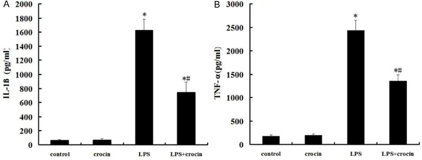
Effect of crocin on the concentrations of IL-1β and TNF-α in BALF of mice with ALI
The concentrations of IL-1β and TNF-α in BALF were significantly increased at 12 h after LPS
administration (Figure 3A and 3B). Crocin
pre-treatment efficiently reduced the production of IL-1β and TNF-α (Figure 3A and 3B).
Effect of crocin on the lung edema of mice with ALI
Compared with the control and crocin groups,
the lung wet/dry weight ratios were significantly increased after LPS administration. The
increase of the lung wet/dry weight ratios was
significantly reduced by crocin administration
(Figure 4).
Effect of crocin on the expression of iNOS in lung tissues of mice with ALI
After LPS administration, the expression of
iNOS in lung tissues markedly increased (Figure 5). However, the pretreatment of crocin signifi -cantly suppressed LPS-induced activation of
iNOS (Figure 5). There were no significant changes in the expression of iNOS in control
and crocin groups (Figure 5). Discussion
LPS are major components of the outer mem-brane of gram-negative bacteria, and in vivo
could trigger potent inflammatory responses. In
gram-negative bacteria sepsis and pneumonia,
LPS is capable of inducing ALI. The animal mod -els of ALI induced by LPS administration are
easy to establish and very reproducible. The intratracheal injection of LPS induces signifi
-cant increase of proinflammatory cytokines in BALF. In the present study, the concentrations of TNF-α and IL-1β in BALF increased markedly in mice treated by LPS. Elevated levels of TNF-α and IL-1β play a key role in the progression of ALI. Because TNF-α and IL-1β can stimulate
production of a host of other cytokines, they have earned a position of prominence at the
head of the inflammatory cytokine cascade
Figure 4. Effect of crocin on the lung edema of mice with ALI. After LPS administration, pretreatment of crocin decreased lung wet/dry ratios markedly. Data are expressed as mean±S.D, *P<0.05 vs. control and crocin groups; #P<0.05 vs. LPS group.
Figure 5. Effect of crocin on the expression of iNOS
in lung tissues of mice with ALI. A. A representative
Western blot showed the expression of iNOS in lung tissues in different groups. B. iNOS optical densitom -etry from different groups. Pretreatment of crocin
sig-nificantly repressed the expression of iNOS after LPS
[15]. Therefore, the inhibition of TNF-α and IL-1β showed the reduction of pulmonary injury
in ALI induced by LPS in mice [16, 17]. In the
present study, crocin pretreatment significantly reduced the concentrations of TNF-α and IL-1β in BALF in mice received intratracheal injection
of LPS.
Neutrophils are an important component of the
inflammatory response that characterizes ALI and are considered to be the final effector cell
responsible for lung injury, due to their ability to express multiple cytotoxic products [18, 19]. In endotoxemia-induced ALI, the neutrophils
accu-mulated in the lungs, express proinflammatory cytokines, such as IL-1β and TNF-α, and finally lead to the pulmonary injury [20]. MPO is a
major constituent of neutrophil cytoplasmic
granules. The total activity of MPO in a tissue
is therefore a direct measure of neutrophil sequestration in that tissue [21]. In the present
study, we found the MPO activity increased evi -dently in lung tissues after LPS exposure. As
expected, crocin pretreatment significantly decreased the MPO activity in lung tissues. In
addition, histopathological study also indicated that crocin pretreatment markedly attenuated
the neutrophil infiltration in lungs.
NO is a pleiotropic mediator, which acts in a
variety of physiological and pathophysiological
processes. Increased levels of NO, measured as nitrite, were present in BALF from
LPS-treated rat lungs and LPS-elicited BAL
leuko-cytes produced increased NO in culture [22]. In humans either at risk of ARDS or with confirmed ARDS, the level of NO in their BALF increased significantly [23]. Moreover, the increased NO
production is associated with evidence for
increased iNOS expression in lung tissue [24, 25]. iNOS is calcium-independent and can be induced by pro-inflammatory agents, such as LPS, IL-1β, TNF-α and interferon-γ (INF-γ), in endothelial and smooth-muscle cells, in macro-phages and in other cell types [26]. In the
pres-ent study, the production of NO and the expres
-sion of iNOS in lung tissues increased significantly after LPS administration, which
were markedly inhibited by crocin
pretreat-ment. It has been demonstrated that iNOS
inhibitor prevents the lung injury associated
with inflammation [27, 28]. Therefore, our results suggest that the attenuation of inflam -matory responses in ALI by crocin were partially
due to the suppression of iNOS expression by
crocin.
Pulmonary edema is a life-threatening condi-tion that frequently leads to acute respiratory failure. Injury to the alveolar epithelium can dis-rupt the integrity of the alveolar barrier or down-regulate ion transport pathways, thus, reducing
net alveolar fluid reabsorption and enhancing
the extent of alveolar edema [29]. Here, we
observed a significant reduction of pulmonary
injury and edema in lungs of ALI mice
pretreat-ed by crocin. Therefore, crocin possesspretreat-ed the
protective effect on ALI, which implied the clini-cal use of crocin in future.
In conclusion, our study shows that crocin can attenuate the pulmonary histological changes,
lung edema, reduce the neutrophils infiltration in lung, and inhibit the release of inflammatory cytokines in BALF. These protective effects of crocin may involve the suppression of iNOS
expression. Although crocin exerts its
anti-inflammatory effect in our study, further and
comprehensive studies are still needed before clinical application.
Disclosure of conflict of interest
None.
Address correspondence to: Drs. Xiaofei Li and
Yunfeng Ni, Department of Thoracic Surgery, Tangdu Hospital, The Fourth Military Medical University,
Xi’an 710038, China. E-mail: lxfchest@fmmu.edu.cn
(LXF); niyunfng@fmmu.edu.cn (NYF)
References
[1] Wheeler AP, Bernard GR. Acute lung injury and the acute respiratory distress syndrome: a clin-ical review. Lancet 2007; 369: 1553-1565. [2] Worthen GS, Haslett C, Rees AJ, Gumbay RS,
Henson JE, Henson PM. Neutrophil-mediated pulmonary vascular injury. Synergistic effect of trace amounts of lipopolysaccharide and neu-trophil stimuli on vascular permeability and neutrophil sequestration in the lung. Am Rev Respir Dis 1987; 136: 19-28.
[3] Rubenfeld GD, Caldwell E, Peabody E, Weaver J, Martin DP, Neff M, Stern EJ, Hudson LD. Inci-dence and outcomes of acute lung injury. N Engl J Med 2005; 353: 1685-93.
Crocin protects lung against lipopolysaccharide induced injury
[5] Wang LF, Patel M, Razavi HM, Weicker S, Jo -seph MG, McCormack DG, Mehta S. Role of inducible nitric oxide synthase in pulmonary microvascular protein leak in murine sepsis. Am J Respir Crit Care Med 2002; 165: 1634-1639.
[6] Lee RP, Wang D, Kao SJ, Chen HI. The lung is
the major site that produces nitric oxide to in-duce acute pulmonary oedema in endotoxin shock. Clin Exp Pharmacol Physiol 2001; 28: 315-320.
[7] Kristof AS, Goldberg P, Laubach V, Hussain SN.
Role of inducible nitric oxide synthase in endo-toxin-induced acute lung injury. Am J Respir Crit Care Med 1998; 158: 1883.
[8] Genovese T, Cuzzocrea S, Di Paola R, Failla M, Mazzon E, Sortino MA, Frasca G, Gili E, Crimi N, Caputi AP, Vancheri C. Inhibition or knock out
of inducible nitric oxide synthase result in re-sistance to bleomycin-induced lung injury. Respir Res 2005; 6: 58.
[9] Wang Y, Han T, Zhu Y, Zheng CJ, Ming QL, Rah
-man K, Qin LP. Antidepressant properties of
bioactive fractions from the extract of Crocus sativus L. J Nat Med 2010; 64: 24-30.
[10] Aung HH, Wang CZ, Ni M, Fishbein A, Mehen
-dale SR, Xie JT, Shoyama CY, Yuan CS. Crocin from Crocus sativus possesses significant
anti-proliferation effects on human colorectal
can-cer cells. Exp Oncol 2007; 29: 175-180.
[11] He SY, Qian ZY, Tang FT, Wen N, Xu GL, Sheng
L. Effect of crocin on experimental atheroscle-rosis in quails and its mechanisms. Life Sci 2005; 77: 907-921.
[12] Lee IA, Lee JH, Baek NI, Kim DH. Antihyperlip-idemic effect of crocin isolated from the fruc-tus of Gardenia jasminoides and its metabolite crocetin. Biol Pharm Bull 2005; 28: 2106-2110.
[13] Nam KN, Park YM, Jung HJ, Lee JY, Min BD, Park SU, Jung WS, Cho KH, Park JH, Kang I,
Hong JW, Lee EH. Anti-inflammatory effects of
crocin and crocetin in rat brain microglial cells. Eur J Pharmacol 2010; 648: 110-116. [14] Xu GL, Li G, Ma HP, Zhong H, Liu F, Ao GZ. Pre
-ventive effect of crocin in inflamed animals
and in LPS-challenged RAW 264.7 cells. J Agric
Food Chem 2009; 57: 8325-8330.
[15] Goodman RB, Pugin J, Lee JS, Matthay MA.
Cytokine-mediated inflammation in acute lung injury. Cytokine Growth Factor Rev 2003; 14:
523-35.
[16] Chen J, Liu X, Shu Q, Li S, Luo F. Ghrelin attenu -ates lipopolysaccharide-induced acute lung
in-jury through NO pathway. Med Sci Monit 2008;
14: 141-6.
[17] Mei SH, McCarter SD, Deng Y, Parker CH, Liles WC, Stewart DJ. Prevention of LPS-induced acute lung injury in mice by mesenchymal stem cells overexpressing angiopoietin 1. PLoS Med 2007; 4: e269.
[18] Abraham E. Neutrophils and acute lung injury. Crit Care Med 2003; 31: 195-199.
[19] Parsey MV, Tuder R, Abraham E. Neutrophils
are major contributors to intraparenchymal
lung IL-1β expression after hemorrhage and
endotoxemia. J Immunol 1998; 160: 1007-1101.
[20] Chen J, Luo J, Li B, Ran P. E1A has no effect on LPS-induced IL-6 secretion in rat alveolar epi-thelial cells. Respiration 2009; 78: 84-92. [21] Wang F, He B. The effect of dithiothreitol on
chemotactic factors in induced sputum of chronic obstructive pulmonary disease pa-tients. Respiration 2009; 78: 217-222. [22] Li XY, Donaldson K, Macnee W.
Lipopolysac-charide-induced alveolar epithelial
permeabil-ity: The role of nitric oxide. Am J Respir Crit
Care Med 1998; 157: 1027-33.
[23] Sittipunt C, Steinberg KP, Ruzinski JT, Myles C,
Zhu S, Goodman RB, Hudson LD, Matalon S,
Martin TR. Nitric oxide and nitrotyrosine in the
lungs of patients with acute respiratory dis-tress syndrome. Am J Respir Crit Care Med 2001; 163: 503-10.
[24] Wang LF, Patel M, Razavi HM, Weicker S, Jo -seph MG, McCormack DG, Mehta S. Role of inducible nitric oxide synthase in pulmonary microvascular protein leak in murine sepsis. Am J Respir Crit Care Med 2002; 165: 1634-9. [25] Ermert M, Ruppert C, Gunther A, Duncker HR,
Seeger W, Ermert L. Cell-specific nitric oxide
synthase-isoenzyme expression and regula-tion in response to endotoxin in intact rat lungs. Lab Invest 2002; 82: 425-41.
[26] Szabó C. Alterations in nitric oxide production in various forms of circulatory shock. New Horiz 1995; 3: 2-32.
[27] Dugo L, Marzocco S, Mazzon E, Di Paola R,
Genovese T, Caputi AP, Cuzzocrea S. Effects of
GW274150, a novel and selective inhibitor of
iNOS activity, in acute lung inflammation. Br J
Pharmacol 2004; 141: 979-87.
[28] Güzel A, Güzel A, Günaydin M, Alaçam H, Saliş O, Sükrü Paksu M, Murat N, Gacar A, Güvenç T. The role of iNOS inhibitors on lung injury in -duced by gastrointestinal decontamination agents aspiration. J Mol Histol 2012; 43: 351-60.
[29] Sartori C, Matthay MA. Alveolar epithelial fluid