LEABHARLANN CHOLAISTE NA TRIONOIDE, BAILE ATHA CLIATH TRINITY COLLEGE LIBRARY DUBLIN
OUscoil Atha Cliath
The University of Dublin
Terms and Conditions of Use of Digitised Theses from Trinity College Library Dublin
Copyright statement
All material supplied by Trinity College Library is protected by copyright (under the Copyright and
Related Rights Act, 2000 as amended) and other relevant Intellectual Property Rights. By accessing
and using a Digitised Thesis from Trinity College Library you acknowledge that all Intellectual Property
Rights in any Works supplied are the sole and exclusive property of the copyright and/or other I PR
holder. Specific copyright holders may not be explicitly identified. Use of materials from other sources
within a thesis should not be construed as a claim over them.
A non-exclusive, non-transferable licence is hereby granted to those using or reproducing, in whole or in
part, the material for valid purposes, providing the copyright owners are acknowledged using the normal
conventions. Where specific permission to use material is required, this is identified and such
permission must be sought from the copyright holder or agency cited.
Liability statement
By using a Digitised Thesis, I accept that Trinity College Dublin bears no legal responsibility for the
accuracy, legality or comprehensiveness of materials contained within the thesis, and that Trinity
College Dublin accepts no liability for indirect, consequential, or incidental, damages or losses arising
from use of the thesis for whatever reason. Information located in a thesis may be subject to specific
use constraints, details of which may not be explicitly described. It is the responsibility of potential and
actual users to be aware of such constraints and to abide by them. By making use of material from a
digitised thesis, you accept these copyright and disclaimer provisions. Where it is brought to the
attention of Trinity College Library that there may be a breach of copyright or other restraint, it is the
policy to withdraw or take down access to a thesis while the issue is being resolved.
Access Agreement
By using a Digitised Thesis from Trinity College Library you are bound by the following Terms &
Conditions. Please read them carefully.
One Step Apexification using two
types of IVIineral Trioxide
Aggregate
A thesis submitted in partial fulfilment of
D.Ch.Dent
2008
Paediatric Dentistry
j^TRiNlTY COLLEG^
G 8 J A ! i 2nC 9
Declaration
I declare that this thesis has not previously been submitted as an
exercise for a degree at this or any other University
I declare that this consists of entirely my own work, except where
references indicate otherwise in the text.
I give the Library permission to lend or copy this thesis on request.
Signed:
Summary
Study Aim
This research investigated the clinical and radiographic success of one
step apexification of non-vital immature permanent incisors in children
using 2 types of white MTA; white MTA Angelus and white MTA
ProRoot®. The objective was to establish the effectiveness of white MTA
as an apexification material for immature permanent incisors and propose
one or both brands as a suitable and time efficient replacement to
calcium hydroxide.
Materials and methods
Ethical approval for this study was obtained from the Faculty of Health
Sciences Research Ethics Committee, Trinity College, Dublin. A total of
21 children with 22 non-vital traumatised immature permanent incisors
were recruited from the Trauma Clinic of Dublin Dental School & Hospital.
Participants were alternatively assigned to Group 1
(white MTA
ProRoot®) or Group 2 (white MTA Angelus). One step apexification was
carried out following a standardised protocol. Clinical and radiographic
review took place at 3, 6, 12 and 18 months. Two calibrated, blinded
examiners evaluated all baseline and follow up radiographs using the
periapical index (PA!) and change in the periapical radiolucency (PARL)
where applicable.
The outcome was recorded as absolute success,
relative success, absolute failure or relative failure according to set
criteria.
The stage of root development, apical anatomy, position of the
MTA plug and timing of apical barrier formation were also recorded by the
examiners.
Inter-examiner and intra-examiner agreement was tested
using Kappa-Cohen tests.
Fisher’s Exact tests were used to assess
relationships between variables and the study groups and also the
healing of PARL over time. A Repeated Measures ANOVA was used to
examine the change in PAI over time.
Results
statistically
significant
differences
were
calculated
between
the
radiographic outcomes of Group 1 and Group 2. A statistically significant
reduction in pathology was seen over time in both PA! and PARL
outcomes. A relative radiographic success rate of 86.4% was recorded
overall, when both the PAI change and PARL healing were considered.
A statistical relationship was established between non-divergent apical
anatomy and ideal positioning of the MTA plug. Cervical discolouration
was observed in 18.2% of teeth after MTA placement, all of these teeth
were in Group 2.
Conclusions
Acknowledgements
To Dr Mary Freda Howley for all her endodontic expertise, advice and
optimism.
To my supervisor. Dr Anne O ’Connell for all her help along the way and
invaluable assistance with pulling it all together.
Professor Claffey for all his kindness, patience and assistance
with the
statistical side of things, without whom I would have been truly
lost in a
sea of num bers and tables.
To my parents who have always provided encouragem ent and support.
To Rona Leith, not only for being an excellent calibrated examiner but
also a constant help, with an endless capacity to listen.
Table of Contents
D eclaration I
Sum m ary Iii
A cknow ledgem ents v
Table of C ontents I
Table of Figures III
Table of Tables V
1 Introduction 1
2 Literature Review 5
2.1 Causes of pulpal necrosis 5
2.2 Root development 7
2.3 Diagnosing pulp status in immature teeth 10
2.4 Apexification 10
2.5 Calcium Hydroxide as an Apexification Material 13
2.6 One visit apexification 28
2.7 Mineral Trioxide Aggregate 28
2.8 Existing literature 46
2.9 Aims and objectives 51
3 M aterials and M ethods 53
3.1 Study population 53
3.2 Inclusion and exclusion criteria 53
3.3 Study design 54
3.4 Baseline Data Capture 56
3.5 Apexification Proceedure 57
3.6 Evaluation 63
4 Results 69
4.1 CONSORT diagram 69
4.2 Epidemiological Data 70
4.3 Treatment Data 72
5 Discussion 111
6 C onclusions 127
7 A ppendices 131
7.1 Appendix 1: Recrutiment letter 131
7.2 Appendix 2: Copy o f Ethical Approval 132
7.3 Appendix 3: Information Sheet 133
7.4 Appendix 4: Consent Forms 135
7.5 Appendix 5: Data Capture Forms 137
7.6 Appendix 6: Radiographic evaluation Forms 140
Table of Figures
Figure 2.2.1 Diagram of root form ation. A, early stage. B, later stage... 8
Figure 2.2.2 Diagram show ing thin w alls of an im m ature tooth com pared with thick walls of a m ature t o o t h ... 9
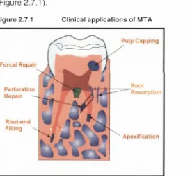
Figure 2.7.1 Clinical applications of M T A ... 41
Figure 3.5.1 Irrigation of the root c a n a l...58
Figure 3.5.2 Drying of the root c a n a l... 58
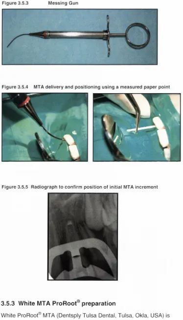
Figure 3.5.3 Messing G un... 60
Figure 3.5.4 MTA delivery and positioning using a m easured paper p o in t... 60
Figure 3.5.5 Radiograph to confirm position of initial M TA in c rem e n t... 60
Figure 3.5.6 W hite M TA P r o R o o t... 61
Figure 3.5.7 Mixing of MTA A n g e lu s ... 62
Figure 3.5.8 Injection and condensation of gutta p e rc h a ... 63
Figure 3.6.1 Stages of root d evelo pm en t... 64
Figure 3.6.2 Diagram of Apical A n a to m y ... 65
Figure 3.6.3 Reference radiographs, corresponding line draw ings and associated PAI score... 66
Figure 3.6.4 Radiographs showing position of the MTA p lu g ... 66
Figure 4.1.1 Flow of participants through the s tu d y ... 69
Figure 4.2.1 Distribution of referral s o u rc e ... 70
Figure 4.2.2 Distribution of affected to o th ...71
Figure 4.2.3 Distribution of age of patient at tim e of tra u m a ... 71
Figure 4.2.4 Distribution of type of traum a e x p e rie n c e d ... 72
Figure 4.3.1 Distribution of W M T A ... 72
Figure 4.3.2 Distibution of follow up tim e s ...73
Figure 4.4.1 Baseline Sinus or Sw elling with relation to PAI o u tc o m e ... 75
Figure 4.4.2 Clinical Param eters at baseline and follow u p ...75
Figure 4.4.3 Distribution of tooth colour at b as elin e ...76
Figure 4.4.4 Tooth colour change from baseline to 6 m o n th s ... 76
Figure 4.5.1 Distribution of Root D evelop m en t... 81
Figure 4.5.2 Sim plified Distribution Of Root D evelop m en t... 81
Figure 4.5.3 Apical Anatom y Distribution within the G ro u p s ... 84
Figure 4.5.4 Distribution of sim plified apical anatom y within the study groups....85
Figure 4.5.5 Presence of a PARL at b a s e lin e ... 88
Figure 4.5.6 Distribution of M TA Plug Position... 90
Figure 4.5.7 Distribution of Sim plified MTA Plug P o s itio n ... 91
Figure 4.5.8 Tim ing of apical barrier fo rm atio n ... 94
Figure 4.5.9 Baseline PAI S c o r e ...96
Figure 4.5.10 PAI outcom e at 6 m onth re v ie w ...96
Figure 4.5.11 Graph to show change in PAI score over tim e ... 98
Figure 4.5.14 R adiographs of C ase # 4 ... 104
Figure 4.5.15 R adiographs of C ase # 8 ... 105
Figure 4.5.16 R adiographs of Cases #15 and # 1 6 ... 106
Figure 4.5.17 R adiographs of C ase # 3 ...107
Figure 4.5.18 R adiographs of C ase # 2 1 ...108
Figure 7.1.1 R ecruitm ent letter to ju n io r hospital s ta ff... 131
Figure 7.2.1 Ethical A p p ro v a l... 132
Figure 7.3.1 Inform ation Sheet Page 1 ...133
Figure 7.3.2 Inform ation Sheet Page 2 ...134
Figure 7.4.1 C onsent Form Page 1 ...135
Figure 7.5.1 Data capture form: Initial d a ta ... 137
Table of Tables
Table 2.1.1 Oehlers Classification of Dens In v a g in a tu s ... 6
Table 2.4.1 Techniques for achieving apical c lo s u r e ... 11
Table 2.4.2 Materials to induce apical c lo s u re ... 12
Table 2.5.1 Studies of Calcium Hydroxide A pexification O u tco m e... 18
Table 2.5.2 Com ercial brands of calcium hydroxide p reparatio n s ... 22
Table 2.5.3 D isadvantages of calcium h yd ro xid e...23
Table 2.6.1 M aterials for an apical end s e a l... 28
Table 2.7.1 Requirem ents of an ideal root end fillin g ...42
Table 2.7.2 A dvantages of M TA as an apexification m a te ria l... 42
Table 2.7.3 Disadvantages of M TA as an apexification m a te ria l...43
Table 2.7.4 Role for calcium hydroxide a p e xifica tio n ... 45
Table 2.8.1 Clinical studies on M TA a p e x ific a tio n ... 47
Table 3.2.1 Inclusion and Exclusion C rite ria ...53
Table 3.3.1 Study Group A llo c atio n ...54
Table 3.3.2 FrankI Behaviour S c a le ... 55
Table 3.4.1 Clinical data re c o rd in g ... 56
Table 3.6.1 Clinical E v alu atio n ... 63
Table 3.6.2 Stage of root d evelo pm en t... 64
Table 3.6.3: Description of PA! categ o rie s ... 66
Table 3.6.4 Criteria of Clinical and Radiographic s u c c e s s ... 68
Table 4.2.1 Distribution of sex in the study g ro u p s ... 70
Table 4.3.1 Distribution of total treatm ent tim e ... 73
Table 4.3.2 Distribution of follow up tim e ... 73
Table 4.4.1 Table showing Clinical Success R ate... 77
Table 4.5.1 Kappa-Cohen values of inter-exam iner ag ree m en t... 77
Table 4.5.2 Root developm ent scores: Exam iner 1 vs. Exam iner 2 ... 78
Table 4.5.3 Apical A natom y scores: Exam iner 1 vs. Exam iner 2 ...78
Table 4.5.4 Apical barrier form ation: Exam iner 1 vs. Exam iner 2 ... 78
Table 4.5.5 Kappa-Cohen Values of intra-exam iner a g re e m e n t... 79
Table 4.5.6 Root developm ent scores:Exam iner 1 tim e 1 vs. tim e 2 ...79
Table 4.5.7 Root developm ent scores: Exam iner 2 tim e 1 vs. tim e 2 ... 79
Table 4.5.8 Apical Anatom y scores: Exam iner 1 tim e 1 vs. tim e 2 ...80
Table 4.5.9 Apical Anatom y scores: Exam iner 2 tim e 1 vs. tim e 2 ... 80
Table 4.5.10 Relationship of Root D evelopm ent to Age of T ra u m a ... 81
Table 4.5.11 Relationship of Root Developm ent to Apical A n a to m y ... 82
Table 4.5.12 R elationship between Root D evelopm ent and M TA Plug P o s itio n 82 Table 4.5.13 Relationship of stage of root developm ent with apical barrier p re s e n c e ... 83
Table 4.5.14 Relationship between Root Developm ent and PAI O u tc o m e ... 83
Table 4.5.17 R elationship of apical anatom y to M TA plug p o s itio n ... 86
Table 4.5.18 R elationship betw een the apical anatom y and the presence of an apical b arrier...86
Table 4.5.19 R elationship betw een Apical A natom y and PAI o u tc o m e ... 87
Table 4.5.20 R elationship betw een Apical A natom y and PARL o u tc o m e ...87
Table 4.5.21 R elationship betw een MTA plug position and baseline P A R L... 89
Table 4.5.22 Apical b arrier form ation with relation to baseline PARL s ta tu s ...89
Table 4.5.23 PAI outcom e in relation to baseline PARL s ta tu s ... 90
Table 4.5.24 R elationship of M TA plug position to apical barrier p re s e n c e ... 92
Table 4.5.25 R elationship betw een MTA plug position and PAI o u tc o m e ...92
Table 4.5.26 R elationship betw een MTA plug position and PARL o u tc o m e ... 93
Table 4.5.27 R elationship betw een presence of an apical barrier and PAI o u tc o m e. ... 95
Table 4.5.28 Apical b arrier form ation in relation to PARL o u tc o m e ... 95
Table 4.5.29 Success Rates using PAI s c o re ...97
Table 4.5.30 C om parison of PAI outcom e between G roup 1 and Group 2 ...97
Table 4.5.31 Data of PARL and PAI score change over t im e ...99
Table 4.5.32 Success rates using PARL outcom e at 6 m o n th s ... 101
Table 4.5.33 C om parison of PARL outcom e over tim e between the study groups.... 102 Table 4.5.34 Correlation betw een PAI outcom e and PARL o u tc o m e ...102
1 Introduction
Root development and apical closure are normally completed within 3-4
years of tooth eruption (Moorrees
et al.
1963). If pulp death occurs
before apical closure, further root developm ent may be arrested. Most
com m only pulp death is caused by traum atic injuries but may also be
due to caries, a structural defect or an anom aly of the tooth.
When a non-vital tooth is immature, endodontic therapy will be
challenging due to the lack of apical stop, divergent apical anatomy,
thin dentinal walls and young age of the patient. It may not be possible
to
achieve
an
adequate
three-dim ensional
apical
seal
using
conventional root canal preparation and obturation. An apical barrier is
needed to provide a stable platform against which gutta percha can be
compacted. A procedure known as apexification has been developed
to assist completion of the root canal treatm ent by form ing an apical
barrier.
Many medicaments have been proposed in times past to
achieve an apical barrier. Apexification using calcium hydroxide was
introduced by Frank in 1966. Calcium hydroxide has been the most
widely used and clinically accepted for over 40 years. This technique is
easy and clinically acceptable, with high clinical success rates reported
(Sheehy and Roberts 1997). The treatm ent time frame is however
unpredictable and often prolonged, risking loss of patient compliance.
The presence of a temporary coronal restoration for increased lengths
of time leaves these teeth at risk of bacterial contamination. There have
also been reports of an increased risk of root fracture in teeth with long
term calcium hydroxide dressings (Andreasen
et al.
2002, Andreasen
et al.
2006). The disadvantages of the calcium hydroxide apexification
technique may be overcome by using a one step apexification
technique. As the name suggests, this entails placement of an artificial
apical stop in one visit, without the need for awaiting biological apical
closure.
This allows prompt obturation and completion of the
Mineral Trioxide Aggregate (MTA) has been introduced as the material
of choice in the one step apexification technique (Torabinejad
et al.
1995a).
There have been many
in vitro studies investigating the
properties of this material, but very few clinical trials using the cement
for apexification (Camilleri and Pitt Ford 2006). Existing clinical trials
have shown favourable results, com parable or superior to apexification
with calcium hydroxide. Since the introduction of grey MTA in 1993, a
more aesthetic white version has been developed.
W hite MTA
ProRoot® (Dentsply Tulsa Dental, Tulsa, Okla, USA) has gained
popularity following reports of coronal discolouration with grey MTA
(Sarris
et al. 2001). The composition of white MTA ProRoot® has been
shown to differ from grey MTA ProRoot® only in the metal oxides
content (Asgary
et al. 2005). Even more recently, another commercial
brand of MTA cement, MTA Angelus (Angelus Dental Solutions,
Industria de Produtos Odontologicos Ltda, Londrina, Parana, Brazil)
has entered the market. MTA Angelus has a faster setting time and it
is also claimed to possess improved handling characteristics.
It is
available as a grey or white cement. MTA Angelus has been shown
in
vitro to be very similar in chemical composition and biocompatibility to
MTA ProRoot® cement (Song
e ta l. 2006).
There is limited current research using white MTA ProRoot® as an
apexification material.
To date, there are no clinical trials or case
reports involving the use of MTA Angelus for apexification. There is
therefore, an immense gap in the literature of
in vivo research involving
white MTA ProRoot® and white MTA Angelus. It is important that more
clinical trials using these newer materials are carried out to assess the
clinical success and efficacy of one-step apexification.
Research
should aim to compare the clinical outcom es of apexification using MTA
with outcom es using the traditional calcium hydroxide apexification
technique.
This will enable clinicians to make an evidence based
choice regarding apexification material and technique.
2 Literature Review
2.1 Causes of pulpal necrosis
Pulpal necrosis may be a consequence of dental trauma, dental caries
or a defect in tooth development.
2.1.1 Trauma
Trauma is the most frequent cause of pulp necrosis in immature
anterior permanent teeth. Worldwide, 20-30% of 12 year-old children
have experienced dental trauma. Boys suffer approximately one third
more injuries than girls. The peak age for anterior trauma is 9-10 years
old, which coincides with root development of permanent incisors. The
most common aetiologies of trauma are falls, sports injuries, road traffic
accidents and violence (Andreasen
et al.
2003).
Loss of vitality following trauma is dependent upon:
i)
The stage of root development
ii)
The type and severity of traumatic injury
iii)
The presence of bacteria (Barnett 2002).
fractures carry a minimal risk of vitality loss but this increases with
additional luxation injury and pulp exposure (Barnett 2002).
2.1.2 Dental Caries
Caries may lead to loss of vitality in antehor permanent teeth. Caries in
newly erupted anterior teeth in children is not commonly seen, the
disease tends to be seen in an older population, when root
developm ent is complete. Due to the visible position of anterior teeth,
treatm ent may be sought before pulp necrosis occurs. Some factors
may predispose children to aggressive anterior carious lesions such as
special medical or intellectual needs, sugary medications or an
excessively high frequency of consumption of cariogenic substances.
Reduced salivary flow may predispose teeth to decay, in children this
may be due to medications, radiation and chem otherapy or salivary
gland surgery.
2.1.3 Defects of tooth development
Dens invaginatus has been defined by The American Association of
Endodontists (2003) as “a defect in tooth development, characterised
by invagination of the enamel organ before the calcification phase.”
Oehlers in 1957 classified dens invaginatus into three categories (Table
2
.1.1).
Table 2.1.1 O ehlers Classification of Dens Invaginatus
TYPE 1
•
enamel lined minor invagination within crown
•
does not extend past the CEJ
Type 2
•
enamel lined, invades past the CEJ into the root
•
remains as a blind sac
•
may or may not connect with the pulp
Type 3
•
enamel or cementum lined and penetrates the root
•
perforates apical area and form s a second foramen
•
no immediate connection with the pulp
commonly affected with a reported prevalence of 9% (Hamasha and
Alomari 2004). There is a risk of bacterial invasion, and subsequent
necrosis in cases where the enamel lining of the invagination is thin or
incomplete or the channels extend into the pulp. Early necrosis before
root development completion may leave the tooth with an open apex
and further complicate its management (Subay and Kayatas 2006).
Various enamel and dentine defects may predispose immature teeth to
weakness and infection risk.
Am elogenesis imperfecta, enamel
hypoplasia
or
hypomineralisation,
dentinogenesis
imperfecta,
hereditary opalescent
dentine,
dentine
dysplasia
and
regional
odontodysplasia may all lead to pulpal disturbances (Witkop 1988,
Weerheijm
et al. 2001, Hamdam
et al. 2004). These defects may allow
more rapid bacterial invasion of the tooth, either due to attrition of
weakened structure or increased porosity.
Penetration of the pulp
leads to increased risk of vitality loss (Witkop 1988).
2.2 Root development
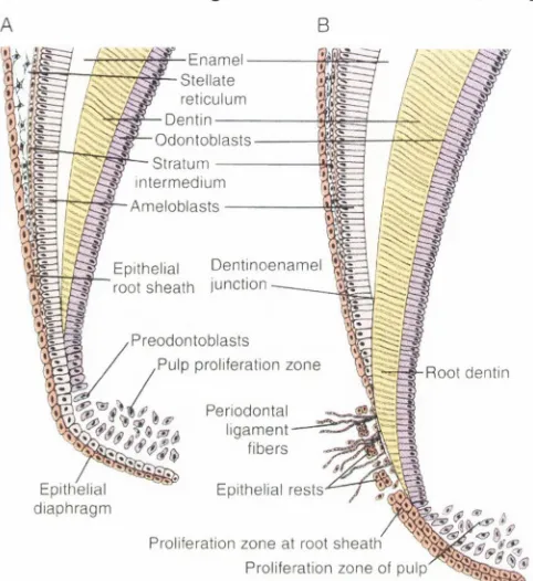
M W — Enamel Stellate reticulum Dentin O dontoblasts Stratum nterm edium Am eloblasts
Figure 2 Diagram of root form ation. A, early stage. B, later stage. 8
Epithelial D entinoenam el ' root sheath junction
Epithelial diaphragm
Root dentin P reodontoblasts
Pulp proliferation zone
Periodontal ligam ent fibers Epithelial rests
Proliferation zone at root sheath P roliferation zone of i
(R eproduced from A very and Cheigo 2006)
Mesenchymal cells from the follicle then move between the rest cells
and differentiate into cem entoblasts secreting cementoid on the root
surface. This cementoid eventually calcifies into mature cementum. At
the apical end the root sheath bends at an almost 45° angle to form the
epithelial diaphragm which encircles the apical opening of the dental
pulp during root development. These cells proliferate and cause root
growth. Dentinogenesis continues from the crown into the root. The
pulp adjacent to the dentine becomes the pulp proliferation zone, which
is though to provide new cells needed for root growth. Once root length
is com plete the dentine walls thicken and the apical opening closes to
1-3mm. This is normally com plete 3-4 years after tooth eruption (Avery
and Chiego 2006).
[image:21.529.63.304.61.324.2]and Andreasen1992, Rafter 2005).
Some authors have noted
continued apical root development in non-vital, immature teeth
following dressing with various antiseptic and antibiotic pastes (Cooke
and Robotham 1960, Ball 1964, Frank 1966, Sato
et al. 1996,
Thibodeau and Trope 2007). Cooke and Robotham (1960) speculated
that even in necrotic cases the root sheath can continue to organise
mesodermal tissue into root components, once apical trauma is
minimised. This apical mesodermal tissue is now considered as the
apical papilla. Stem cells from this apical papilla (SCAP) may survive
infection and allow root maturation (Sonoyama
et al. 2008).
Complete destruction of the root sheath and apical papilla will halt
normal developm ent of the root. In these cases there may still be hard
tissue formation by fibroblasts and cementoblasts of the dental follicle
and periodontal ligament (Torneck 1982).
Cessation of root
developm ent results in an immature root.
An incomplete root was
defined by Moorrees
et al. (1963) as, a quarter to full root length with a
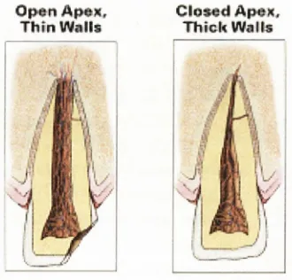
fully or half open apex. Immature teeth have blunt ends and wide open
apices. Incomplete dentinogenesis may render the dentinal walls thin
(Figure 2.2.2), predisposing them to fracture (Andreasen
et al. 2002).
Fig ure 2.2.2 Diagram showing thin walls of an im m ature tooth com pared with thick walls of a m ature tooth
The immature apical anatomy may be divergent, making an optimal
apical seal difficult to achieve. Sealing the root canal using lateral
condensation techniques is challenging due to the absence of an apical
Open Apex Thin Walls
[image:22.529.213.378.511.669.2]stop (Walton and Torabinejad 2002). Consequently, the combination of
these factors, pose a difficult situation for the clinician.
2.3 Diagnosing pulp status in immature teeth
The diagnosis of pulpal necrosis in immature teeth is challenging. A full
and comprehensive history must be taken prior to clinical and
radiographic assessment. Careful note must be made of any history of
traum a or pain in the tooth. Prolonged or spontaneous pain may be
indicative of irreversible pulpitis as it arises from stimulation of
unmyelinated C fibres.
Clinical observations of tenderness to
percussion, mobility and presence of a swelling or sinus may also
indicate necrosis.
Discolouration is not always associated with
necrosis so should be considered in conjunction with other factors
(Jacobsen 1980). Sensibility testing using thermal and electrical tests
is unreliable and erratic in immature teeth as the subodontoblastic
nerve plexus is undeveloped. Trauma itself may cause pressure or
tension on nerve fibres resulting in inconclusive pulp testing for four-six
weeks following traum a (Skieller 1960, Andreasen
et al. 2003). Laser
Doppler Flowmetry, which measures blood flow seems to be the most
reliable recent method of testing pulp status but is still not widely
available (StrobI
et al. 2004).
Radiographic examination may also be difficult to decipher. Healthy
developing teeth often have radiolucencles around the apex, which
may resemble pathology. Transient apical breakdown may resemble
pathological changes radlographically (Andreasen
et al. 2003).
Regular radiographic review Is recommended to monitor for occurrence
of periapical pathology, continued root development and to enable
comparison with the contralateral tooth (Rafter 2005). A combination of
the history with frequent clinical and radiographic investigations will
allow an accurate diagnosis of pulp status in these difficult cases.
2.4 Apexification
‘the process o f creating an environm ent wittiin the root canal and
periapical tissues after pulp death that allows a calcific barrier to form
across the open apex. The barrier has been characterized as dentine,
cementum, bone and osteodentine. ’’
The aim of apexification is to achieve apical closure with a hard tissue
barrier enabling condensation of gutta percha. The success of
apexification depends on the migration of cells from healing peri-
radicular tissues to the root apex capable of producing a cementum,
osteocementum or osteodentine organic matrix (Ripamonti 1997).
Specific cellular signals, including growth factors and cytokines enable
cell differentation. Bone morphogenetic proteins (BMPs) belong to the
superfamily of transforming growth factors (TGF). BMPs can induce
osteogenesis and cementogenesis at ectopic sites and provide a
scaffolding for bone regeneration (Ham
et al. 2005).
Over the years various techniques have been proposed to achieve
apical closure of immature teeth (Table 2.4.1).
Table 2.4.1 Techniques for achieving apical closure
Customised gutta percha point
Stewart
1963
Clot induction
Ham
1972
Surgical + oxidised
regenerated cellulose
Dimashkieh
1977
Control of infection
Das
1980
No treatment
Lieberman and Trowbridge
1983
Surgical + therm oplasticised
GP
Dawood and Pitt Ford
1989
preparation of the thin walls and optimise access for proper
condensation of a root end filling (Dawood and Pitt Ford 1989). The
disadvantage is a reduction in the crown root ratio, which may further
com prom ise the longevity of the tooth (Rafter 2005). Compliance with
invasive surgery is also an issue in this young patient group (Frank
1966, W itherspoon and Ham 2001).
Periapical clot induction was
explored in monkey teeth by Ham
et al. (1972) as a means of inducing
apical closure and interestingly, a case report where no treatm ent was
provided yet apical closure was achieved was described by Lieberman
and Trowbridge in 1983. Endodontic therapy and control of infection
has been found adequate to promote apical barrier formation (Das
1980). Minimal or no root canal instrumentation has been advised to
avoid interfering with root developm ent in necrotic teeth (Das 1980,
Das and Das 1997).
[image:25.529.40.423.370.629.2]Many techniques have described first stage endodontics followed by
the placement of various dressings to induce an apical hard tissue
barrier (Table 2.4.2).
Table 2.4.2 M aterials to induce apical closure
Antiseptic paste
Cooke and Robotham
1960
Antibiotic paste
Ball
1964
Calcium & CMCP
Kaiser
1964
Calcium hydroxide
Frank
1966
Calcium & CMCP
Dylewski
1971
Tricalcium phosphate
Roberts and Brilliant
1975
Collagen-calcium
phosphate gel
Nevins
et al.
1978
2.5 Calcium Hydroxide as an Apexification Material
Despite early success of other materials, it is calcium hydroxide (CH)
that has been the most commonly used medicament for over 40 years.
In 1964, Kaiser was the first to propose the use of calcium hydroxide
combined with camphorated monochlorophenol (CMCP), to induce
apical barrier formation.
In 1966 Frank described an apexification
technique using calcium hydroxide, which still forms the basis of
apexification methods to the present day.
The clinical success of
calcium hydroxide and CMCP for barrier induction was confirmed by
Dylewski (1971). The cytoxicity of CMCP has led to further
investigations using plain calcium hydroxide mixed with saline, sterile
water or distilled water and showed similar clinical results with reduced
cytoxicity (Rafter 2005).
2.5.1 Antibacterial Properties
The antibacterial properties of calcium hydroxide have been well
documented (Foreman and Barnes 1990).
Bacteria and their
subproducts stimulate organic reactions, which are the main cause of
pathological alterations in root canals and the periapical area (Sjogren
et al. 1991). If bacteria persist in the root canal and dentinal tubules,
following trauma, inflammatory root resorption may occur which can
destroy significant amounts of tooth structure in relatively short time
periods (Sjogren
et al. 1991, Trope 2002).
The primary cause of
failure of root canal therapy is the persistence of bacteria (Sundqvist
et
al. 1998). The complex anatomy of root canals provides an excellent
opportunity for the growth and proliferation of microorganisms.
Brystrom and Sundqvist (1981) demonstrated that not all bacteria are
removed by instrumentation alone, therefore an adjunct such as
calcium hydroxide is necessary to com plem ent mechanical cleaning.
Apical barrier formation is much more likely once bacterial levels have
decreased (Ham
et al. 1972).
by osteoclasts, preventing further destruction of mineralised tissues
(Heithersay 1975, Foreman and Barnes 1990).
The alkaline
environm ent created is also unfavourable to the majority of aciduric
endopathogens (Sjogren
et al. 1991). It has also been postulated that
calcium ions can stimulate the enzyme pyrophosphatase, which
denatures pyrophosphates and leads to increased repair mechanisms
(Heithersay 1975).
An
in vitro study by Estrela in 1998, examined the direct antimicrobial
effect of calcium hydroxide, and dem onstrated that the ionic
dissociation of calcium hydroxide into
and OH” imparted
antimicrobial and mineralising properties.
The ions were shown to
inhibit cytoplasm ic membrane enzymes of bacteria, denature bacterial
proteins, damage bacterial DNA and activate alkaline phosphatases
within 3 days. Further research by Estrela (1995) led to the hypothesis
that the high pH of calcium hydroxide causes reversible and irreversible
bacterial enzymatic inactivation. Sjogren
et al. (1991) investigated the
effect of calcium hydroxide on bacterial samples from 30 root canals
with pulp necrosis and apical periodontitis. The results showed 100%
of bacteria that survived biomechanical preparation were eliminated by
calcium hydroxide in 7 days. Sjogren
et al. proposed that 7 days was
adequate to allow m ovement of the free hydroxyl ions into the dentine
tubules by diffusion gradient.
traumatised teeth that lose vitality.
Inflammatory root resorption is
defined as
“an unprotected or altered tooth surface attracting resorbing
cells and an Inflammatory response m aintained by Infection’’
(Trope
2000).
The initial damage to the protective root cementoid can be
further propagated by bacteria from a necrotic pulp (Trope 2002). The
denuded root is exposed to resorption by clastic cells and products of
inflammation as long as stimulation exists from the pulp bacteria (Trope
et al. 1995, Trope 2002).
A dressing of calcium hydroxide placed one week following injury has
been shown to be effective at removing bacteria and halting
inflammatory root resorption (Trope
et al. 1992). It is not recommended
to place calcium hydroxide earlier than 1 week due to potential
cytoxicity and damage of cementum and PDL cells (Foreman and
Barnes
1990,
AAE
Guidelines
2004).
Cvek
(1992)
looked
retrospectively at 885 luxated mature and immature maxillary incisors.
It was found that 97% of inflammatory root resorption was arrested
after dressing with calcium hydroxide and filling with gutta percha.
Another property, which enhances the ability of calcium hydroxide to
cleanse a root canal system is a proteolytic action. This Is thought to
help canal debridement by dissolving necrotic tissue remnants
(Andersen
et al. 1992). On the negative side this proteolytic effect may
weaken circumpulpal dentine and result in increased weakening of an
immature tooth (Cvek 1992).
2.5.2 Apical Barrier Form ation
low grade inflammatory reaction which stimulates calcification. Some
dispute exists as to where the calcium ions incorporated into the hard
tissue barrier originate. Pisanti and Sciaky (1964) demonstrated that
the calcium ions in apical barriers of dog teeth originated from the
calcium reserves of the animal. This would suggest the role of calcium
hydroxide as an initiator but not a substrate (Foreman and Barnes
1990).
The calcific barrier shape has been described as a cap, bridge or
ingrown wedge, a cap being the most frequent finding (Ghose
et al.
1987). The apical shape has been described as either physiological,
rounded or a straight bridge.
In a recent prospective study by
Dominguez Reyes
et al. (2005), physiological apical shape was most
frequently observed (73.11%), followed by a rounded form (19.2%) and
a straight form or bridge (7.7%). This differs from Ghose
et al. (1987)
who reported rounded apical closure in 65% and straight bridges in
24% cases.
The composition of the calcific barrier has been described as
osteocementum (Chosack
et al. 1997), mineralised scar tissue
(Foreman and Barnes 1990), and cementum like tissue with loose
connective tissue inclusions (Heithersay 1975). Baldassari-Crus
et al.
(1998) examined the barrier under a scanning electron microscope
(SEM). The findings were a 2-layered porous and irregular cap-like
structure. The dense outer layer was acellular-like cementum and the
dense inner layer consisted of irregular fibrous collagen connective
tissue with granular inclusions of foreign material and irregular calcified
fragments.
Steiner and Van Hassel (1971) histologically examined
sections of apical barriers in primate teeth. They described the barrier
com position as cementum-like, and formation proceeding from the
periphery of the apex towards the centre in concentric circles.
Abbott 1998).
Abbott (1998) does not agree that radiographs can
reliably assess the quality of calcium hydroxide dressing or barrier
formation and recommends regular dressing change at two or three
month intervals to allow clinical barrier assessment. W alia
et al. (2000)
reported a porous structure despite radiographic and clinical evidence
of a complete barrier.
The position of apical stop seems to be variable and may be related to
presence of calcium hydroxide in the apical area.
Mitchell and
Skankwalker (1958) emphasised the necessity of direct calcium
hydroxide contact with the periapical tissues for apical barrier
formation. Finucane and Kinirons (1999) found most barriers formed at
the radiographic apex but some more coronally. A relationship was
found between more apically positioned barriers and increased
frequency of calcium hydroxide dressing changes (<2 monthly),
suggesting that dissolution of calcium hydroxide may allow ingrowth of
apical tissue and result in a more coronal barrier. Conversely, Kinirons
et al. (2001) also recorded the majority of barriers were formed at or
near the radiographic apex, but were unable to confirm a relationship
with frequency of calcium hydroxide dressing, with a variety of dressing
protocols resulting in an acceptable barrier position. Dissolution (wash
out) may be affected by pH of the periapical tissues, fluid exudates and
the repeated pumping effects of mastication (Metzger
et al. 2001).
Wash out is more likely in wide open apices immediately following
placement (Yates 1988).
Table 2.5.1 Studies of Calcium Hydroxide Apexification Outcome
Heithersay 1970
21 CH &Methylcellulose
15-75 90%
Cvek et al. 1972
55 CH powder & saline 18.2 mean 90%Winter et al. 1977
34 Reogan Rapid* =27CH powder & sterile water=7
Not stated 74%
Chawla et al. 1986
26 Reogan Rapid* 6 (65%) 12 (35%)100%
Ghose et al. 1987
51 Calasept 3-10 96Thater et al. 1988
34 Pulpdent Not stated 74%Mackie et al. 1988
112 Reogan Rapid 10.3 mean 96%Yates et al. 1988
22 Study protocol 26 control (no protocol)CH powder & saline Hypocal
9 (study)
20.2 (control)
100%
Kleier et al.1991
48 CH paste & CMCP Pulpdent12 + /-7 100%
Mackie et al. 1994
19 Reogan Rapid 6.8 mean 100%19 Hypocal 5.1 mean 100%
Finucane &
Kinirons 1999
44 Not stated 7.8 mean
(parallel) 11 mean (divergent)
100% 100%
Kinirons et al.2001
107 Not stated 10.8 mean 100%Dominguez Reyes
et al.2005
26 CH & distilled water 12.19 mean 100%
* Reogan Rapid: see Table 2.5.2
2.5.3 Factors Influencing barrier formation
Much controversy exists amongst the literature as to the desirable
frequency of calcium hydroxide dressing change.
Some authors
advocate a single placement while some recommend once monthly or
three monthly changes, and others believe change should only occur
when radiographs indicate resorption of the apical third of dressing.
A single application is favoured by Felippe
et al. (2005) who recently
investigated the influence of renewing calcium hydroxide paste on
apexification and periapical healing in 40 dog teeth. The conclusion
was that replacement of calcium hydroxide paste is not necessary for
apexification to occur but did decrease periapical inflammation.
Similarly, Chosack
et al. (1997) examined the development of apical
osteodentine in monkey teeth and concluded that nothing was to be
gained by repeated dressing with calcium hydroxide either monthly or 3
monthly for at least 6 months.
Gupta
et al. (1999) reported on a case
of completed root development 7 months following a single application
of calcium hydroxide. It was proposed that root development continued
because necrosis had not involved the root sheath or odontoblasts.
Morse
et al. (1990) postulated that CH is necessary to promote initial
healing and canal disinfection, but multiple visits re-accessing a canal
with repeated cleansing may disturb the process of apexification.
44 teeth that had undergone apexification and found increased
frequency of dressing change had a positive relationship with speed of
apical barrier formation. A multicenter study of 107 teeth by Kinirons
et
al. (2001) supports this finding that barrier detection was significantly
earlier in more frequently dressed teeth.
Replacement of the calcium hydroxide dressing only when a radiograph
indicates resorption of medicament in the apical third or if symptoms
develop is also an option (Walia
et al. 2000, Yates 1988, Ghose
et al.
1987). Yates (1988) examined at the success rates of apexification in
22 teeth following a strict protocol, compared to 26 teeth that had been
treated without any protocol.
Radiographs were used to assess
calcium hydroxide dressing quality at 3 and 6 monthly intervals and
calcium hydroxide dressing change was carried out if found
inadequate. Interesting it was noted that more calcium hydroxide was
resorbed after 1 month in teeth with wide open apices and often on
changing a dressing a barrier was palpated clinically although not
evident radiographically.
The initial size and anatomy of the apical foramen has been shown to
affect the speed of barrier formation. Apical closure was a significant
factor in relation to speed of barrier formation, with divergent apices
requiring significantly more time in some reports (Yates 1988, Finucane
and Kinirons 1999, Domingues Reyes
et al. 2005).
However other
authors did not find apical opening significant (Ghose
et al. 1987, Kleier
and Barr 1991).
Age may be inversely related to barrier formation time, with children
older than eleven needing significantly less treatment time (Mackie
et
a/.1988).
This is likely to be a consequence of more mature root
development.
advantage of diagnosing pulp necrosis early to avoid the development
of infection and ensure a good coronal seal between appointments,
preventing bacterial ingress from compromising barrier formation.
Other studies found infection to be a weak predictor of the speed of
barrier formation (Finucane and Kinirons 1999, Mackie
et al. 1988,
Yates 1988). Kleier and Barr (1991) found that teeth with a sizeable
periapical radiolucency (>5mm) were at significantly more risk of inter
appointment symptoms.
Inter-appointment symptoms were found to
delay apexification by 5 months while periapical infection was found to
delay or reverse barrier formation and healing.
The type of trauma is also thought to influence treatment time, as some
injuries will inflict more damage on the tooth and periodontium than
others.
Finucane and Kinirons (1999) found that degree of
displacement by trauma increased the time taken for apexification and
Mackie
et al. (1988) observed more failed of apexification in teeth that
had suffered displacement injuries.
on the available proprietary brands to help clinicians make an evidence
based decision on material choice.
Table 2.5.2 C om ercial brands of calcium hydroxide preparations
Pulpdent
(Pulpdent Corporation, Watertown, MA, USA)
CH
Methycellulose
Reogan Rapid
(Vivadent, Liechtenstein)
CH
Casein paste
Hypocal
(Ellman, New York, USA)
98% CH paste
Calasept
(Nordiska Dental, Angelholm, Sweden)
Pure CH powder
Isotonic saline
Ultracal XS
(Ultradent Products Inc., South Jordan, UT, USA)
CH
Hydroxyapatitie
Thickeners +
water
canals prepared with a wider taper and apical size showed few er voids.
They also favoured paste delivery using lentulo-spiral fillers over paste
injection to reduce voids.
This study did not exam ine hand
condensation.
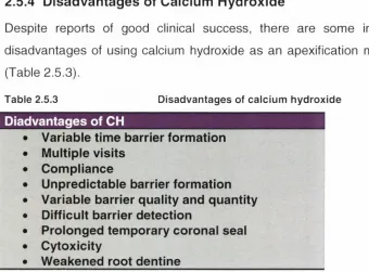
2.5.4 D isadvantages of Calcium H ydroxide
Despite reports of good clinical success, there are some inherent
disadvantages of using calcium hydroxide as an apexification material
(Table 2.5.3).
Table 2.5.3 Disadvantages of calcium hydroxide
•
Variable time barrier formation
•
Multiple visits
•
Compliance
•
Unpredictable barrier formation
•
Variable barrier quality and quantity
•
Difficult barrier detection
•
Prolonged temporary coronal seal
•
Cytoxicity
•
Weakened root dentine
Although rates of barrier formation are good, the time taken to achieve
this ranges from 5-20.2 months (Sheehy and Roberts 1997).
This
variation prevents the clinician from being able to give patients a
definitive tim e scale of treatment completion (Steinig
et al.
2005). It
may be difficult to maintain positive morale when patients must attend
indefinitely.
Multiple visits and extended treatm ent times may be
problematic and it is crucial that these teeth are not lost to follow up.
The development of a hard tissue barrier is unpredictable. In some
cases, despite prolonged treatment with calcium hydroxide, failure to
achieve an apical stop is noted (Maroto
et al.
2003, Karp
et
a/.2006).
Diagnosing the presence of an apical stop can be challenging. Often
radiographic examination does not reveal a barrier palpated clinically
(Yates 1988, Sheehy and Roberts 1997, Abbott 1998). The quality of
[image:36.529.100.440.171.422.2]barrier and examination under SEM has also described a porous
structure (Baldassari-Crus
et al. 1998).
The importance of a coronal seal in endodontically treated teeth has
been well documented (Ray and Trope 1995, Tronstad
et a/.2000).
Often between visits a tem porary restoration is used to seal the access
cavity, with a limited life span (Abbott 1998). Invasion of a root canal
with bacteria is the most frequent cause of periapical pathology and
root canal failure (Sjogren
et al. 1991).
Ray and Trope (1995)
examined
1010
endodontically treated
teeth.
Good
coronal
restorations resulted in significantly less periapical inflammation than
good endodontic obturation. A combination of good coronal restoration
and good endodontic obturation showed 91.4% absence of periapical
inflammation compared to 18.1% of teeth with both poor coronal and
apical restorations.
treatment provided may not be perceived by the patient (Witherspoon
and Ham 2001).
The cytotoxity of calcium has been questioned when placed
immediately in a traumatised tooth. PDL cell death, cementum damage
and acceleration of replacement root resorption have been reported
(Foreman and Barnes 1990, Lengheden
et al. 1991). It has been
recommended to delay the placement of calcium hydroxide dressing for
7-10 days following trauma when pulp extripation is required to avoid
further inflammation of the traumatised periodontal tissues (AAE
Traumatic Dental Injuries Guidelines 2004).
demonstrates the high risk of root fractures in immature teeth following
calcium hydroxide therapy, therefore it would be judicious to warn
patients and parents of the risk of this complication. The retrospective
nature, multiple operators of varying skill levels and inconsistent
treatment protocol weakens this study, however it is strong on numbers
allowing for meaningful statistical analysis and has an excellent follow
up time.
Mackie
et at.
(1993) followed up 93 non-vital immature incisor treated
with calcium hydroxide apexification and gutta percha root filling. Life
table analysis suggested that 86% of teeth treated by this modality
would be present after 5 years. 7% (6) of the teeth had been lost at
minimum 6 month follow up, half the losses (3) were due to fracture
following further trauma, 1 from an attempt at post and core placement
and 2 from progressive resorption.
The authors concluded that
immature teeth may be weak and brittle and prone to loss from even
minor trauma or operative procedures.
days (3 months).
Teeth in the prolonged calcium hydroxide group
showed a significant reduction (22%) in fracture strength resistance
compared to the other groups.
This study confirms the weakening
effect of long-term calcium hydroxide on root dentine.
The authors
promoted short-term calcium hydroxide to dissolve pulp remnants,
disinfect the canal and remove apical infection before MTA barrier
placement with less detrimental effect of root strength. This
in vitro
study is well explained and executed, but is weak in numbers, with less
than 10 teeth in each group.
Clinical tnals in this area would be
beneficial in order to make evidence based treatment decisions,
however this would pose ethical issues.
An
in vitro
study by Rosenburg
et ai.
(2007) measured the effect of
calcium hydroxide root filling on the microtensile fracture strength
(MTFS) of 40 extracted human teeth.
Teeth matched for size and
dentine thickness, were filled with calcium hydroxide for 7, 28 or 84
days or with gutta percha (control group) and sealed. Study teeth were
placed in a humidor to mimic the oral environment for the specified time
period then removed for fracture testing. The results and statistical
analysis showed intracanal placement of calcium hydroxide significantly
weakened the teeth by 0.157 MPa/day.
There was a significant
difference between the MTFS of the control group and 84-day calcium
hydroxide group. Overall the root dentine in the calcium hydroxide
groups was weakened by 23-43.9%, leading the authors to suggest re-
evaluation of the use of calcium hydroxide in clinical practice.
2.6 One visit apexification
One visit apexification has been defined by Morse
et al.
(1990) as
“non-surglcal condensation o f a biocom patible m aterial into the apical portion
o f a root canal”.
The rationale is that an immediate apical stop is
created allowing prompt canal obturation and definitive coronal
restoration. It is unnecessary to wait for a biological response before
obturation can proceed, the biological response occurs after the
artificial barrier has been placed. The barrier formation would be of
predictable thickness and quality, facilitating adequate condensation of
gutta percha, and ensuring 3-dimensional obturation of the canal. This
technique would allow predictable completion of treatment in a shorter
time frame requiring fewer visits, decreasing the risk of attendance
failure and compliance loss. Tem porary coronal restorations are
in situ
for a short period, reducing the risk of coronal restoration leakage and
canal re-infection.
Numerous materials have been proposed to achieve an end seal
(Table 2.6.1).
Table 2.6.1 M aterials for an apical end seal