Review Article
Application of three-dimensional visualization
technology in hepatectomy: a systematic review
Liang Jiang, Yi-Biao He, Gang Yao, Zhi-Peng Wang, Lei Bai, Tao Li, Hao Wen, Jin-Ming Zhao
Department of Liver and Laparoscopic Surgery, Digestive and Vascular Surgery Center, The First Affiliated Hospi -tal of Xinjiang Medical University, Urumqi 830054, Xinjiang Uyghur Autonomous Region, China
Received January 10, 2017; Accepted February 3, 2017; Epub May 15, 2017; Published May 30, 2017
Abstract: Objective: This study systematically reviewed the global application of 3D visualization technology in liver surgery. Methods: PubMed database was searched for English language reports regarding the application of 3D visualization technology in hepatectomy published from 2000 to 2016. The included articles were classified and deeply analyzed. Results: 34 articles were included and reported 1,553 cases. Thirteen studies assessed the ac-curacy of 3D techniques to estimate for resection volume and margin. The 3D estimated values revealed a good correlation with actually measured values. The coefficient range was 0.874-0.995 (P < 0.001) for the resected liver volume and 0.702-0.967 (P < 0.01) for the resection margin. Five studies compared differences in liver volume es-timation between 3D and conventional two-dimensional (2D) imaging technologies. These 3D-estimated values had significantly smaller errors than 2D-estimated values, as compared with the actual measured volumes (P < 0.05). Five studies evaluated the effects of 3D and 2D technologies on surgical procedure and efficacy. 3D technology was associated with shorter surgical duration and less intraoperative blood loss (P < 0.05), and was also superior based on other indicators including incidence for postoperative complications and laboratory test results. Conclusion: 3D visualization technology can clearly display the anatomical structures of the liver and features of the lesions. 3D visualization can accurately estimate liver volume and the resection margin, and is more accurate than traditional 2D imaging. This technology plays an important role in preoperative evaluation and surgical planning, and is also helpful during surgery and postoperative recovery. The application of 3D technology improves the efficacy and safety of hepatectomy.
Keywords: Three dimensional visualizations, hepatectomy, liver resection, liver disease
Introduction
Three-dimensional (3D) visualization technolo-gy has been used in medical field for many years and has been extensively used for the treatment of liver diseases[1-4], especially with hepatectomy. During the preoperative planning of hepatectomy, surgeons should clearly under-stand the liver anatomy and lesion characteris-tics. However, conventional two-dimensional (2D) images provide limited information of the resection range and path for detailed surgical planning. With the rapid development of digital medical technology, 3D visualization technolo-gy is now more applied in liver resection sur-gery. 3D technology can intuitively and clearly display the liver anatomy, as well as the routes and variations of the intrahepatic vascular sys-tem with multiple angles. This technology can
accurately localize lesions, estimate the liver volume and surgical margins, and simulate sur-gery. Furthermore, it also plays important roles in preoperative evaluation and surgical plan-ning and implementation. Many researchers have reported the application of 3D technology in the surgical treatment of a variety of liver dis-eases. However, these surgical approaches for various diseases differ, and study methods and results also have unique characteristics. Therefore, the aim of the present study was to systematically and comprehensively review the latest applications of 3D techniques in the field of liver surgery.
Materials and methods
clinical studies published in English between October 2000 and October 2016. The search terms used alone or combined included: (1) hepatectomy and liver resection; (2) three di- mensional, visualization, reconstruction, com-puter simulation, comcom-puter-assisted and virtu-al; and (3) hepatic, liver and liver disease. All references of the retrieved articles and related publications, as well as related supplemented
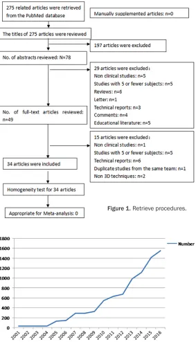
diversity of included liver diseases; (2) different study designs; (3) use of a variety of surgical approaches; and (4) various observational indi-cators. Therefore, only a systematic review was conducted in the present study. From 2001 to 2016, the number of the reports that applied 3D technology in hepatectomy steadily increased (Figure 2). These included articles reported 1,553 cases of 3D visualization-guid-Figure 1. Retrieve procedures.
Figure 2. Increasing trend of the application of 3D techniques in the treat-ment of liver diseases by hepatectomy.
studies, were also reviewed manually. The study designs of these included articles included clinical trials and observational studies. Case reports, studies with < 5 sub-jects, review articles, com-ments, articles used for edu-cational purpose, technical articles, and duplicated stud-ies were excluded. Each arti-cle was carefully reviewed to exclude the study data from the same team and potential duplications. After screening, a total of 34 articles were included; and the data was extracted and classified. Th- ese studies were described, analyzed and reviewed. Then, associated figures and tables were plotted.
Results
Systematic search and de-scription of included studies
[image:2.612.94.377.71.565.2]Table 1. Application of 3D technology in the treatment of liver disease by hepatectomy (ordered by publications year)
Author Country Published year Published Journal periodStudy Disease of cases Male Female Age (years)Number
Wigmore et al. [5] Scotland 2001 Annals of Surgery NR Liver tumor 27 13 14 68
Lang et al. [6] Germany 2005 Arch Surg NR Liver tumor 25 14 11 52
Saito et al. [7] Japan 2005 Hepatology 2001-2004 Liver tumor 72 51 21 62
Kamiyama et al. [8] Japan 2006 World J Surg 2002-2003 Liver disease 17 11 6 57.82
Yamanaka et al. [9] Japan 2007 World J Surg y 2001-2005 Hepatocellular carcinoma 113 81 32 65+9
Endo et al. [10] Japan 2007 Surgery 2003-2006 Hilar cholangiocarcinoma 15 NR NR 65
Dong et al. [11] China 2007 Pediatr Surg Int 1999-2005 Children liver tumor 18 8 10 4.2
Yamanaka et al. [12] Japan 2009 J Hepatobiliary Pancreat Surg 1993-2008 Liver tumor 35 25 10 63+9
Radtke et al. [13] Germany 2010 Ann Surg 1999-2007 Liver disease 157 117 85 56+12
Chen et al. [14] China 2010 International Journal of Surgery 2006-2008 Liver tumor 38 NR NR 39
Fang et al. [15] China 2010 Chinese Medical Journal NR Liver tumor 17 8 9 NR
Lamata et al. [16] England 2010 Surg Endosc 2008 Liver tumor 7 NR NR NR
Sasaki et al. [17] Japan 2011 The American Journal of Surgery 2004-2008 Hilar cholangiocarcinoma 19 13 6 67
Pianka et al. [18] Germany 2011 Arch Surg NR Liver tumor 13 10 3 60
Mise et al. [19] Japan 2011 British Journal of Surgery 2004-2009 Liver tumor 55 43 12 64
Wang et al. [20] China 2012 Dig Surg 2007-2009 Hepatocellular carcinoma 13 12 1 55
Stavrou et al. [21] Germany 2012 Advances in Medical Sciences 2002-2009 Liver tumor 29 NR NR NR
Ariizumi et al. [22] Japan 2013 J Hepatobiliary Pancreat Sci 2010-2011 Liver tumor 92 65 27 69
Fang et al. [23] China 2013 J Am Coll Surg 2005-2012 Intrahepatic bile duct stone 56 28 28 50.6+11.5 Takamoto et al. [24] Japan 2013 The American Journal of Surgery 2009-2012 Liver tumor 83 61 22 65 Tang et al. [25] China 2013 Asian Pacific J Cancer Prev 2009-2010 Hepatocellular carcinoma 22 NR NR NR
Kingham et al. [26] America 2013 J Gastrointest Surg 2008-2011 Liver tumor 64 44 20 58.5
Be’gin et al. [27] Canada 2014 Surg Endosc 2006-2009 Liver tumor 36 NR NR 56
Xie et al. [28] China 2014 Journal of Gastroenterology and Hepatology 2010-2011 Relapsed intrahepatic bile duct stone 20 7 13 NR
Simpson et al. [29] America 2014 J Am Coll Surg 2008-2010 Liver tumor 66 33 33 54
Okuda et al. [30] Japan 2015 Surgery 2009-2014 Cholangiocarcinoma 49 34 15 64+11
He et al. [31] China 2015 World J Gastroenterol 2011-2015 Hepatic alveolar hydatid disease 59 32 27 41.4+13.1 Fang et al. [32] China 2015 J Am Coll Surg 2008-2014 Central hepatocellular carcinoma 60 52 8 47.5+13.8 Tian et al. [33] China 2015 World J Gastroenterol 2013-2014 Central hepatocellular carcinoma 39 34 5 54.3+12.1
Oshiro et al. [34] Japan 2015 World J Gastroenterol 2010-2013 Liver tumor 99 78 21 65
Su et al. [35] China 2016 Pediatr Surg Int 2012-2015 Children liver tumor 16 10 6 15.12+13.16
Warmann et al. [36] Germany 2016 Journal of Pediatric Surgery 2004-2016 Liver tumor 18 12 12 33 Guan et al. [37] China 2016 Biomed Res Int 2006-2010 Hepatocellular carcinoma 92 75 17 52.48+8.36
Zygomalas et al. [38] Greece 2016 Med Biol Eng Comput 2013-2014 Liver tumor 12 6 6 54.2
ed hepatectomy for a variety of liver diseases. Among these cases, liver malignancies were predominant. The date of publication ranged from 1993 to 2016. The study subjects includ-ed adults and children (Table 1) [5-38]. 3D printing and intraoperative real-time navigation are an extended utility of 3D visualization in the investigation stage in liver surgery. However, there were few related articles and most were case reports with < 5 subjects. Moreover, the contents were often the introduction of new technologies and sharing of experience. Therefore, these related articles were not included in the present study. The two types of technologies are described in the “Discussion” section.
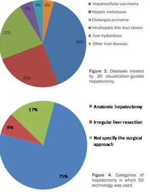
Diseases treated by 3D visualization-guided hepatectomy
Liver malignancies were predominant in cases treated by 3D visualization-guided
hepatecto-tectomy; which accounted for 75% of cases., Irregular liver resection accounted for 8% of cases; and the remaining 17% did not specify the surgical approach (Figure 4). Anatomic hep-atectomy included major hephep-atectomy (n = 213), bisegmentectomy (n = 61), segmentecto-my (n = 183), limited resection (n = 166), sec-tionectomy (n = 71), mesohepatectomy (n = 23), left trisectionectomy (n = 33), right trisec-tionectomy (n = 28), left hemihepatectomy (n = 97), right hemihepatectomy (n = 150), extend-ed left hemihepatectomy (n = 47), and extend-ed right hemihepatectomy (n = 65) (Figure 5).
A total of 13 articles evaluated the accuracy of 3D technology to estimate the liver volume and resection margin
[image:4.612.94.389.74.450.2]The evaluation method was chosen to calculate the correlation or difference between 3D-esti- mated values and the actual measured values. Results revealed a high correlation and small Figure 3. Diseases treated
by 3D visualization-guided hepatectomy.
Figure 4. Categories of hepatectomy in which 3D technology was used.
my. Among these reports, hepatocellular carcinoma ac- counted for 40% of cases, cholangiocarcinoma account-ed for 22% of cases, hepatic metastases (from colon and rectal cancer, duodenal can-cer, adrenal tumor, etc.) acc- ounted for 25% of cases, intrahepatic bile duct stones accounted for 5% of cases, liver hydatidosis accounted for 4% of cases, and other dis-eases, including liver heman-gioma, gallbladder cancer, liver focal hyperplasia nod-ules, liver adenoma, hepato-blastoma, liver mesenchymal tumor, hepatic hemangioen-dothelioma, liver sarcoma, teratoma, and liver malignant fibrous histiocytoma com-bined with hepatic cystadeno-ma, accounted for 4% of cases (Figure 3).
Categories of hepatectomy where 3D technology was used
hepa-discrepancy between the 3D-estimated resect-ed liver volume, residual liver volume and resection margin; and these actual values were measured intra- or post-operatively (Table 2). In addition, these results revealed that 3D tech-nology could accurately estimate the liver vol-ume and resection margin. Ariizumi et al. [22] calculated the accuracy of 3D estimated values in different surgical approaches: for sectionec-tomy, the correlation between the estimated liver volume and actual value was relatively high (R = 0.985, P < 0.0001), and the median error was 26 mL; for hemihepatectomy, the R
was 0.967 (P < 0.0001) and the median error was 38 mL. In addition, the estimated and actual values in non-cancerous liver lesions also revealed a good correlation. For example, in cirrhosis, chronic hepatitis and normal liver tissue, the coefficients (R) for resected liver vol-ume were 0.984 (P < 0.0001), 0.988 (P < 0.0001) and 0.9777 (P < 0.0001), respectively. Zygomalas et al. [38] compared the estimated residual liver volumes with the actual intraop-eratively measured values and found a signifi-cant correlation. For residual liver volume, R = 0.99 (P < 0.0001). Simpson et al. [29] com-pared 3D-estimated values with postoperative CT results, and for residual liver volume, R = 0.941 (P < 0.001).
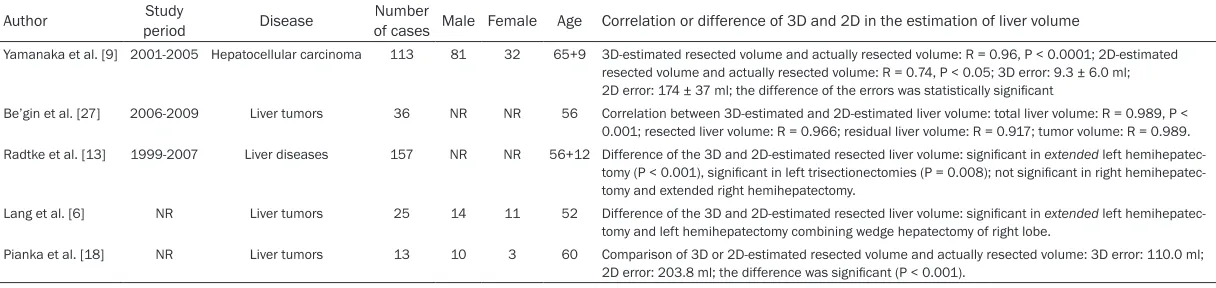
A total of five articles evaluated the correla -tions and differences between 3D and tradi-tional 2D technologies to estimate liver volume
By calculating the correlations and differences between 3D and 2D technologies to estimate
tomy between the 3D and 2D groups. Com- parisons with actually measured values sug-gest that 3D estimation was more accurate. For example, Yamanaka et al. [9] and Pianka et al.
[18] compared resected liver volume estimated by 2D and 3D technologies and found that the estimation errors by 3D technology were signifi-cantly smaller from the estimation errors by 2D technology (P < 0.01) (Table 3).
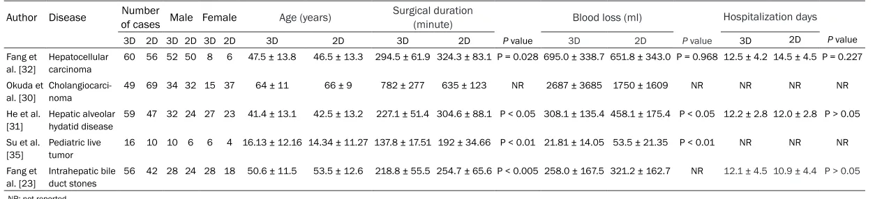
Five clinical studies evaluated the impact of 3D technology on surgical procedure and
ef-ficacy
[image:5.612.93.372.70.242.2]The associated indicators of surgical procedure and efficacy were compared among patients who underwent liver resection with and without preoperative planning, and the assessment of 3D technology. These results revealed that these disease conditions were similar, but the surgical duration was shorter and blood loss was less with 3D-guided hepatectomy. Furth- ermore, there was no significant difference in the length of hospitalization (Table 4). In terms of postoperative complications, including bili-ary fistula, ascites, incision infection, pleural effusion and liver failure, there was no signifi-cant difference in most results between the 3D and 2D groups. However, two teams reported that the incidences of biliary fistula, ascites and liver failure were lower in the 3D group than in the 2D group (P = 0.04) [30, 32]. Fang et al.[23, 32] reported that postoperative serum bilirubin level in the 2D group was significantly higher than in the 3D group (P = 0.032), while the hemoglobin level was significantly lower in the Figure 5. Surgical approaches for anatomical hepatectomy.
hemihepatec-Table 2. Studies investigating the accuracy of 3D-estimated liver volume or resection margin
Author Disease Number of cases (N) Male Female Age (years) Volume measurement (Correlation or difference between 3D-estimated value and actual value) Margin (Correlation or difference between 3D-estimated value and actual value)
Yamanaka et al. [9] Hepatocellular carcinoma 113 81 32 65+9 Resected liver volume: R = 0.96, P < 0.0001 R = 0.84, P < 0.01, error: 1.6 ± 2.6 mm.
Be’gin et al. [27] Liver tumor 36 NR NR 56 Resected liver volume: R = 0.874, P < 0.001. Tumor
volume: R = 0.758 NR
Tian et al. [33] Central liver cancer 39 34 5 54.3+12.1 No significant difference in tumor volume: P = 0.910 No significant difference: P = 0.488
Chen et al. [14] Liver tumor 38 NR NR NR No significant difference in resected liver volume: P > 0.05 NR
Takamoto et al. [24] Liver tumor 83 61 22 65 Resected liver volume: R = 0.9942, P < 0.01 NR
Yamanaka et al. [12] Liver tumor 35 25 10 63+9 Resected liver volume: R = 0.995, P < 0.0001 R = 0.702, P < 0.01, error: 1.3 ± 4.8 mm
Saito et al. [7] Liver tumor 72 51 21 62 Resected liver volume: R = 0.96, P < 0.0001 R = 0.84, P < 0.01, error: 1.6 ± 2.6 mm
Ariizumi et al. [22] Liver tumor 92 65 27 69 Resected liver volume: R > 0.9, P < 0.0001 NR
Wigmore et al. [5] Liver tumor 27 13 14 68 Resected liver volume: R = 0.94, P < 0.0001, NR
Kingham et al. [26] Liver tumor 64 42 22 58.5 NR Error: 6.5 ± 3.7 mm
Simpson et al. [29] Liver tumor 66 33 33 54 Residual liver volume: R = 0.941, P < 0.001 NR
Zygomalas et al. [38] Liver tumor 12 6 6 54.2 Residual liver volume: R = 0.99, P < 0.0001 NR
Wang et al. [20] Hepatocellular carcinoma 13 12 1 55 Resected liver volume: R = 0.995, P < 0.0001 R = 0.967, P < 0.0001
[image:6.792.91.710.85.270.2]NR: not reported.
Table 3. Correlation or difference in the estimation of liver volume by 3D and 2D technologies
Author periodStudy Disease Number of cases Male Female Age Correlation or difference of 3D and 2D in the estimation of liver volume
Yamanaka et al. [9] 2001-2005 Hepatocellular carcinoma 113 81 32 65+9 3D-estimated resected volume and actually resected volume: R = 0.96, P < 0.0001; 2D-estimated resected volume and actually resected volume: R = 0.74, P < 0.05; 3D error: 9.3 ± 6.0 ml;
2D error: 174 ± 37 ml; the difference of the errors was statistically significant
Be’gin et al. [27] 2006-2009 Liver tumors 36 NR NR 56 Correlation between 3D-estimated and 2D-estimated liver volume: total liver volume: R = 0.989, P < 0.001; resected liver volume: R = 0.966; residual liver volume: R = 0.917; tumor volume: R = 0.989. Radtke et al. [13] 1999-2007 Liver diseases 157 NR NR 56+12 Difference of the 3D and 2D-estimated resected liver volume: significant in extended left
hemihepatec-tomy (P < 0.001), significant in left trisectionectomies (P = 0.008); not significant in right hemihepatec -tomy and extended right hemihepatec-tomy.
Lang et al. [6] NR Liver tumors 25 14 11 52 Difference of the 3D and 2D-estimated resected liver volume: significant in extended left
hemihepatec-tomy and left hemihepatechemihepatec-tomy combining wedge hepatechemihepatec-tomy of right lobe.
Pianka et al. [18] NR Liver tumors 13 10 3 60 Comparison of 3D or 2D-estimated resected volume and actually resected volume: 3D error: 110.0 ml;
2D error: 203.8 ml; the difference was significant (P < 0.001).
[image:6.792.91.699.313.457.2]Table 4. Difference in surgical duration, blood loss, and length of hospitalization between the 3D and 2D groups
Author Disease of cases Male FemaleNumber Age (years) Surgical duration (minute) Blood loss (ml) Hospitalization days 3D 2D 3D 2D 3D 2D 3D 2D 3D 2D P value 3D 2D P value 3D 2D P value Fang et
al. [32] Hepatocellular carcinoma 60 56 52 50 8 6 47.5 ± 13.8 46.5 ± 13.3 294.5 ± 61.9 324.3 ± 83.1 P = 0.028 695.0 ± 338.7 651.8 ± 343.0 P = 0.968 12.5 ± 4.2 14.5 ± 4.5 P = 0.227 Okuda et
al. [30]
Cholangiocarci-noma
49 69 34 32 15 37 64 ± 11 66 ± 9 782 ± 277 635 ± 123 NR 2687 ± 3685 1750 ± 1609 NR NR NR NR
He et al. [31]
Hepatic alveolar hydatid disease
59 47 32 24 27 23 41.4 ± 13.1 42.5 ± 13.2 227.1 ± 51.4 304.6 ± 88.1 P < 0.05 308.1 ± 135.4 458.1 ± 175.4 P < 0.05 12.2 ± 2.8 12.0 ± 2.8 P > 0.05
Su et al.
[35] Pediatric live tumor 16 10 10 6 6 4 16.13 ± 12.16 14.34 ± 11.27 137.8 ± 17.51 192 ± 34.66 P < 0.01 21.81 ± 14.05 53.5 ± 21.35 P < 0.01 NR NR NR Fang et
al. [23] Intrahepatic bile duct stones 56 42 28 24 28 18 50.6 ± 11.5 53.5 ± 12.6 218.8 ± 55.5 254.7 ± 65.6 P < 0.005 258.0 ± 167.5 321.2 ± 162.7 NR 12.1 ± 4.5 10.9 ± 4.4 P > 0.05
2D group than in the 3D group (P = 0.033). In a study conducted by Okuda et al. [30], there was a significant difference in tumor stage between the 2D and 3D groups, since there were more T4-stage cases in the 3D group (P = 0.025). The incidence of portal vein reconstruc-tion was also higher in the 3D group than in the 2D group (P = 0.002). Since the conditions of patients were rather different between these two groups, surgical duration and blood loss were both higher in the 3D group than in the 2D group. However, when limited to the initial resection, the proportion of negative resection margins and invasive cancer-free ratio were both higher in the 3D group than in the 2D group (P = 0.03 and P = 0.02, respectively). Moreover, for intrahepatic bile duct stones, the stone recurrence rate was also significantly lower in the 3D group than in the 2D group (P < 0.004). These results suggest that 3D technol-ogy may be helpful to improve the efficiency and safety of liver resection surgery. However, without adequate follow-up data, the effect of 3D technology on the efficacy of hepatectomy could not be evaluated.
Impact of 3D technology on surgical strategy
Few studies have reported that the use of 3D technology led to the change in surgical strate-gy based on 2D results [6, 10, 13, 14]. A total of 70 cases were involved, including 43 cases for which the resection range was expanded, 17 cases of vascular reconstruction, and four cases that were unfit for surgical treatment due to inadequate estimated residual liver volume. In these three cases, 2D assessment revealed no surgical opportunity. However, after 3D reassessment, it was decided that surgery was viable.
Other special reports regarding the application of 3D visualization
The application of 3D technology in the treat-ment of pediatric liver tumors has been report-ed. [11, 35]3D technology has significant value in preoperative planning of various pediatric-specific liver tumors. 3D-assisted surgery was associated with shorter surgical duration and less intraoperative blood loss. Guan et al. [37] compared the efficacy of 3D-assisted hepatec-tomy to radiofrequency ablation for the treat-ment of small liver cancer. They found that when radiofrequency ablation can also be used
as an alternative, and that 3D-assisted hepa-tectomy should be the first choice. The applica-tion of 3D technology in relapsed intrahepatic bile duct stones and the resection of liver metastasis of colon cancer have also been reported to achieve satisfactory efficacy. Sta- vrou et al. [21] reported that 3D technology was adequate for planning Associated Liver Partition and Portal vein ligation for Staged hepatectomy (ALPPS).
Discussion
With the development of digital medical tech-nology, 3D visualization technology has increas-ingly been used in liver surgery. 3D visualiza-tion technology can reconstruct 3D liver models from computed tomography images. Further- more, 3D visualization not only displays the blood vessels and bile ducts in the liver more clearly, but also has the ability to estimate liver volume and simulate the surgery. 3D visualiza-tion plays an important role in preoperative planning, and is ever more commonly used in liver surgery. In 2000, Lamade et al. [39] report-ed that 3D technology was superior to tradition-al 2D imaging for preoperative tumor localiza-tion and preoperative planning. In 2001,
the parenchyma and vascular variation, which is common [42]. The biological characteristics of liver cancer determine the close relationship between tumors and liver tissues. Ultrasono- graphy, computed tomography (CT), and mag-netic resonance imaging are the most com-monly used imaging technologies for the preop-erative evaluation of liver cancer [43]. However, in many cases, 2D imaging is insufficient to clearly display the tumor and important vessels or their relationships, which creates a certain difficulty and risk of surgery. 3D imaging can clearly display the anatomical structures of the liver, accurately localize tumors, and show the relationship between the tumor and blood ves-sels. This technology can also be used for simu-lating the liver resection and designing a ratio-nal surgical plan to improve the safety and effectiveness of surgery. Therefore, 3D technol-ogy has been widely used in surgical treatment of liver malignancies. Lang et al. [44] performed a feasibility assessment of 3D technology for the resectability of relapsed liver metastases of colon cancer. The residual liver volume could be accurately predicted, which resulted in an improvement in the success rate of reopera-tions. The 3D reconstruction of intrahepatic and extrahepatic bile ducts can fully display the
anatomical relationship between the bile ducts, blood vessels, and tumors, and has certain advantages over traditional 2D imaging. There- fore, 3D technology is very helpful in the surgi-cal treatment of cholangiocarcinoma. Okuda et al. [26] reported that for the first resection, the ratio of the negative resection margin and inva-sive cancer-free ratio were significantly greater in the 3D group than in the 2D group. Accurate localization of intrahepatic bile duct stones has consistently posed a problem to liver surgeons. 3D reconstruction images of the bile duct sys-tem clearly display the bile duct branches at all levels, as well as the size and location of stones, which provides key information for planning the surgical approach. Various surgical paths can be simulated using a 3D model to develop a surgical plan with minimum trauma, while the problems of excessive resection and stone resi-due can be largely avoided. Fang et al. [19] reported that the application of 3D technology in the treatment of intrahepatic bile duct stones was associated with a relatively low recurrence rate. He et al. [27] investigated the application of 3D technology in hepatic resection of hepat-ic alveolar echinococcosis, and found that liver
morphology and structure often change in response to disease-induced damage. Further- more, intrahepatic multiple lesions are also common. Assisted by 3D technology, personal-ized surgical approaches can be planned to successfully implement the surgical plan and achieve satisfactory efficacy. 3D technology also plays an important role in the treatment of pediatric liver tumors, which are characterized by the diversity and complexity of pathological changes. In addition, the relatively small liver volume and large tumor volume of pediatric patients complicate surgical intervention. Surgeons can use 3D images to elucidate these lesions more clearly and design a safe and rational operation plan to reduce the surgical duration and intraoperative blood loss. Fuchs
et al. [45] applied 3D technology in the surgical treatment of pediatric hepatic vascular malfor-mations, and their results support the impor-tant role of 3D technology in the diagnosis and preoperative planning of liver diseases.
and vein reflux of liver segments. Ueno et al.
[46] used 3D techniques to display the anatom-ical structures of liver segments and perform precise laparoscopic anatomic hepatectomy, which significantly reduced surgical trauma. Associating liver partition and portal vein liga-tion for staged hepatectomy (ALPPS) is one of the most important technical innovations in the field of hepatobiliary surgery in recent years. ALPPS can be applied to liver cancer patients with a relatively small residual liver volume, and who cannot tolerate extensive liver resection [47]. ALPPS is increasingly used in clinic as a new surgical strategy [48, 49]. 3D visualization is well suited for ALPPS, as this technique can simulate the ligation of the portal vein, adjust the resection plane, estimate the residual liver volume, predict liver regeneration, and provide a feasibility analysis for second-stage liver resection. Together, these features can facili-tate the successful implementation of ALPPS. Accurate preoperative assessment and surgi-cal strategies are of great significance for hepa-tectomy. However, traditional 2D images some-times fail to provide adequate anatomical information and pathological features, which may lead to an irrational surgical strategy, resulting in severe liver damage. For instance,
inadequate resection increases the risk of recurrence, while excessive resection results in inadequate residual liver volume and postop-erative liver failure. 3D technology can improve preoperative planning. A study conducted by Hansen et al. [50] revealed that preoperative risk analysis based on 3D-modelling could increase risk awareness and assist surgeons to design surgical strategies with a safer resec-tion margin, minimizing the loss of liver volume. These reported strategy modifications based on 3D visualization include changes to the extent of resection, vascular reconstruction, and resectability evaluation. 3D visualization displays the relationship between lesions and liver structures more clearly, thereby helping the surgeons define the extent of resection more accurately. This favors the curative resec-tion of tumors and prevents liver failure due to the inadequate remnant liver volume. In addi-tion, by clearly displaying the important vessels and their branches in the liver, 3D visualization can also be used to assess the impact of ves-sel ligation or the resection of the remaining liver tissues, as well as the necessity of
vascu-lar reconstruction. Mise et al. [15] applied 3D technology to observe the route and supply region of the hepatic vein, and to assess the impact of reconstruction of the hepatic vein on the blood supply of the involved liver tissues. A study conducted by Lamade et al. [39] high-lighted the value of 3D technology in preopera-tive planning by comparing differences bet- ween 3D and 2D technologies in preoperative assessment. 3D technology revealed more accurate tumor localization and selection of the extent of resection. These accuracies increased by 37% and 31%, as compared with 2D technology. Tang et al. [25] conducted surgi-cal planning through the 3D-assisted estima-tion of resected liver volume, and determined whether hepatectomy was feasible according to the proportion of the resected liver. The sur-gical plan and actual operation showed good consistency.
The estimation of liver volume and resection margin is critical to surgical planning, and 3D technology can improve the accuracy of liver volume estimation [5, 14, 51]. Our study re- vealed a strong correlation between 3D-esti- mated value and the actual measured liver vol-ume, suggesting that 3D technology can accu-rately estimate the liver volume and resection margin. A comparison with 2D technology re- vealed a good consistency between 3D and 2D technologies for the estimation of liver and tumor volumes, suggesting that 3D technology has a similar reliability as traditional 2D tech-nology; while a comparison of actual measured values revealed greater accuracy with 3D tech-nology, especially for the estimation of the resection margin. The features of 3D technolo-gy facilitate the precision of preoperative assessment and planning, and improve intra-operative efficacy and safety.
promot-ing postoperative recovery. Although there was no significant difference in the length of hospi-talization between these 3D and 2D groups, the 3D group had certain advantages in terms of postoperative complications, as demonstrat-ed by the lower incidence of biliary fistula, asci-tes and liver failure in the 3D group. Studies have also shown that 3D-assisted hepatectomy resulted in a higher proportion of negative resection margins, which is critical for progno-sis [26]. In addition, the application of 3D tech-nology reduces intraoperative blood loss in pediatric hepatectomy, which is of great clinical significance in pediatric liver resection [31]. Laboratory examinations revealed that postop-erative serum bilirubin and hemoglobin levels were superior in the 3D group. All of these find-ings indicate that 3D technology is helpful for the implementation of an operation and post-operative recovery, which may have potential impacts on prognosis. However, more clinical trials and long-term follow-up data are required to more fully evaluate the effect of 3D technol-ogy on the efficacy of hepatectomy.
3D printing technology and intraoperative real-time navigation technology are extensions of 3D visualization. However, the application of these advanced digital medical technologies in hepatectomy remains in the exploratory stage. Igami et al. [52] used a 3D-printed transparent liver model to find small liver tumors, which were undetectable with intraoperative ultra-sound, and found that the application of 3D printing in the surgical treatment of small liver cancer achieved satisfactory effects. Xiang et al. [53] applied 3D printing technology-assisted hepatectomy to treat a complex massive hepa-tocarcinoma with variations of the portal vein. The printed liver model was used for preopera-tive planning, as well as intraoperapreopera-tive naviga-tion. At present, the high cost of 3D technology remains an obstacle to its clinical application. Oshiro et al. [54] invented a new 3D-printed liver model for hepatectomy, which reduced cost, shortened production time and improved visualization. 3D technology translates abstract 3D images in the surgeon’s brain to intuitive 3D images on the computer, while 3D printing technology transforms virtual 3D images to an actual solid model [55]. 3D-printed models offer greater advantages in the display of ana-tomical structures and lesions, the estimation of liver volume and resection margin, as well as
operational simulation. Hence, this technology plays an important role in preoperative plan-ning and intraoperative navigation.
Real-time intraoperative navigation is one of the most challenging techniques in the field of surgical research. With the development of digi-tal medical technology, a variety of navigation techniques have been applied in hepatectomy. One of these applications is the use of intraop-erative ultrasound for real-time navigation [56, 57]. Some researchers have reported the transformation of 3D information into a liver “risk map”, which was applied for intraopera-tive navigation to reduce surgical risk [12, 58]. Ntourakis et al. [59] introduced augmented reality guidance technology for the treatment of minimal residual liver metastases of colon can-cer after chemotherapy. This technique can dis-play the liver structure in real time, and detect small lesions by combining preoperative 3D reconstruction images and intraoperative real-time patient images. Hannes et al. [60] report-ed the use of a real-time navigation system based on intraoperative CT imaging. Intraope- rative CT images can be simultaneously trans-formed into 3D images to realize real-time navi-gation. Aoki et al. [61] reported the application of 3D virtual endoscopy in the navigation of laparoscopic hepatectomy, which increased surgical precision. Liu et al. [62] applied an indocyanine green-mediated infrared fluores-cence detection technique for intraoperative navigation, which helped to determine the re- section margin and guide hepatectomy. Buchs
increases the safety and efficacy of hepatecto-my, and may have a potential impact on dis-ease prognosis.
Disclosure of conflict of interest
None.
Address correspondence to: Jin-Ming Zhao, Depart- ment of Liver and Laparoscopic Surgery, Digestive and Vascular Surgery Center, The First Affiliated Hospital of Xinjiang Medical University, No. 137, Liyushan Road, Xinshi District, Urumqi 830054, Xinjiang Uyghur Autonomous Region, China. Tel: +86 13899881677; Fax: 0991-4364529; E-mail: [email protected]
References
[1] Banz VM, Baechtold M, Weber S, Peterhans M, Inderbitzin D and Candinas D. Computer planned, image-guided combined resection and ablation for bilobar colorectal liver metas-tases. World J Gastroenterol 2014; 20: 14992-14996.
[2] Qin JP, Tang SH, Jiang MD, He QW, Chen HB, Yao X, Zeng WZ and Gu M. Contrast enhanced computed tomography and reconstruction of hepatic vascular system for transjugular intra-hepatic portal systemic shunt puncture path planning. World J Gastroenterol 2015; 21: 9623-9629.
[3] Radtke A, Sgourakis G, Sotiropoulos GC, Mol-menti EP, Saner FH, Timm S, Malagó M and Lang H. Territorial belonging of the middle he-patic vein in living liver donor candidates eval-uated by three-dimensional computed tomo-graphic reconstruction and virtual liver resection. Br J Surg 2009; 96: 206-213. [4] He YB, Bai L, Jiang Y, Ji XW, Tai QW, Zhao JM,
Zhang JH, Liu WY and Wen H. Application of a three-dimensional reconstruction technique in liver auto transplantation for end-stage hepat-ic alveolar echinococcosis. J Gastrointest Surg 2015; 19: 1457-1465.
[5] Wigmore SJ, Redhead DN, Yan XJ, Casey J, Madhavan K, Dejong CH, Currie EJ and Garden OJ. Virtual hepatic resection using three-di-mensional reconstruction of helical computed tomography angioportograms. Ann Surg 2001; 233: 221-226.
[6] Lang H, Radtke A, Hindennach M, Schroeder T, Frühauf NR, Malagó M, Bourquain H, Peitgen HO, Oldhafer KJ and Broelsch CE. Impact of vir-tual tumor resection and computer-assisted risk analysis on operation planning and intra-operative strategy in major hepatic resection. Arch Surg 2005; 140: 629-638; discussion 638.
[7] Saito S, Yamanaka J, Miura K, Nakao N, Nagao T, Sugimoto T, Hirano T, Kuroda N, Iimuro Y and Fujimoto J. A novel 3D hepatectomy simulation based on liver circulation: application to liver resection and transplantation. Hepatology 2005; 41: 1297-1304.
[8] Kamiyama T, Nakagawa T, Nakanishi K, Kama-chi H, Onodera Y, Matsushita M and Todo S. Preoperative evaluation of hepatic vasculature by three-dimensional computed tomography in patients undergoing hepatectomy. World J Surg 2006; 30: 400-409.
[9] Yamanaka J, Saito S and Fujimoto J. Impact of preoperative planning using virtual segmental volumetry on liver resection for hepatocellular carcinoma. World J Surg 2007; 31: 1249-1255.
[10] Endo I, Shimada H, Sugita M, Fujii Y, Morioka D, Takeda K, Sugae S, Tanaka K, Togo S, Bour-quain H and Peitgen HO. Role of three-dimen-sional imaging inoperative planning for hilar-Cholangiocarcinoma. Surgery 2007; 142: 666-675.
[11] Dong Q, Xu W, Jiang B, Lu Y, Hao X, Zhang H, Jiang Z, Lu H, Yang C, Cheng Y, Yang X and Hao D. Clinical applications of computerized to-mography 3-Dreconstruction imaging for diag-nosis and surgery in childrenwith large liver tumors or tumors at the hepatic hilum. Pediatr Surg Int 2007; 23: 1045-1050.
[12] Yamanaka J, Okada T, Saito S, Kondo Y, Yoshi-da Y, Suzumura K, Hirano T, Iimuro Y and Fuji-moto J. Minimally invasive laparoscopic liver resection: 3D MDCT simulation for preopera-tive planning. J Hepatobiliary Pancreat Surg 2009; 16: 808-815.
[13] Radtke A, Sotiropoulos GC, Molmenti EP, Schroeder T, Peitgen HO, Frilling A, Broering DC, Broelsch CE and Malago’ M. Computer-as-sisted surgery planningfor complex liver resec-tionswhen is it helpful? A single-center experi-ence over an 8-year period. Ann Surg 2010; 252: 876-883.
[14] Chen G, Li XC, Wu GQ, Wang Y, Fang B, Xiong XF, Yang RG, Tan LW, Zhang SX and Dong JH. The use of virtual reality for the functional sim-ulation of hepatic tumors (case control study). Int J Surg 2010; 8: 72-78.
[15] Fang CH, Huang YP, Chen ML, Lu CM, Li XF and Qiu WF. Digital medical technology based on 64-slice computed tomography in hepatic sur-gery. Chin Med J (Engl) 2010; 123: 1149-1153.
[17] Sasaki R, Kondo T, Oda T, Murata S, Waka-bayashi G and Ohkohchi N. Impact of three-di-mensional analysis of multidetectorrow com-puted tomography cholangioportography ino- perative planning for hilar cholangiocarcino-ma. Am J Surg 2011; 202: 441-448.
[18] Pianka F, Baumhauer M, Stein D, Radeleff B, Schmied BM, Meinzer HP and Müller SA. Liver tissue sparing resection using a novel planning tool, Langenbecks. Langenbecks Arch Surg 2011; 396: 201-208.
[19] Mise Y, Hasegawa K, Satou S, Aoki T, Beck Y, Sugawara Y, Makuuchi M and Kokudo N. Ve-nous reconstruction based on virtual liver re-section to avoidcongestion in the liver rem-nant. Br J Surg 2011; 98: 1742-1751.
[20] Wang Y, Zhang Y, Peitgen HO, Schenk A, Yuan L, Wei G and Sun Y. Precise local resection for hepatocellular carcinoma based on tumor-sur-rounding vascular anatomy revealed by 3D analysis. Dig Surg 2012; 29: 99-106.
[21] Stavrou GA, Donati M, Ringe KI, Peitgen HO and Oldhafer KJ. Liver remnant hypertrophy induction-How often do we really use it in the time of computer assisted surgery? Adv Med Sci 2012; 57: 251-258.
[22] Ariizumi S, Takahashi Y, Kotera Y, Omori A, Yoneda G, Mu H, Katagiri S, Egawa H and Ya-mamoto M. Novel virtual hepatectomy is use-ful for evaluation of the portal territory for ana-tomical sectionectomy, segmentectomy, and hemihepatectomy. J Hepatobiliary Pancreat Sci 2013; 20: 396-402.
[23] Fang CH, Liu J, Fan YF, Yang J, Xiang N and Zeng N. Outcomes of hepatectomy for hepato-lithiasis based on 3-dimensional reconstruc-tion technique. J Am Coll Surg 2013; 217: 280-288.
[24] Takamoto T, Hashimoto T, Ogata S, Inoue K, Maruyama Y, Miyazaki A and Makuuchi M. Planning of anatomical liver segmentectomy and sub segmentectomy with 3-dimensional simulation Software. Am J Surg 2013; 206: 530-538.
[25] Tang JH, Yan FH, Zhou ML, Xu PJ, Zhou J and Fan J. Evaluation of computer-assisted quanti-tative volumetric analysis for poperative re-sectability assessment of huge hepatocellular carcinoma. Asian Pac J Cancer Prev 2013; 14: 3045-3050.
[26] Kingham TP, Jayaraman S, Clements LW, Scherer MA, Stefansic JD and Jarnagin WR. Evolution of image-guided liver surgery: transi-tion from open to laparoscopic procedures. J Gastrointest Surg 2013; 17: 1274-1282. [27] Bégin A, Martel G, Lapointe R, Belblidia A,
Lep-anto L, Soler L, Mutter D, Marescaux J and Vandenbroucke-Menu F. Accuracy of preopera-tive automatic measurement of the liver
vol-ume by CT-scan combined to a 3D virtual surgi-cal planning. software (3DVSP). Surg Endosc 2014; 28: 3408-3412.
[28] Xie A, Fang C, Huang Y, Fan Y, Pan J and Peng F. Application of three-dimensional reconstruc-tion and visible simulareconstruc-tion technique in reop-eration of hepatolithiasis. J Gastroenterol Hep-atol 2013; 28: 248-254.
[29] Simpson AL, Geller DA, Hemming AW, Jarnagin WR, Clements LW, D’Angelica MI, Dumpuri P, Gönen M, Zendejas I, Miga MI and Stefansic JD. Liver planning software accurately predicts postoperative liver volume and measures early regeneration. J Am Coll Surg 2014; 219: 199-207.
[30] Okuda Y, Taura K, Seo S, Yasuchika K, Nitta T, Ogawa K, Hatano E and Uemoto S. Usefulness of operative planning based on 3-dimensional CT cholangiographyfor biliary malignancies. Surgery 2015; 158: 1261-1271.
[31] He YB, Bai L, Aji T, Jiang Y, Zhao JM, Zhang JH, Shao YM, Liu WY and Wen H. Application of 3D reconstruction for surgical treatment of hepat-ic alveolar echinococcosis. World J Gastroen-terol 2015; 21: 10200-10207.
[32] Fang CH, Tao HS, Yang J, Fang ZS, Cai W, Liu J and Fan YF. Impact of three-dimensional re-construction technique in the operation plan-ning of centrallylocated hepatocellular carci-noma. J Am Coll Surg 2015; 220: 28-37. [33] Tian F, Wu JX, Rong WQ, Wang LM, Wu F, Yu
WB, An SL, Liu FQ, Feng L, Bi C and Liu YH. Three-dimensional morphometric analysis for hepatectomy of centrally located hepatocellu-lar carcinoma: a pilot study. World J Gastroen-terol 2015; 21: 4607-4619.
[34] Oshiro Y, Yano H, Mitani J, Kim S, Kim J, Fuku-naga K and Ohkohchi N. Novel 3-dimensional virtual hepatectomy simulation combined with real-time deformation. World J Gastroenterol 2015; 21: 9982-9992.
[35] Su L, Dong Q, Zhang H, Zhou X, Chen Y, Hao X and Li X. Clinical application of a three-dimen-sional imaging technique in infants and young children with complex liver tumors. Pediatr Surg Int 2016; 32: 387-395.
[36] Warmann SW, Schenk A, Schaefer JF, Ebinger M, Blumenstock G, Tsiflikas I and Fuchs J. Computer-assisted surgery planning in chil-dren with complex liver tumors identifies vari-ability of the classical Couinaud classification. J Pediatr Surg 2016; 51: 1801-1806.
[38] Zygomalas A, Karavias D, Koutsouris D, Marou-lis I, Karavias DD, Giokas K and Megalooikono-mou V. Computer-assisted liver tumor surgery using a novel semiautomatic and a hybrid semiautomatic segmentation algorithm. Med Biol Eng Comput 2016; 54: 711-721.
[39] Lamadé W, Glombitza G, Fischer L, Chiu P, Cárdenas CE Sr, Thorn M, Meinzer HP, Gre-nacher L, Bauer H, Lehnert T and Herfarth C, Meinzer HP, Grenacher L. The impact of 3-di-mensional reconstructions on operation plan-ning in liver surgery. Arch Surg 2000; 135: 1256-1261.
[40] Torre LA, Bray F, Siegel RL, Ferlay J, Lortet-Tieu-lent J and Jemal A. Global cancer statistics, 2012. CA Cancer J Clin 2015; 65: 87-108. [41] Torre LA, Siegel RL, Ward EM and Jemal A.
Global cancer incidence and mortality rates and trends-an update. Cancer Epidemiol Bio-markers Prev 2016; 25: 16-27.
[42] Fang CH, You JH, Lau WY, Lai EC, Fan YF, Zhong SZ, Li KX, Chen ZX, Su ZH and Bao SS. Ana-tomical variations of hepatic veins: three-di-mensional computed tomography scans of 200 subjects. World J Surg 2012; 36: 120-124.
[43] Maluccio M and Covey A. Recent progress in understanding, diagnosing, and treating hepa-tocellular carcinoma. CA Cancer J Clin 2012; 62: 394-399.
[44] Lang H, Radtke A, Liu C, Sotiropoulos GC, Hin-dennach M, Schroeder T, Peitgen HO and Bro-elsch CE. Improved assessment of functional resectability in repeated hepatectomy by com-puter-assistedoperation planning. Hepatogas-troenterology 2005; 52: 1645-1648.
[45] Fuchs J, Warmann SW, Sieverding L, Haber HP, Schäfer J, Seitz G, Hofbeck M, Bourquain H and Peitgen HO. Impact of virtual imaging pro-cedures on treatment strategies in children with hepatic vascular malformations. J Pediatr Gastroenterol Nutr 2010; 50: 67-73.
[46] Ueno S, Sakoda M, Kurahara H, Iino S, Minami K, Ando K, Mataki Y, Maemura K, Ishigami S, Takumi K, Fukukura Y and Natsugoe S. Preop-erative segmentation of the liver, based on 3D CT images, facilitates laparoscopic anatomic hepatic resection for small nodular hepatocel-lular carcinoma in patients with cirrhosis. Hepatogastroenterology 2010; 57: 807-812. [47] Narita M, Oussoultzoglou E, Ikai I, Bachellier P
and Jaeck D. Right portal vein ligation com-bined with in situ splitting induces rapid left lateral liver lobe hypertrophy enabling 2-staged extended right hepatic resection in small-for-size settings. Ann Surg 2012; 256: e7-8; au-thor reply e16-7.
[48] Tanaka K, Matsuo K, Murakami T, Kawaguchi D, Hiroshima Y, Koda K, Endo I, Ichikawa Y,
Taguri M and Tanabe M. Associating liver parti-tion and portal vein ligaparti-tion for staged hepa-tectomy (ALPPS): short-term outcome, func-tional changes in the future liver remnant, and tumor growth activity. Eur J Surg Oncol 2015; 41: 506-512.
[49] Hernandez-Alejandro R, Bertens KA, Pineda-Solis K and Croome KP. Can we improve the morbidity and mortality associated with the as-sociating liver partition with portal vein ligation for staged hepatectomy (ALPPS) procedure in the management of colorectal liver metasta-ses? Surgery 2015; 157: 194-201.
[50] Hansen C, Zidowitz S, Preim B, Stavrou G, Old-hafer KJ and Hahn HK. Impact of model-based risk analysis for liver surgery planning. Int J Comput Assist Radiol Surg 2014; 9: 473-480. [51] DuBray BJ Jr, Levy RV, Balachandran P, Conzen
KD, Upadhya GA, Anderson CD and Chapman WC. Novel three-dimensional imaging tech-nique improves the accuracy of hepatic volu-metric assessment. HPB (Oxford) 2011; 13: 670-674.
[52] Igami T, Nakamura Y, Hirose T, Ebata T, Yokoya-ma Y, Sugawara G, Mizuno T, Mori K and Nagi-no M. Application of a three-dimensional print of a liverin hepatectomy for small tumors invis-ible by intraoperative ultrasonography: prelimi-nary experience. World J Surg 2014; 38: 3163-3166.
[53] Xiang N, Fang C, Fan Y, Yang J, Zeng N, Liu J and Zhu W. Application of liver three-dimen-sional printing in hepatectomy for complex massive hepatocarcinoma with rare variations of portal vein: preliminary experience. Int J Clin Exp Med 2015; 8: 18873-18878.
[54] Oshiro Y, Mitani J, Okada T and Ohkohchi N. A novel three-dimensional print of liver vessels and tumors in hepatectomy. Surg Today 2017; 47: 521-524.
[55] Michalski MH and Ross JS. The shape of things to come: 3D printing in medicine. JAMA 2014; 312: 2213-2214.
[56] Satou S, Aoki T, Kaneko J, Sakamoto Y, Hasega-wa K, SugaHasega-wara Y, Arai O, Mitake T, Miura K and Kokudo N. Initial experience of intraopera-tive three-dimensional navigation for liver re-section using real-time virtua sonography. Sur-gery 2014; 155: 255-262.
[57] Banz VM, Müller PC, Tinguely P, Inderbitzin D, Ribes D, Peterhans M, Candinas D and Weber S. Intraoperative image-guided navigation sys-tem: development and applicability in 65 pa-tients undergoing liversurgery. Langenbecks Arch Surg 2016; 401: 495-502.
[59] Ntourakis D, Memeo R, Soler L, Marescaux J, Mutter D and Pessaux P. Augmented reality guidance for the resection of missingcolorectal liver metastases: an initial experience. World J Surg 2016; 40: 419-426.
[60] Kenngott HG, Wagner M, Gondan M, Nickel F, Nolden M, Fetzer A, Weitz J, Fischer L, Speidel S, Meinzer HP, Böckler D, Büchler MW and Müller-Stich BP. Real-time image guidance in laparoscopic liver surgery: first clinical experi-ence with a guidance system based on intraop-erative CT imaging. Surg Endosc 2014; 28: 933-940.
[61] Aoki T, Murakami M, Koizumi T, Fujimori A, Ga-reer H, Enami Y, Koike R, Watanabe M and Ot-suka K. Three-dimensional virtual endoscopy for laparoscopic and thoracoscopic liverre sec-tion. J Am Coll Surg 2015; 221: e21-26.
[62] Liu Y, Bauer AQ, Akers WJ, Sudlow G, Liang K, Shen D, Berezin MY, Culver JP and Achilefu S. Hands-free, wireless goggles for near-infrared fluorescence and real-time image-guided sur-gery. Surgery 2011; 149: 689-698.