Int J Clin Exp Med 2016;9(3):5413-5425
www.ijcem.com /ISSN:1940-5901/IJCEM0020337
Original Article
Role of bone marrow-derived mesenchymal stem cells
in a rat model of sepsis-induced acute lung injury
Mingli Yao1*, Kejing Tang2*, Shunwei Huang1
Departments of 1Critical Care Medicine, 2Pulmonary Medicine, The First Affiliated Hospital of Sun Yat-sen
University, Guangzhou, China. *Equal contributors.
Received November 23, 2015; Accepted February 10, 2016; Epub March 15, 2016; Published March 30, 2016
Abstract: Background: Acute Respiratory Distress Syndrome (ARDS) is a life-threatening condition in patients with predisposing factors, among which sepsis is a common cause. There are no effective strategies to treat this syn-drome except supportive therapies. Some preclinical studies suggest a potential therapeutic effect of mesenchymal
stem cells (MSCs) on ARDS. However, data specifically evaluating the impact of MSCs therapy on development of
sepsis-induced ARDS are still limited. Methods: There were 25 rats in each group, including sepsis group, sepsis +
antibiotic group, sepsis + antibiotic + MSCs group and sham group, twenty of which being sacrificed at 12 h, 18 h, 24 h and 48 h after surgery, respectively, five for survival analysis, and 5 normal controls. Survival rate, pulmonary physiological function, alveolar capillary barrier, lung inflammatory reaction and pathological injury were measured.
Results: ① at 18 h, total nucleated cell count of bronchoalveolar lavage fluid was 12.29±7.03 in the sepsis + antibi
-otic + MSCs subgroup, as compared with 42.3±8.18 in the sepsis subgroup (P=0.007), 35.77±16.80 in the sepsis
+ antibiotic subgroup (P=0.016) and 37.80±22.96 in the normal group (P=0.025). ② The protein concentration of bronchoalveolar lavage fluid in the sepsis + antibiotic + MSCs subgroup was less than that in the sepsis subgroup at 24 h, (0.41±0.24 vs. 1.17±0.57 mg/ml, P=0.004). Conclusion: Intravenous injection of allogeneic MSCs is safe for rats with sepsis-induced acute lung injury. To be administrated in early stage of sepsis, MSCs improve alveolar
inflammatory cells infiltration and protein exudation, as well as alveolar congestion and hemorrhage. However, there
is a potential risk of oxygenation impairment and lung water increase with intravenous injection of MSCs.
Keywords: Sepsis, Acute Respiratory Distress Syndrome (ARDS), bone marrow derived mesenchymal stem cells (BMSCs)
Introduction
Acute Respiratory Distress Syndrome (ARDS) remains a serious life-threatening condition, usually develops in patients with predisposing
conditions that induce systemic inflammatory
response, such as sepsis, pneumonia, major trauma, multiple transfusions, aspiration and acute severe pancreatitis [1]. The mainstay of ARDS treatment is supportive care. Some po-
tential treatments may show benefits in improv
-ing oxygenation or accelerat-ing alveolar fluid
clearance. However, none of them has been proven to reduce mortality or improve any other fatal clinical outcomes [2-5].
Mesenchymal stem cells (MSCs) have received increasing interest for treatment of ARDS [6]. Although the pathogenesis of ARDS remains
unclear, well accepted hypotheses include alve-olar epithelial and vascular endothelial injury, increased permeability of the endothelial and epithelial barriers, imbalance in coagulation
and fibrinolysis systems, combined with infla-mmatory and anti-inflainfla-mmatory reactions [3,
7-10]. Most current researches demonstrate
that MSCs potentially improve major abnormali-ties on ARDS based on a variety of mechanisms
[11]. The concordance between ARDS patho-physiology and regulatory effects of MSCs ther-apy makes MSCs a promising option for treat-ment of ARDS [12]. However, application of
MSCs for treatment of ARDS is still a novel field
5414 Int J Clin Exp Med 2016;9(3):5413-5425 ARDS, etc [13]. Sepsis is one of the most
com-mon causes of ARDS [2]. Sepsis-induced ARDS has a higher overall disease severity and higher mortality than non-sepsis-induced ARDS [1]. This study was undertaken to examine the effects of BMSCs injection on sepsis-induced ARDS in a rat abdominal sepsis model.
Materials and methods
Animals
Male Sprague-Dawley (SD) rats (n=105, body
weight 200±20 g) and neonatal SD rats (n=5, 2-3 wk old) in specific pathogen free (SPF)
grade were obtained from animal center of Sun Yat-sen University (SCXK 2011-0029). Animals were maintained under standard laboratory
conditions of 12-hour light/dark cycles, 23±3°C
room temperature, 40%-70% environment hu- midity, and were given free access to standard rat chow and water though the whole experi-ment. The animals were allowed to adapt to laboratory conditions for at least 2 days. The animal experiment was approved by the Animal Care and Use Ethics Committee of Sun Yat-sen University.
Experimental grouping
Male SD rats (n=105) were randomly allocated to the sepsis group (n=25), the sepsis + antibi-otic group (n=25), the sepsis + antibiantibi-otic + MSCs group (n=25) , the sham group (n=25),
twenty of which being sacrificed at 12 h, 18 h, 24 h and 48 h after surgery, respectively, five
for survival analysis, and normal controls (n=5). Flow diagram of study design is as follows (Figure 1).
Sepsis model
The procedure was carried out under 10% chlo-ral hydrate anesthesia (0.4 ml/100 g body weight, i.p.). After disinfection, a 2-cm long median laparotomy was performed. The ileoce-cus was retracted out of abdominal cavity. Then an approximately 5-mm-long venous indwelling cannula (16 G) was inserted approximately 1 cm distal to the ileocecal valve at the antimes-enteric site. The venous indwelling cannula was
fixated to the wall with sutures. By careful pal -pation of the cecum with cotton swabs, the
[image:2.612.95.521.74.341.2]stent was filled with feces. The layers of the abdomen wall were sutured, followed by fluid
MSCs for treatment of ARDS
supplement with 2 ml saline solution. Animals in sham group underwent the same surgery
except that the stent was fixed outside cecum.
Six hours after operation, animals in the sepsis + antibiotic + MSCs group were given one dose of Imipenem/Cilastatin (30 mg/0.4 ml) by intra-peritoneal injection, and P4 or P5 MSCs (1 × 10⁶/0.2 ml) though tail vein. Then Imipenem/
Cilastatin was given until sampling (i.e., 30 mg/0.4 ml i.p., twice a day). Animals in other three groups received corresponding
interven-tion. Imipenem/cilastatin is an optimal antibi-otic for enterogenous peritonitis. Drug dose is
determined based on conversion coefficient
method referring to “Basic Knowledge of Laboratory Animal Science and Experimental Technique Course”.
Survival analysis
[image:3.612.90.524.72.524.2]Animals in the sepsis + antibiotic + MSCs group, sepsis + antibiotic group, sepsis group Figure 2. Abdominal cavity presentation in rat model of sepsis. A. At 12 h, model rats revealed obvious peritonitis. C
and D. At 24 h and 48 h, the whole abdominal cavity was fixed. B. At 18 h, the severity of peritonitis was in between. Red arrows represent dilatation of bowel loops, yellow arrows represent fiber formation, green arrows represent as
5416 Int J Clin Exp Med 2016;9(3):5413-5425 and sham group (n=5 in each group) were
fol-lowed up every 12 h until death.
Sampling handling
Survived animals were anesthetized and lapa-rotomized at predetermined time points. Ab- dominal cavities were fully exposed. Severity of
peritonitis was assessed and qualified animals
were included. After that, exposed the abdomi-nal aorta, extracted 1 ml of arterial blood for
gas analysis, and then sacrificed the animal.
Subsequently separated the trachea, perfo- rmed a transverse incision between the third and fourth trachea ring, carried out
endotra-cheal intubation with an outer diameter of 1.8 mm endotracheal tube, and fixed the tube with
silk sutures, then performed thoracotomy and ligation of right lung lobes. Three aliquots (2 ml each) of sterile normal saline were instilled and aspirated 10 seconds later, repeated 3 times for each aliquot. The bronchoalveolar lavage
fluid (BALF) was pooled in ice-cold tubes for
[image:4.612.92.524.69.520.2]testing. Separated the right upper lobe, blotted Figure 3. Lung histopathology for rat model of sepsis. Lung tissues were obtained at 12 h (A), 18 h (B), 24 h (C) and 48 h (D) after surgery (H&E, × 100 magnifications). Green arrows represent aggregation of inflammatory cells and
MSCs for treatment of ARDS
it up and weighed, then kept in a thermostatic
drying chamber of 65°C for 48 hours, weighed
the dried tissue again, thus calculated the ratio of Wet/Dry weight. The right lower lobe was
immediately fixed in 10% formalin for histo -pathological analysis.
Histopathological evaluation
Lung injury was scored by experienced investi-gator blinded to the groups according to the fol-lowing parameters: a) alveolar congestion and
hemorrhage, b) infiltration or aggregation of inflammatory cells in airspace or interstitium, c)
thickness of alveolar wall, d) hyaline membrane formation, e) atelectasis. Each parameter was scored on a 4-point scale as follows: 0 minimal
damage, 1 mild damage, 2 moderate damage, 3 severe damage.
Isolation, culture and identification of BMSCs
MSCs derived from bone marrow of rats were isolated as Houlihan etc [14] described. SD neonatal rats (n=5, 2-3 wk old) were chosen for BMSCs isolation. The cells applied in subse-quent experiments were of passage 4 or 5,
identified by detecting surface markers expres
-sion by flow cytometry.
Statistical analysis
[image:5.612.88.522.68.520.2]The SPSS software package (version 13.0) was used for the statistical tests. Continuous data
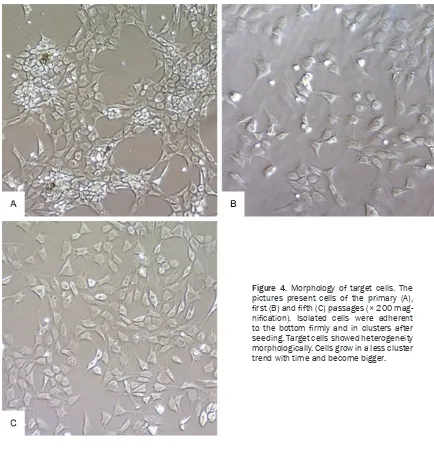
Figure 4. Morphology of target cells. The pictures present cells of the primary (A),
first (B) and fifth (C) passages (× 200 mag
-nification). Isolated cells were adherent to the bottom firmly and in clusters after
5418 Int J Clin Exp Med 2016;9(3):5413-5425 Figure 5. Surface markers of Pas-sage 5 target cells: the mesen-chymal stem cell markers: CD29
87.51%, CD44 95.9%, CD90 95.79%,
and hematopoietic lineage mark-ers: CD34 0.41%, CD45 0.31%.
were expressed as mean ± SD and categorical
data as the median with interquartile ranges (IQRs). The One-Way ANOVA test and least
sig-nificant difference t test (LSD-t) were used for
multiple comparisons if data met correspond-ing conditions, otherwise Kruskal-Wallis test was done and Bonferroni method was applied for multiple comparisons. A P value of <0.05
was considered to be significant.
Results
Preparation of sepsis-induced acute lung in-jury model
At 12 h after surgery, the rats revealed conges-tion and edema of both parietal peritoneum and intestinal walls, dilatation of bowel loops, slight adhesion, a small amount of exudates
and fiber formation (Figure 2A). At 24 h, the rats demonstrated extensive dilatation of intes-tines, obvious adhesion formation, moderate
amount of bloody as cites and denser fiber for -mation (Figure 2C). One model rat died between
36 h and 48 h after surgery, thus survival rate in a week was 80% (4/5).
There was a significant increase in hyaline
membrane formation in the model group at 24 h compared to normal controls (P=0.005) and
a more serious atelectasis at 48 h (P=0.014).
All of the four model groups presented a signifi
-cant increase in alveolar congestion and hem-orrhage compared to the normal, (P<0.05) (Figure 3).
Identification of target cells
The target cells were uneven in shape (Figure 4). To further characterize these cells, surface markers of Passage 5 cells were examined by
flow cytometry, showing the mesenchymal stem
cell markers: CD29 87.51%, CD44 95.9%, CD90 95.79%, and the hematopoietic lineage mark-ers: CD34 0.41%, CD45 0.31% (Figure 5).
The effect of MSCs transplantation on sepsis-induced acute lung injury
One rat in the sepsis group died between 36 h
and 48 h after surgery, thus survival rate in a week was 80% (4/5). All animals in other three
groups survived the observation period of 7 days.
In the sepsis group, obvious alveolar conges-tion and hemorrhage was detected starting from 12 h compared to normal control, (P=0.03,
0.048, 0.001, 0.001, respectively), significant
hyaline membrane formation observed in the 24 h subgroup and more atelectasis
demon-strated in the 48 h subgroup compared to nor -mal control, (P=0.005, 0.014, respectively). All
MSCs for treatment of ARDS
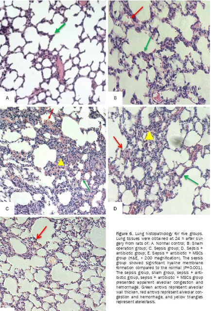
Figure 6. Lung histopathology for five groups.
Lung tissues were obtained at 24 h after sur-gery from rats of: A. Normal control; B. Sham operation group; C. Sepsis group; D. Sepsis + antibiotic group; E. Sepsis + antibiotic + MSCs
group (H&E, × 200 magnification). The sepsis group showed significant hyaline membrane
5420 Int J Clin Exp Med 2016;9(3):5413-5425
within 48 h in sham group. In the sepsis + anti -biotic group, obvious alveolar congestion and
hemorrhage was detected in 12 h, 18 h, 24 h subgroups, significant hyaline membrane for
-mation in 18 h subgroup compared to normal
control, (P=0.01, 0.001, 0.001 and P=0.041). In the sepsis + antibiotic + MSCs group, appar-ent alveolar congestion and hemorrhage was
observed at 18 h compared to normal control
(P=0.03) (Figure 6 and Table 1).
At 12 h after surgery, PH in normal control was
7.315±0.062, as compared with 7.215±0.013
in the sepsis subgroup (P=0.003) and 7.234±
[image:8.612.93.529.85.320.2]0.029 in the sepsis + antibiotic + MSCs sub-group (P=0.009). At 18 h, SaO2 in the sepsis +
Table 1. Lung histopathology of different subgroups
Groups Thickness ofalveolar wall inflammatory cellsAggregation of Hyaline membrane Atelectasis Congestion and hemorrhage
Sepsis 12 h n=3 0.7 (0.5, 0.7) 1.7 (1.7, 1.7) 0.1 (0.1, 0.1) 1.2 (0.9, 1.2) 1.6 (0.9, 1.6)*
18 h n=3 1.2 (1, 1.2) 1.5 (1.4, 1.5) 0.1 (0.1, 0.1) 0.3 (0.3, 0.3) 1.3 (1.2, 1.3)*
24 h n=9 0.9 (0.35, 1.4) 1.7 (1.3, 2) 0.6 (0.35, 0.65)* 0.9 (0.6, 1.1) 1.4 (1.3, 1.75)*
48 h n=5 1 (0.25, 1.25) 1.8 (1.45, 2.05) 0.1 (0, 0.6) 1.4 (1.05, 2.25)* 1.5 (1.15, 2.3)*
Sham 12 h n=8 0.9 (0.33, 1.6) 1.5 (1.3, 1.73) 0.05 (0, 0.18) 1.25 (0.85, 2.05)* 0.55 (0.15, 1.20)
18 h n=4 0.35 (0.23, 0.93) 1.2 (1.05, 1.5) 0.05 (0, 0.25) 0.95 (0.75, 1.68) 0.75 (0.33, 2) 24 h n=4 0.45 (0.18,0.73) 1.18 (1.03, 1.36) 0.2 (0.03, 0.45) 1.35 (1, 1.6) 1.65 (1.6, 1.80)*
48 h n=2 0.65 (0.6, 0.65) 1.3 (1.3, 1.3) 0.1 (0, 0.1) 1.3 (1.3, 1.3) 1.85 (1.8, 1.85)*
Sepsis + 12 h n=4 0.4 (0.08, 0.8) 1.2 (1.05, 1.65) 0.1 (0, 0.35) 1.2 (0.65, 1.3) 1.2 (1.03, 1.75)*
Antibiotic 18 h n=8 0.7 (0.4, 0.8) 1.5 (1.33, 1.8) 0.35 (0.13, 0.55)* 1.05 (0.78, 1.18) 1.25 (1.03, 1.8)*
24 h n=4 0.15 (0.1, 1.25) 1.4 (1.15, 2.03) 0.4 (0.175, 0.48) 0.7 (0.7, 1.6) 1.5 (1.3, 2.15)*
48 h n=3 0.5 (0, 0.5) 1.5 (1.1, 1.5) 0 (0, 0) 0.6 (0.5, 0.6) 1.1 (1.1, 1.1)
Sepsis + 12 h n=3 0.9 (0.9, 0.9) 1.4 (1.4, 1.4) 0 (0, 0) 1.4 (1.3, 1.4) 1.1 (0.8, 1.1)
Antibiotic 18 h n=3 1.2 (0.6, 1.2) 1.5 (1.4, 1.5) 0 (0, 0) 1.1 (0.8, 1.1) 1.4 (1.2, 1.4)*
+ MSCs 24 h n=4 1.5 (1.2, 1.58) 1.85 (1.63, 2) 0.15 (0.03, 0.28) 1.65 (1.12, 1.88)* 1.9 (1.15, 2.28)*
48 h n=3 0.6 (0.1, 0.6) 1.4 (0.9, 1.4) 0 (0, 0) 1 (0.5, 1) 1.4 (1, 1.4)
Normal n=5 0.8 (0.45, 1.15) 1.6 (1.1, 1.7) 0 (0, 0) 0.5 (0.3, 0.65) 0 (0, 0.6)
The data are expressed as the median (P25, P75); *Significant difference compared with normal controls (P<0.05), #Significant difference between groups (P<0.05).
Figure 7. Inferential error bars for PH and PaO2. Error bars indicate 95% confidence interval. Asterisks indicate sta
[image:8.612.96.523.364.574.2]MSCs for treatment of ARDS
antibiotic + MSCs subgroup (81.1±18.63%) was lower than the sepsis subgroup (95.75±
1.26%, P=0.022), sepsis + antibiotic sub-
group (94.4±7.73%, P=0.028), sham subgroup
(94.5±2.52%, P=0.034) and normal control
(96.33±2.34%, P=0.012) (Figure 7).
At 12 h after surgery, total nucleated cell co-
unt of BALF in the sepsis subgroup (78.05± 8.68) was significantly higher than normal con
-trol (37.80±22.96, P=0.001), sham subgroup
(25.3±8.61, P<0.001), sepsis + antibiotic
sub-group (30.78±9.80, P<0.001) and sepsis +
antibiotic + MSCs subgroup (45.88±16.28,
P=0.005). At 18 h, cell count was 12.29±7.03
in sepsis + antibiotic + MSCs subgroup, as
compared with 42.3±8.18 in sepsis subgroup
(P=0.007), 35.77±16.80 in sepsis + antibiotic
subgroup (P=0.016) and 37.80±22.96 in nor -mal controls (P=0.025) (Figure 8).
Protein concentration of BALF did not differ
among the four subgroups (0.64±0.38 mg/ml in sepsis subgroup, 0.65±0.52 mg/ml in sham subgroup, 0.30±0.34 mg/ml in sepsis + antibi
-otic subgroup, 0.31±0.16 mg/ml in sepsis +
antibiotic + MSCs subgroup) and normal
con-trol (0.23±0.09 mg/ml) at 12 h. At 24 h, protein concentration in the sepsis subgroup (1.17±
0.57 mg/ml) and the sepsis + antibiotic
sub-group (0.73±0.37 mg/ml) were higher than
normal control (P=0.001, 0.047, respectively), the sepsis subgroup higher than sepsis +
anti-biotic + MSCs subgroup (0.41±0.24 mg/ml,
P=0.004). At 48 h, the protein concentration of the sepsis + antibiotic subgroup (0.45±0.11
mg/ml) and sepsis + antibiotic + MSCs
sub-group (0.45±0.20 mg/ml) were higher than that of the sepsis subgroup (0.18±0.18 mg/ml,
[image:9.612.93.304.72.466.2]P=0.025, 0.019, respectively) and the sham Figure 8. Inferential error bars for total nucleated cell count and protein concentra-tion of BALF, the ratio of W/D lung weight.
5422 Int J Clin Exp Med 2016;9(3):5413-5425
subgroup (0.16±0.18 mg/ml, P=0.013, 0.009, respectively) (Figure 8).
No significant difference in the ratio of W/D
lung weight was detected among subgroups
(1.47±0.47 in sepsis subgroup, 1.84±1.37 in sham subgroup, 1.96±1.86 in sepsis + antibi
-otic subgroup and 3.61±1.67 in sepsis + antibi -otic + MSCs subgroup) and normal control
(1.21±0.39) 12 hours after surgery. At 18 h and 24 h, the W/D ratios were 4.45±1.41 and 4.12±2.16 in the sepsis + antibiotic + MSCs
subgroups respectively, as compared with
1.09±0.03 and 1.53±0.66 in the sepsis sub -groups (P=0.002 and 0.005, respectively),
1.10±0.03 and 1.41±0.62 in the sepsis + anti -biotic subgroups (P=0.001 and 0.008, respec -tively) (Figure 8).
Discussion
Acute Respiratory Distress Syndrome (ARDS) is
well defined in humans. However, no
agreed-upon diagnostic criteria of different lung injury entities (ALI vs. ARDS) in animal models of lung injury have been made [15]. It is obviously not
practical to use the Berlin definition in animal
studies. For this consideration, ALI was adopt-ed in the whole study to keep consistent with previous research.
In this study, we induced polymicrobial acute diffuse peritonitis by establishing a surgical connection between the cecum and the
perito-neal cavity (by placing a tube with specific
diameter across the cecum), allowing the leak-age of faeces into the peritoneal cavity. We
made several modifications to the previously
described Colon Ascendens Stent Peritonitis (CASP) model in which the tube is placed in the ascending colon, the diameter of which is mu- ch narrower than cecum, leading to obstruc- tion rather than diffuse peritonitis. The Cecum Stent Peritonitis model applied in our study constructed acute diffuse peritonitis stably and induced lung injury in rats successfully. In addi-tion, operaaddi-tion, foreign body stimulation and other stimulations may also play important roles in the pathophysiological process of inju-ry. The facts that sham group in our experiment presented signs of peritonitis including
perito-neal hyperemia and edema, fibrous exudation,
intestinal dilatation and as cites formation, and demonstrated obvious alveolar congestion and hemorrhage at 24 h after surgery are in support
of this standpoint. A potential explanation is that stimulations of operation, trauma, circula-tory disorder, hypoxia and foreign body have the potential to induce gastrointestinal disor-ders, mucosal barrier damage, followed by in- testinal bacteria and endotoxin translocation, SIRS and organ damage [16].
Sepsis-induced ARDS is a major cause of mor-bidity and mortality in critically ill patients in spite of appropriate antimicrobial therapy [17] and significant progress in intensive care thera -py. Innovative therapeutic strategies for sepsis-induced ARDS are needed to improve clinical outcome. MSCs have received increasing inter-est for ARDS treatment [11]. To adequately mimic the actual clinical practice, as recom-mended by Deitch [18], new therapeutic app- roaches must be administered in combination with standard clinical therapies, antibiotic as a critical component of sepsis therapy is the
basic therapy in our experiment. Our findings
indicate that MSCs administrated in early stage
of sepsis improve alveolar inflammatory cells infiltration and protein exudation, as well as
alveolar congestion and hemorrhage, which are in line with previous research [19].
The mechanisms of different risk factors induc-ing ARDS remain largely unknown. It’s well accepted that ARDS is a type of acute diffuse
inflammatory lung injury associated with differ -ent predisposing risk factors. Pulmonary and extra-pulmonary insults lead to alveolar epithe-lial and vascular endotheepithe-lial injury, resulting in increased permeability of the endothelial and epithelial barriers, and then accumulation of
protein rich and highly cellular edema fluid in
interstitium and alveoli, coupled with
coagula-tion cascade activacoagula-tion and inflammatory medi -ators’ release. Profound imbalance in systemic
and local coagulation and fibrinolysis systems, combined with inflammatory and anti-inflam -matory reactions result in lung injury [2, 3, 7-10]. Given this, it is perhaps not surprising that strategies targeted at one aspect of the disease process have been unsuccessful [12]. Most current researches demonstrate that
beneficial effects of MSCs are based on a vari -ety of mechanisms [11, 20], among which para-crine soluble factors appear to play a major role
che-MSCs for treatment of ARDS
mokine induced migration, then interact with injured host cells directly and secrete multiple
paracrine factors including anti-inflammatory
cytokines, growth factors and antimicrobial pe- ptides, which potentially improve major abnor-malities on ARDS [11, 20]. Further researches suggest that MSCs may enhance repair by mitochondrial transfer of material from one cell to another (transport of intracellular substanc-es including protein, nucleic acid, cytoplasmic organoids to damaged tissue cells) [21-24]. MSCs have been widely accepted as a potential therapy for numerous diseases [25]. Meanwhile, security issues attract considerable attention, including transfusion safety, delayed toxicity and oncogenicity etc. At present, most of the experiments and clinical studies regarding use of autologous or allogeneic MSCs present no obvious delayed toxicity. The most controver-sial safety concern with MSCs is tumorigenicity.
Novelty of this research field, coupled with
scarcity of clinical long-term follow-up, lead to uncertainty on this issue. Obtained data
nei-ther confirm nor exclude the risk for tumorige -nicity in patients [26]. In aspect of infusion security, researches focus on hemodynamic changes during and within a short time after MSCs transplantation. In spite of size variation associated with sources, culture conditions, number of generation and other factors, the diameters of MSCs have been shown generally larger than capillaries and precapillary vessels of receptors. Therefore, some systemically delivered MSCs may be entrapped in lung [27]. In addition, as Kohei Tatsumi [28] reported, tis-sue factor as a triggering factor in procoagula-tive cascade is highly expressed at the level of mRNA and localized to cell surface of cultured mouse or human adipose-derived MSCs. The possibility exists that when dose, frequency and other infusion conditions meet particular condition, entrapment of MSCs in lung micro-vascular and subsequent microthrombosis
for-mation may result in significant hemodynamic
changes. Rats administrated with MSCs in our study presented a decrease in oxygen satura-tion and PH, accompanied with an increase of lung water within 24 h after MSCs transplanta-tion. One possible explanation is that a large number of infused MSCs detained in pulmo-nary microcirculation and related microthrom-bosis formation result in respiratory and circu-latory changes. S. Schrepfer etc [29] also ob-
served episodes of tachypnea, apnea, and hemodynamic alterations characteristic of pul-monary embolism after MSCs transplantation in their study. Additional studies are needed
to confirm the safety of MSCs for patients
wi-th ARDS and determine an optimal infusion strategy.
We recognize several limitations of this study. First, the failure to surgically treat the focus of infection results in this model clinically resem-bling incompletely treated peritonitis. Moreover, efforts to provide organ support and recreate the clinical environment of patients with sepsis are limited, creating a gap between this model of bacterial peritonitis and septic humans. Second, we cannot determine with certainty whether a particular sepsis model has been
successfully modeled without sacrificing it, which weakens the efficiency of survival analy -sis. Third, differentiation assays and other
bio-logical properties identification of cultured cells will be done to further confirm viability and
functioning of these cells. Additionally, some therapeutic effects of MSCs reversed with time. The fact that we prescribed just one dose of MSCs in the early stage of disease prevents us from being able to identify the exact cause of this reverse effect. One possibility is that therapeutic effects of MSCs attenuate 24 ho- urs after transplantation.
In summary, our findings indicate that intrave -nous injection of ex vivo cultured allogeneic MSCs is safe for rats with sepsis induced acute lung injury. MSCs administrated in early stage
of sepsis improve alveolar inflammatory cells infiltration and protein exudation, as well as
alveolar congestion and hemorrhage. However, some therapeutic effects reverse with time. In addition, there is a potential risk of oxygenation impairment and lung water increase with intra-venous injection of MSCs. Further studies are
needed to confirm the therapeutic potential
and safety of MSCs in sepsis-induced ARDS.
Acknowledgements
This project was supported by Guangdong Na- tural Science Foundation (S2011040004796 and 2016A030313256).
Disclosure of conflict of interest
5424 Int J Clin Exp Med 2016;9(3):5413-5425 Address correspondence to: Shunwei Huang, De-
partment of Critical Care Medicine, The First
Affi-liated Hospital of Sun Yat-sen University, Guang-
zhou, China. Tel: 8620 87755766-6200; Fax: 8620 87331008; E-mail: huangshunwei@163.com
References
[1] Sheu CC, Gong MN, Zhai R, Chen F, Bajwa EK, Clardy PF, Gallagher DC, Thompson BT and Christiani DC. Clinical characteristics and out-comes of sepsis-related vs non-sepsis-related
ARDS. Chest 2010; 138: 559-567.
[2] Matthay MA and Zemans RL. The acute respi-ratory distress syndrome: pathogenesis and treatment. Annu Rev Pathol 2011; 6: 147-163. [3] Cepkova M and Matthay MA. Pharmacotherapy
of acute lung injury and the acute respiratory distress syndrome. J Intensive Care Med 2006; 21: 119-143.
[4] Dushianthan A, Grocott MP, Postle AD and Cusack R. Acute respiratory distress syndrome and acute lung injury. Postgrad Med J 2011;
87: 612-622.
[5] Calfee CS and Matthay MA. Nonventilatory treatments for acute lung injury and ARDS. Chest 2007; 131: 913-920.
[6] Lee JW, Fang X, Krasnodembskaya A, Howard JP and Matthay MA. Concise review: Mesen- chymal stem cells for acute lung injury: role of paracrine soluble factors. Stem Cells 2011; 29: 913-919.
[7] Matthay MA and Zimmerman GA. Acute lung injury and the acute respiratory distress syn-drome: four decades of inquiry into pathogen-esis and rational management. Am J Respir Cell Mol Biol 2005; 33: 319-327.
[8] Wheeler AP and Bernard GR. Acute lung injury and the acute respiratory distress syndrome: a clinical review. Lancet 2007; 369: 1553-1564. [9] Idell S. Coagulation, fibrinolysis, and fibrin de -position in acute lung injury. Crit Care Med 2003; 31: S213-S220.
[10] Liu KD and Matthay MA. Advances in critical care for the nephrologist: acute lung injury/
ARDS. Clin J Am Soc Nephrol 2008; 3: 578-586.
[11] Lee JW, Fang X, Krasnodembskaya A, Howard JP and Matthay MA. Concise review: mesen-chymal stem cells for acute lung injury: role of paracrine soluble factors. Stem Cells 2011; 29: 913-919.
[12] Hayes M, Curley G, Ansari B and Laffey JG. Clinical review: Stem cell therapies for acute lung injury/acute respiratory distress syn-drome-hope or hype? Crit Care 2012; 16: 205. [13] Matthay MA, Thompson BT, Read EJ, McKenna
DJ, Liu KD, Calfee CS and Lee JW. Therapeutic potential of mesenchymal stem cells for
se-vere acute lung injury. Chest 2010; 138:
965-972.
[14] Houlihan DD, Mabuchi Y, Morikawa S, Niibe K, Araki D, Suzuki S, Okano H and Matsuzaki Y. Isolation of mouse mesenchymal stem cells on the basis of expression of Sca-1 and PDGFR-alpha. Nat Protoc 2012; 7: 2103-2111.
[15] Matute-Bello G, Downey G, Moore BB, Groshong SD, Matthay MA, Slutsky AS and
Kuebler WM. An official American Thoracic
Society workshop report: features and mea-surements of experimental acute lung injury in animals. Am J Respir Cell Mol Biol 2011; 44:
725-738.
[16] Choudhry MA, Rana SN, Kavanaugh MJ, Kovacs EJ, Gamelli RL and Sayeed MM. Impaired intestinal immunity and barrier func-tion: a cause for enhanced bacterial transloca-tion in alcohol intoxicatransloca-tion and burn injury.
Alcohol 2004; 33: 199-208.
[17] Doerschug KC, Powers LS, Monick MM, Thorne PS and Hunninghake GW. Antibiotics delay but do not prevent bacteremia and lung injury in
murine sepsis. Crit Care Med 2004; 32:
489-494.
[18] Deitch EA. Rodent models of intra-abdominal infection. Shock 2005; 24 Suppl 1: 19-23. [19] Zhang S, Danchuk SD, Imhof KM, Semon JA,
Scruggs BA, Bonvillain RW, Strong AL, Gimble JM, Betancourt AM, Sullivan DE and Bunnell BA. Comparison of the therapeutic effects of human and mouse adipose-derived stem cells in a murine model of lipopolysaccharide-in-duced acute lung injury. Stem Cell Res Ther 2013; 4: 13.
[20] Maron-Gutierrez T, Laffey JG, Pelosi P and Rocco PR. Cell-based therapies for the acute respiratory distress syndrome. Curr Opin Crit Care 2014; 20: 122-131.
[21] Islam MN, Das SR, Emin MT, Wei M, Sun L, Westphalen K, Rowlands DJ, Quadri SK, Bhattacharya S and Bhattacharya J. Mito- chondrial transfer from bone-marrow-derived stromal cells to pulmonary alveoli protects
against acute lung injury. Nat Med 2012; 18:
759-765.
[22] Zhu YG, Feng XM, Abbott J, Fang XH, Hao Q, Monsel A, Qu JM, Matthay MA and Lee JW. Human mesenchymal stem cell microvesicles for treatment of Escherichia coli endotoxin-in-duced acute lung injury in mice. Stem Cells 2014; 32: 116-125.
[23] Islam MN, Otsu K, Houser SD, Lindert J and Bhattacharya J. Mitochondrial donation by mesenchymal stromal cells rescues alveolar surfactant secretion in sepsis. FASEB J 2010; 24.
MSCs for treatment of ARDS
Auber F, Yiou R, Dubois-Rande JL and Rodriguez AM. Human mesenchymal stem cells reprogram adult cardiomyocytes toward a progenitor-like state through partial cell fusion and mitochondria transfer. Stem Cells 2011;
29: 812-824.
[25] Le Blanc K, Frassoni F, Ball L, Locatelli F, Roelofs H, Lewis I, Lanino E, Sundberg B, Bernardo ME, Remberger M, Dini G, Egeler RM, Bacigalupo A, Fibbe W and Ringden O. Mesenchymal stem cells for treatment of ste-roid-resistant, severe, acute graft-versus-host
disease: a phase II study. Lancet 2008; 371: 1579-1586.
[26] Barkholt L, Flory E, Jekerle V, Lucas-Samuel S, Ahnert P, Bisset L, Buscher D, Fibbe W, Foussat A, Kwa M, Lantz O, Maciulaitis R, Palomaki T, Schneider CK, Sensebe L, Tachdjian G, Tarte K, Tosca L and Salmikangas P. Risk of tumori-genicity in mesenchymal stromal cell-based
therapies--bridging scientific observations and
regulatory viewpoints. Cytotherapy 2013; 15: 753-759.
[27] Furlani D, Ugurlucan M, Ong L, Bieback K, Pittermann E, Westien I, Wang W, Yerebakan C, Li W, Gaebel R, Li RK, Vollmar B, Steinhoff G and Ma N. Is the intravascular administration of mesenchymal stem cells safe? Mesenchymal stem cells and intravital microscopy. Microvasc Res 2009; 77: 370-376.
[28] Tatsumi K, Ohashi K, Matsubara Y, Kohori A, Ohno T, Kakidachi H, Horii A, Kanegae K, Utoh R, Iwata T and Okano T. Tissue factor triggers procoagulation in transplanted mesenchymal stem cells leading to thromboembolism. Bio- chem Biophys Res Commun 2013; 431: 203-209.


