Original Article
Sub-classification of BI-RADS by MRI dynamic enhanced
vascular imaging and diffusion weighted imaging
Feixue Zhang1*, Zhaogui Ba2*, Yonghua Zhang3, Zhenqiang Wang2, Dandan Liu2, Xiaoli Ni2, Lei Song1
1Department of Radiology, The Second Hospital of Shandong University, Jinan, P. R. China; 2Department of
Radiology, Laigang Hospital Affiliated to Taishan Medical University, Laiwu, P. R. China; 3Department of Radiology,
People’s Hospital of Yutian County, Yutian, P. R. China. *Equal contributors.
Received March 17, 2017; Accepted April 28, 2017; Epub July 15, 2017; Published July 30, 2017
Abstract: Aims: To promote the classification of BI-RADS by MRI dynamic enhanced vascular imaging and diffusion
weighted imaging (DWI). Methods: A total of 143 patients with 150 breast lesions (confirmed by operation or biopsy) received dynamic contrast enhancement and DWI. The cut-off of the receiver operating characteristics curve was the threshold of apparent diffusion coefficient. BI-RADS Category four breast lesions were subdivided by combining Fischer’s scoring system and DWI. The BI-RADS-MRI 4A, 4B and 4C were defined as BI-RADS-MRI 4A(-), 4A(+), 4B(-), 4B(+), 4C(-), and 4C(+) by combining the enhanced 3D maximum intensity projection images of MRI. The positive predicted value of each category of lesions was analyzed and clinical treatment strategies for the lesions were de -termined accordingly. Results: Among the 150 breast lesions, there were 62 BI-RADS-MRI 4 lesions. The positive
predicted values of 4A, 4B and 4C lesions were 7.7%, 52.4% and 89.3%, respectively. Those values of the 4A(-), 4A(+), 4B(-), 4B(+), 4C(-) and 4C(+) lesions were 0, 50%, 33.3%, 66.7%, 66.7% and 100%, respectively. Accord
-ingly, new sub-classification could be as follows: 4A(-) could be re-classified as the 3rd class of lesions and avoided
unnecessary puncture biopsy of 11 benign cases; 4C(+) re-classified as the 5th class of lesions, 4A(+) and 4B(-)
re-classified as the 4B class of lesions, and 4B(+) and 4C(-) re-re-classified as the 4C class of lesions. Conclusions: DWI
can be used to subdivide BI-RADS-MRI four classes of lesions and combining the MRI dynamic enhanced vessel imaging and the sub-classification improves the accuracy of BI-RADS classification.
Keywords: Breast MR imaging, contrast-enhanced MRI, diffusion-weighted imaging, breast biopsy
Introduction
Breast magnetic resonance imaging (MRI) is the most sensitive approach to detect
mam-mary cancer; however, the diagnostic specifici
-ty is low, and the false positive rate is high, which further results in the over treatment of
breast lesions and high puncture biopsy rate
[1-4]. BI-RADS is the most widely used guide for describing and categorizing these abnormali
-ties. The BI-RADS Category 4 is defined as a lesion that is suspicious for malignancy, with a malignancy possibility of 2-95% [5]. There are
high overlaps between benign and malignant
lesions, which are the most important cause of
the high puncture biopsy rate. Reasonably
sub-classification of the 4 classes of lesions in BI-RADS-MRI will be helpful for the choice of correct clinical diagnostic plan, reduction of the
puncture biopsy rate.
Many studies [5, 6] reported that BI-RADS Category 4 could be subdivided into 4A, 4B and 4C with mammography and sonography.
Category 4A has low suspicion for malignancy (> 2% but ≤ 10% likelihood of malignancy). Category 4B is moderate suspicion for malig
-nancy (> 10% but ≤ 50% likelihood of malignan
-cy), and Category 4C is high suspicion for malig
-nancy (> 50% but ≤ 95% likelihood of malig-nancy). No sub-categorization has been estab
-lished for suspicious abnormalities found with
MRI, because it is unclear whether particular imaging characteristics will provide enough
information for the sub-classification. Diffusion weighted imaging (DWI) with the advantage of simplicity, is considered as a useful adjuvant method for the enhanced characterization of
breast lesions. Studies [7-11] have reported
the raising diagnostic specificity of breast dis
-dards are still missing, and how to sub-classify the 4 classes of lesions in BI-RADS-MRI still
needs to be investigated.
Dynamic contrast-enhanced MRI (CE-MRI) 3D
vascular subtraction could reflect the blood supply of tumors, clearly show the anatomic structure and spatial distribution of blood ves -sels in breast, and explain the vascular
abnor-mality and microcirculation characteristics of lesion areas [12-14]. Thus, it has great signifi -cance in breast disease diagnosis. The purpose
of this paper is to investigate whether Fischer’s scores combined with DWI are adequate for
sub-dividing BI-RADS category 4 in accordance
with the BI-RADS criteria. We also sought to fur
-ther classify the 4A, 4B and 4C categories and to increase the classification accuracy of
BI-RADS through combining the dynamic CE-MRI blood vessel imaging.
Materials and methods
Patients
A total of 150 patients with breast lesions who
received breast MRI examination between June 2013 and March 2016 in the hospital were recruited. All the patients received puncture
biopsy and the diagnosis of the breast lesions was further confirmed by operation and pathol
-ogy. The exclusion criteria were as follows: with -out a DWI sequence and those in which
move-ment and field inhomogeneity artifacts prese-nted diagnostic difficulties to the reviewers; lesions considered foci or not consistently
de-picted by DWI; lesions without pathologic
evalu-ation. Among them, 7 patients with insufficient fat saturation and rendering DWI images non-diagnostic were excluded from further evalua -tion. Finally, 143 patients were involved in the
study. All patients were female with an average age of 46 years old, ranging from 28 to 82
years old. Among them, 73 patients had mam-mary cancer (77 lesions in total), and 70 were benign (73 lesions in total). Thus, there were a
total of 150 lesions including 115 mass-like
enhancement and 35 non-mass-like enhance
-ment lesions. No patient had received any
med-ical treatment before the MRI examination, and
the operation and puncture biopsy were
per-formed within 2 weeks after the MRI examina
-tion. Prior written and informed consent was obtained from every patient and the study was
approved by the ethics review board of Taishan
Medical University.
Magnetic resonance imaging (MRI)
All patients received breast MRI scan, multi-phase dynamic enhanced scan and DWI
exami-nation before operation. A 1.5 T HDe type
superconducting MR imaging system (GE
Healthcare, US) and 4-channel special surface coil (GE Healthcare, US) for mammary gland were used for the MRI scan.
The multi-phase dynamic enhanced scan was
performed after the fat suppression T2WI and
DWI examination. In the DWI examination, the single shot echo technique was used, with
rep-etition time (TR) of 8400 ms, echo time (TE) of 93.8 ms, a parallel acquisition factor of 2, b
value of 0.800 s/mm2, a matrix of 128 mm ×
128 mm and the number of excitation (NEX) of
2. Fat-suppression used T2WI: Fast spin echo
(FSE) Asequence. In chemical frequency, fat
saturation was selected, and TR was 4660 ms,
TE was 89.2 ms, matrix was 320 mm × 256 mm, and NEX was 2. Axial scan was used for
both the DWI sequence and T2WI sequence, and the position was identical, with a visual
field of 320 mm × 320 mm, a layer thickness of 4 mm, an interval of 1 mm, and 32 layers to
cover the whole breast. The VIBRANT sequence was used in dynamic enhanced scan, which is a three dimensional disturbed phase gradient
echo sequence, with TR of 4.7 ms, a matrix of 320 mm × 320 mm, a layer thickness of 1.0
mm. The scan covered breast and axilla. The
12 periods of scan were performed with each period lasting 41 s. The first period of scan was pre-scan, and then 0.1 mmol/kg contrast medi -um gadopentetate megl-umine was injected
through the elbow vein group with a flow rate of
2 mL/s. The same sequence and parameter as the above dynamic enhanced scan were used
for TIWI.
Post-imaging process and analysis
All captured images were transferred to an
ADW4.3 workstation (Advantage Windows ver
-sion 4.3, GE Healthcare, US) and processed.
The Regions of Interests (ROIs) were placed in
the most obvious area in the dynamic enhanced
curve test. The test was performed in triplicate,
and the worst curve was chosen as the
lesions. The early intensity rate of the second period of enhanced scan was also calculated. The after-subtraction images of the second
period of enhanced scan were 3D MIP recon
-structed, and the number of the mammary ves
-sels of two sides was counted. The vessel length ≥ 3 cm and the maximum diameter ≥ 2 mm were used as the selection criteria. If the difference between the counts of two sides of
mammary vessels was no less than 2, the
asymmetry of mammary blood supply was con
-sidered increased [10]. The condition of the
blood vessels around the lesions was observed.
If there were one or more blood vessels enter -ing the lesion, the adjacent blood vessel sign was determined as positive [11]. The location
of ROIs measured by the apparent diffusion coefficient (ADC) value was determined based on the image of the dynamic enhancement test. The ADC values of the lowest three ROIs
were selected, and the average value was
cal-culated. The morphological characteristics of
lesions were determined based on the dynamic
enhanced images. All the characteristics of lesions and data were captured after the con
-sultation of two doctors with many years of
breast MRI experience.
Sub-classification of BI-RADS-MRI 4
BI-RADS-MRI classification of breast diseases was performed based on the Fischer scoring criteria [15]. Score 0-1 was classified as the
BI-RADS-MRI 1st class, score 2 as the 2nd class,
score 3 as the 3rd class and score 4-5 as the 4th
class. If the Fischer’s score is 4 and the ADC
value is more than the optimal diagnostic th-
reshold, the disease is classified into BI-RADS-MRI 4A class. If the Fischer’s score is 5 and
the ADC value is not more than the optimal
diagnostic threshold, the disease is classified into BI-RADS-MRI 4C class. If the Fischer’s
score is 4 and the ADC value is not more than
the optimal diagnostic threshold, or if the
Fischer’s score is 5 and the ADC value is more than the optimal diagnostic threshold, the
dis-ease is classified into the BI-RADS-MRI 4B
class (Table 1). If there is no increase of the asymmetry of blood supply and no adjacent blood vessel sign, the lesions of BI-RADS-MRI 4A, 4B and 4C classes are further defined as
BI-RADS-MRI 4A(-), 4B(-) and 4C(-); otherwise,
they are defined as BI-RADS-MRI 4A(+), 4B(+) and 4C(+).
Statistical analysis
The SPSS 17.0 software was used for statisti
-cal analysis. The classification accuracy of ADC value for the distinguishment of benign and
malignant lesions was established using Re-
ceiver Operating Characteristics (ROC) analy
-sis, with ADC value as the classification vari
-ables and histological lesion type as the refer -ence variable. The areas under the curves were calculated. By varying the diagnostic ADC th-
reshold to differentiate benign and malignant
lesions, the rule-in and rule-out criteria was set
as greater than 95%. At appropriate ADC cut-off values, sensitivity, specificity, and positive and
negative predictive values were computed. The
positive predicted values of BI-RADS-MRI 4A, 4B, 4C and 4A(-), 4A(+), 4B(-), 4B(+), 4C(-) and 4C(+) classes were calculated. The P-value <
0.05 was considered as statistically
signifi-cant. Results
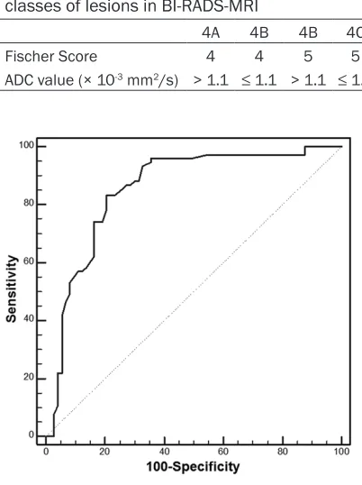
ROC curves of ADC
To determine the optimal diagnosis threshold
of the ADC value, the ROC analysis was per
[image:3.612.90.291.86.355.2]-formed. Based on the analysis of the ROC
Table 1. Criteria for the sub-classification of 4 classes of lesions in BI-RADS-MRI
4A 4B 4B 4C
Fischer Score 4 4 5 5
ADC value (× 10-3 mm2/s) > 1.1 ≤ 1.1 > 1.1 ≤ 1.1
curves, ADC=1.08 × 10⁻³ mm2/s was used as
the threshold to determine whether the breast lesion was benign or malignant. AUC equaled to
0.845 (95% CI: 0.736-0.907), and the sensitiv
-ity, specific-ity, positive and negative predicted values were 82.1%, 77.4%, 79.3% and 80.4%,
respectively (Figure 1). This indicates that
balanced cutoff point of ADC value for separat
-ing non-malignant from malignant lesions was 1.08 × 10⁻³ mm2/s.
BI-RADS-MRI sub-classification
To determine whether the combination of
dynamic CE-MRI blood vessel imaging and DWI
is adequate for the sub-categorization of
BI-RADS category 4, the positive predicted
val-ues of each sub-classification was performed. the 150 cases of lesions, 62 cases belonged to
BI-RADS-MRI 4 class, in which 13 cases were 4A class (12 cases were benign and 1 case was malignant), 21 cases were 4B class (10 cases were benign and 11 cases were malignant), and 28 cases were 4C class (3 cases were benign and 25 cases were malignant). The
pos-itive predicted values of the 4A, 4B and 4C
classes were 7.7%, 52.4% and 89.3%, respec
-tively (Table 2). After a further sub-classifica -tion, it was determined that 11 cases belonged to the 4A(-) class, and all cases were benign; 2
cases belonged to the 4A(+) class, in which one
was benign and the other was malignant; 9 cases belonged to the 4B(-) class, in which 6 cases were benign and 3 cases were
malig-nant; 12 cases belonged to the 4B(+) class, in
which 4 cases were benign and 8 cases were
5th class of lesions (Figure 5). This indicates
that sub-classification of BI-RADS-MRI 4 with
the MRI dynamic enhanced vessel imaging and
DWI is reasonable, which will be helpful for the choice of proper clinical treatment plan.
Discussion
Breast MRI is highly sensitive in showing lesions, and has great advantages in
discover-ing multiple malignant lesions of one or two
breasts. However, the high sensitivity may lead
to the over-treatment of lesions [1-4]. MR-guided biopsy is considered as a fast, safe and accu
[image:4.612.91.329.98.240.2]-rate method. However, CE-MRI of the breast may lead to false-positive results up to 74.1% [1, 16, 17]. This fact increases the demand for improved differentiation between benign and
malignant lesions to reduce unnecessary
biop-sies, and to raise the overall accuracy of breast
CE-MRI. Recently, many researches have shown
that combination of DWI and CE-MRI could decrease the false positive rate of breast MRI
diagnosis and increase the diagnostic accuracy
[1, 18-20]. However, how to effectively combine the two methods still lacks standards to be followed.
The malignant probability of the BI-RADS 4 classes of lesions of breast diseases falls between 2% and 95% [5], so there will be
unavoidable unnecessary puncture biopsies on
many benign cases if it is operated on all the 4 classes of lesions. Besides, the negative result of puncture biopsy always baffles clinical doc
-tors to determine further treatment plan, so
Table 2. The positive predicted value of each sub-class of the BI-RADS-MRI 4 classes
Sub-classification Benign Malignant Positive predicted value (95% CI)
4A 12 1 7.7%
4B 10 11 52.4% (29.8%-74.3%)
4C 3 25 89.3% (71.3%-97.8%)
4A(-) 11 0 0
4A(+) 1 1 50%
4B(-) 6 3 33.3% (7.5%-70.1%)
4B(+) 4 8 66.7% (34.9%-90.1%)
4C(-) 3 6 66.7% (29.9%-92.5%)
4C(+) 0 19 100%
Note: Because the case numbers of 4A, 4A(-), 4A(+) and 4C(+) class
-es were small, the 95% confident interval of their positive predicted
value were unable to calculate.
malignant; 9 cases belonged to the 4C(-) class, in which 3 cases were benign and 6 cases were malignant; 19 cases be-
longed to the 4C(+) class, and all cases
were malignant. The positive predicted
values of the 4A(-), 4A(+), 4B(-), 4B(+), 4C(-) and 4C(+) classes were 0, 50%,
33.3%, 66.7%, 66.7% and 100%, respec
-tively (Table 2). Accordingly, the 4A(-) class could be considered as the 3rd class
of lesions, so the 11 benign cases would
not receive the puncture biopsy (Figure 2). The 4A(+) and 4B(-) classes of lesions could be recognized as the 4B class of
lesions (Figure 3), and the 4B(+), 4C(-) classes of lesions could be recognized as the 4C class of lesions (Figure 4). The
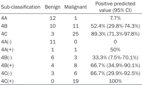
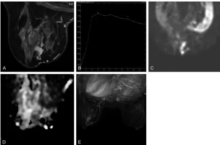
Figure 2. The sub-classification of the breast cancer of a 43 years old female, whose pathological result was
fibro-adenoma in the right breast. A: Dynamic contrast enhanced (DCE) MRI showed irregular nodules in the right breast,
and the edge was clear with homogeneous enhancement. B: The TIC curve showed the shape of flatbed, and the early enhancement rate was 198%. C and D: The Fischer score was assigned as 4 and the ADC value was 1.60 ×
10-3 mm2/s, and the lesion was classified into the BI-RADS-MRI 4A class. E: No increased asymmetric blood supply
and adjacent blood vessel sign was observed, and the lesion was classified into the BI-RADS-MRI 4A(-) class and
that missed diagnosis of breast cancer cannot
be avoided. In mammography and sonography,
there are already standards for the sub-classifi
-cation of BI-RADS 4, including 4A, 4B and 4C. The malignant probability of the 4A class of lesions falls between 2% and 10% [5, 6]. If the biopsy of the 4A class of lesions is benign, rou
-tine follow-up or six months follow-up can be prescribed. The malignant probability of the 4B class of lesions falls between 10% and 50% [5, 6]. If the result of puncture biopsy of the 4B
lesions is exclusively typical benign,
observa-tion is sufficient. However, if the result is papil
-lary tumor or atypical hyperplasia, then further
biopsy is necessary. The malignant probability
of the 4C class of lesions falls between 50% and 95% [5, 6, 21]. For the cases that the result of puncture biopsy is benign, consultation with the pathology department and further analysis
are needed [21, 22]. However, no standards
exist for the sub-classification of BI-RADS 4 categories of lesions in breast MRI. To our knowledge, this work is the first sub-classifica
-tion for BI-RADS-MRI 4 categories based on
DWI.
Assigning the Fisher score as 4, if the ADC
value supports benign lesions, the lesions are
defined as the 4A class lesions. A lower Fischer
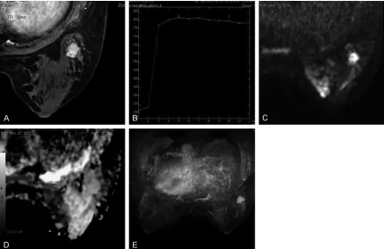
[image:6.612.90.522.152.450.2]score and higher ADC value indicate a very high Figure 3. The sub-classification of the breast cancer of a 69 years old female, whose pathological result was cancer with early soaking in the tubes. A: DCE-MRI showed irregular nodules in the left breast, and the edge was unclear
with homogeneous enhancement. B: The TIC curve showed gradual rising, and the early enhancement rate was
119%. C and D: The Fischer score was assigned as 4 and the ADC value was 1.36 × 10⁻³ mm2/s, and the lesion was
classified into the BI-RADS-MRI 4A class. E: The blood supply asymmetry increased in the right side, and the lesion was classified into the BI-RADS-MRI 4A(+) class and treated as the BI-RADS 4B class.
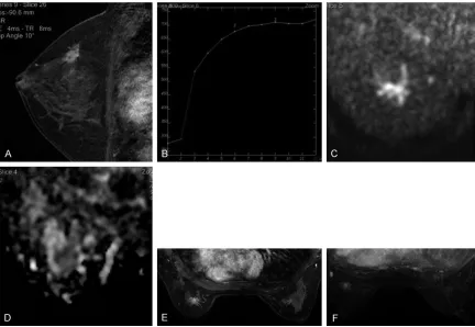
Figure 4. The sub-classification of the breast cancer of a 52 years old female, whose pathological result was soaking lobular cancer in the right breast. A: DCE-MRI showed irregular nodules in the left breast, and the edge was unclear
with inhomogeneous enhancement. B: The TIC curve showed gradual rising, and the early enhancement rate was
95%. C and D: The Fischer score was assigned as 4 and the ADC value was 0.98 × 10⁻³ mm2/s, and the lesion was
classified into the BI-RADS-MRI 4B class. E and F: Two blood vessels entered the lesion, and the lesion was classi
probability of benign lesions. In this study, there
were 13 BI-RADS-MRI 4A lesions, in which 12 were benign and only one was malignant, and
the positive predicted value was 7.7%. Assigning the Fischer score as 5, if the ADC value sup
-ports malignant, the lesions are defined as the
4C class lesions. A higher Fischer score and lower ADC value indicate a very high probability
of malignant lesions. In this study, there were
28 BI-RADS-MRI 4C class lesions, in which 25 were malignant and 3 were benign, and the
positive predicted value was 89.3%. The other 4 categories of lesions were defined as 4B
class. In this study, there were 21 BI-RADS-MRI 4B class lesions, in which 11 were malignant and 10 were benign, and the positive predicted
value was 52.4%. This result was a little higher
than previous reports [5, 6, 21], which might be
caused by the high sensitive nature of breast MRI, but it has no influence on the guidance of
clinical treatment choice. Basically, the
sub-classification method proposed in this paper is
in accordance with the reported BI-RADS 4
sub-classification standards [5, 21, 22]. At the
same time, this method is simple and practical,
easy to operate, and convenient for widespread
clinical application.
In this study, there were 62 lesions belonging to the BI-RADS-MRI 4 classes, in which 25 cases were benign. Puncture biopsy or surgery was
operated on all the cases. If the benign lesions
in the BI-RADS-MRI 4 classes can be detected by MRI, the unnecessary puncture biopsy rate would be dramatically reduced. Combined with MRI dynamic blood vessel imaging, the BI- RADS-MRI 4A, 4B and 4C class lesions were
further sub-classified. If there is no asymmetry
incensement or adjacent blood vessel sign, the
lesions are classified into negative classes indi -cated as BI-RADS-MRI 4A(-), 4B(-) and 4C(-);
[image:7.612.89.522.69.355.2]otherwise, they were classified as positive classes indicated as BI-RADS-MRI 4A(+), 4B(+) and 4C(+). In this study, the 4A(-) lesions were
Figure 5. The sub-classification of the breast cancer of a 52 years old female, whose pathological result was in-tube cancer in the right breast. A: DCE showed enhanced lesions of tube branching in the left breast, and the edge was
clear with homogeneous enhancement. B: The TIC curve showed clearance type, and the early enhancement rate
was 213%. C and D: The Fischer score was assigned as 5 and the ADC value was 1.04 × 10⁻³ mm2/s, and the lesion
was classified into the BI-RADS-MRI 4C class. E: The blood supply asymmetry increased in the right breast, and more than one blood vessel entering the lesion was observed. The lesion was classified into the BI-RADS-MRI 4C(+) class,
all benign. If all these lesions were classified
into the 3rd class, 44% (11/25) of the benign
lesions could avoid unnecessary puncture biopsy.
Several studies indicated that the false nega
-tive rate of breast MRI guided puncture biopsy was 0%-17% [20, 23-28]. Therefore, in clinical analysis, the result of puncture biopsy should be combined with the test of iconography in
order to avoid missed diagnosis, and
BI-RADS-MRI classification with higher accuracy is demanded. In this study, the 27 4C(+) lesions were all malignant, and could be classified as
the 5th class. For the 4C(+) class, surgery or
biopsy can be performed. If the result of the
puncture biopsy is benign, excision biopsy
should be positively performed to avoid missed diagnosis of breast cancer. For there were only two cases of 4A(+) lesions, they were catego
-rized into 4B(-) class and analyzed. If the posi
-tive predicted value is 33.3%, the lesions can be treated as BI-RADS 4B class. If the positive predicted value of the 4B(+) and 4C(-) lesions is 66.7%, the lesions can be treated as BI-RADS
4C class.
The sub-classification of breast cancer by the combination of DWI and CE-MRI has great sig
-nificance for the clinical diagnosis. However, there are some shortcomings of this study.
First, the sample number in this study was rela-tively small, so bias might exist in the selection
of cases. Second, the 3 benign 4C lesions were
all non-mass enhancement lesions, and the one malignant 4A class lesion was also a non-mass enhancement lesion. This suggests that more misdiagnosis can be caused by non-mass enhancement lesions. Thus, it is necessary
to analyze and classify the mass
enhance-ment and non-mass enhanceenhance-ment lesions.
Third, because the malignant possibility of the
5th class lesions is high and all the 5th class
lesions in this study were malignant, the
appli-cation of DWI in the 5th class lesions was not
discussed.
To sum up, reasonable sub-classification of BI-RADS-MRI 4 classes of lesions can be per
-formed with the help of DWI. Combining the
dynamic blood vessel imaging and the results
of sub-classification, the rate of unnecessary
puncture biopsy can be dramatically decreased,
and the misdiagnosis of breast cancer can be
effectively avoided to help design appropriate
clinical treatment plan.
Acknowledgements
This work was supported by the Medical and
Health Science and Technology Development
Project of Shandong Province.
Disclosure of conflict of interest
None.
Address correspondence to: Lei Song, Department
of Radiology, The Second Hospital of Shandong
University, 247 Beiyuan Street, Jinan 250033, P. R. China. Tel: 86-15153169081; E-mail: 19241480- [email protected]
References
[1] Spick C, Pinker-Domenig K, Rudas M, Helbich TH and Baltzer PA. MRI-only lesions: applica
-tion of diffusion-weighted imaging obviates un -necessary MR-guided breast biopsies. Eur Ra-diol 2014; 24: 1204-1210.
[2] Warner E, Messersmith H, Causer P, Eisen A,
Shumak R and Plewes D. Systematic review:
using magnetic resonance imaging to screen
women at high risk for breast cancer. Ann In -tern Med 2008; 148: 671-679.
[3] Peters NH, Borel Rinkes IH, Zuithoff NP, Mali WP, Moons Kg and Peeters PH. Meta-analysis of MR imaging in the diagnosis of breast le -sions. Radiology 2008; 246: 116-124. [4] Heywang-Köbrunner SH, Hacker A and Sed
-lacek S. Magnetic resonance imaging: the evo
-lution of breast imaging. Breast 2013; 22
Suppl 2: S77-82.
[5] Cecilia L. BI-RADS update. Radiol Clin N Am 2014; 52: 481-487.
[6] Torres-Tabanera M, Cárdenas-Rebollo JM,
Vil-lar-Castaño P, Sánchez-Gómez SM, Cobo-Soler J, Montoro-Martos EE and Sainz-Miranda M. Analysis of the positive predictive value of the subcategories of BI-RADS(@) 4 lesions: prelimi-nary results in 880 lesions. Radiologia 2012; 54: 520-531.
[7] Pereira FP, Martins G and Carvalhaes de Olivei
-ra RD. Diffusion magnetic resonance imaging of the breast. Magn Reson Imaging Clin N Am
2011; 19: 95-110.
[8] Dorrius MD, Dijkstra H, Oudkerk M and Sijens PE. Effect of b value and pre-admission of con
-trast on diagnostic accuracy of 1.5-T breast
[9] Kul S, Cansu A, Alhan E, Dine H, Gunes G and Reis A. Contribution of diffusion-weighted im -aging to dynamic contrast-enhanced MRI in
the characterization of breast tumors. AJR Am
J Roentgenol 2011; 196: 210-217.
[10] Baltzer PA, Renz DM, Herrmann KH, Dietzel M, Krumbein I, Gajda M, Camara O, Reichenbach JR and Kaiser WA. Diffusion-weighted imaging
(DWI) in MR mammography (MRM): clinical
comparison of echo planar imaging (EPI) and half-fourier single-shot turbo spin echo (HA-STE) diffusion techniques. Eur Radiol 2009;
19: 1612-1620.
[11] Baltzer A, Dietzel M, Kaiser CG and Baltzer PA. Combined reading of contrast enhanced an diffusion weighted magnetic resonance imag -ing by us-ing a simple sum score. Eur Radiol 2016; 26: 884-891.
[12] Sardanelli F, Iozzelli A, Fausto A, Carriero A and Kirchin MA. Gadobenate dimeglumine en -hanced MR imaging breast vascular maps: association between invasive cancer and ipsi-lateral vascularity. Radiology 2005; 235: 791-797.
[13] Dietzel M, Baltzer PA, Vag T, Herzog A, Gajda M, Camara O and Kaiser WA. The adjacent ves -sel sign on breast MRI: Ner data and a
sub-group analysis for 1084 histologically verified cases. Korean J Radiol 2010; 11: 178-186.
[14] Mahfouz AE, Sherif H, Saad A, Taupitz M, Fili
-monow S, Kivelitz D and Hamm B. Gadolinium-enhanced MR angiography of the breast: is
breast cancer associated with ipsilateral high-er vascularity? Eur Radiol 2001; 11: 965-969. [15] Al-Khawari H, Athyal R, Kovacs A, Al-Saleh M
and Madda JP. Accuracy of the fischer scoring
system and the breast imaging reporting and
data system in identification of malignant breast lesions. Hematol Oncol Stem Cell Ther
2009; 29: 280-287.
[16] Schrading S, Simon B, Braun M, Wardelmann
E, Schild HH and Kuhl CK. MRI-guided breast biopsy: influence of choice of vacuum biopsy syetem on the mode of biopsy of MRI-only sus -picious breast lesions. Am J Roentgenol 2010; 194: 1650-1657.
[17] Perlet C, Heywang-Kobrunner SH, Heinig A, Sit
-tek H, Casselman J, Anderson I and Taourel P.
Magnetic resonance-guided, vacuum-assisted
breast biopsy: results from a European multi
-center study of 538 lesions. Cancer 2006;
106: 982-990.
[18] Pinker K, Bickel H, Helbich TH, Gruber S, Dub
-sky P, Pluschnig U, Rudas M, Bago-Horvath Z,
Weber M, Trattnig S and Bogner W. Combined contrast-enhanced magnetic resonance and
diffusion-weighted imaging reading adapted to
the “breast imaging reporting and data
sys-tem” for multiparametric 3-T imaging of breast
lesions. Eur Radiol 2013; 23: 1791-1802.
[19] Pinker K, Bogner W, Baltzer P, Gruber S, Bickel H, Brueck B, Trattnig S, Weber M, Dubsky P,
Bago-Horvath Z, Bartsch R and Helbich TH. Im-proved diagnostic accuracy with
multiparamet-ric magnetic resonance imaging of the breast
using dynamic contrast-enhanced magnetic
resonance imaging, diffusion-weighted imag -ing, and 3-dimensional proton magnetic reso-nance spectroscopic imaging. Investig Radiol 2014; 49: 421-430.
[20] Parsian S, Rahbar H, Allison KH, Demartini WB, Olson ML, Lehman CD and Partridge SC. Nonmalignant breast lesions: ADCs of benign and high risk subtypes assessed as false-posi -tive at dynamic enhanced MR imaging. Radiol-ogy 2012; 265: 696-706.
[21] D’Orsi CJ, Sickles EA and Mendelson EB. ACR
BI-RADS Atlas, breast imaging reporting and data system. 5th edition. Reston, VA: American
College of Radiology; 2013.
[22] Burnside ES, Sickles EA, Bassett LW, Rubin DL, Lee CH, Ikeda DM, Mendelson EB, Wilcox PA, Butler PF and D’Orsi CJ. The ACR BI-RADS ex
-perience: learning from history. J Am Coll Ra -diol 2009; 6: 851-860.
[23] Spick C, Schernthaner M, Pinker K, Kapetas P, Bernathova M, Polanec SH, Bickel H, Wengert GJ, Rudas M, Helbich TH and Baltzer PA. MR-guided vacuum-assisted breast biopsy of
MRI-only lesions: a single center experience. Eur Radiol 2016; 23: 1791-1802.
[24] Rauch GM, Dogan BE, Smith TB, Liu P and
Yang WT. Outcome analysis of 9-guage
MRI-guided vacuum-assisted core needle breast biopsies. AJR Am J Roentgenol 2012; 198: 292-299.
[25] Imschweiler T, Haueisen H, Kampmann G, Rageth L, Seifert B, Rageth C, Freiwald B and Kubik-Huch RA. MRI-guided vacuum-assisted
breast biopsy: comparision with stereotatically guided and ultrasound-guided techniques. Eur Radiol 2014; 24: 128-135.
[26] Schrading S, Simon B, Braun M, Wardelmann
E, Schild HH, Kuhl CK. MRI-guided breast bi
-opsy: influence of choice of vacuum biopsy sys
-tem on the mode of biopsy of MRI-only suspi -cious breast lesions. AJR Am J Roentgenol 2010; 194: 1650-1657.
[27] Malhaire C, El Khoury C, Thibault F, Athanasiou A, Petrow P, Ollivier L, Tardivon A. Vacuum-as
-sisted biopsies under MR guidance: results of
72 procedures. Eur Radiol 2010; 20: 1554-1562.
[28] Malhaire C, El Khoury C, Thibault F, Athanasiou A, Petrow P, Ollivier L, Tardivon A. MRI-guided