1
Optimisation of the self-assembly process: production of stable, alginate-based polyelectrolytenanocomplexes with protamine
Maria Dula, Krzysztof J. Palucha,b, Anne Marie Healya, Astrid Sassea, Lidia Tajbera*
*To whom correspondence should be addressed: [email protected]
Phone: 00353 1 896 2787 Fax: 00353 1 896 2810
Affiliations:
a. School of Pharmacy and Pharmaceutical Sciences, Trinity College Dublin, Dublin 2, Ireland.
b. Bradford School of Pharmacy, School of Life Sciences, Centre for Pharmaceutical Engineering Science,
2
Abstract:The aim of this work was to investigate the possibility of covalent cross-linker free, polyelectrolyte complex
formation at the nanoscale between alginic acid (as sodium alginate, ALG) and protamine (PROT).
Optimisation of the self-assembly conditions was performed by varying the type of polymer used, pH of
component solutions, mass mixing ratio of the components as well as the speed and order of component addition
on the properties of complexes. Homogenous particles with nanometric sizes resulted when an aqueous
dispersion of ALG was rapidly mixed with a solution of PROT. The polyelectrolyte complex between ALG and
PROT was confirmed by infrared spectroscopy. To facilitate incorporation of drugs soluble at low pH, pH of
ALG dispersion was decreased to 2, however no nanoparticles (NPs) were formed upon complexation with
PROT. Adjusting pH of PROT solution to 3 resulted in the formation of cationic or anionic NPs with a size
range 70-300 nm. Colloidal stability of selected AAL/PROT formulations was determined upon storage at room
temperature and in liquid media at various pH. Physical stability of NPs correlated with the initial surface
charge of particles and was time- and pH-dependent. Generally, better stability was observed for anionic NPs
stored as native dispersions and in liquids covering a range of pH.
3
IntroductionNanoparticles (NPs) have attracted special interests in the drug delivery area, mainly as carriers for
bioactives including peptide drugs (Delie and Blanco-Prieto 2005). NPs formulated from natural polymers have
been extensively studied due to their advantages, including the potential to retain protein stability and activity,
an increase of the duration of therapeutic effects of proteins and the possibility to be administered by
nonparenteral routes (Sarmento et al. 2007). The use of natural polymers as drug carries has also been widely
investigated (Liechty et al. 2010; Ngwuluka et al. 2014; Srivastava et al. 2016).
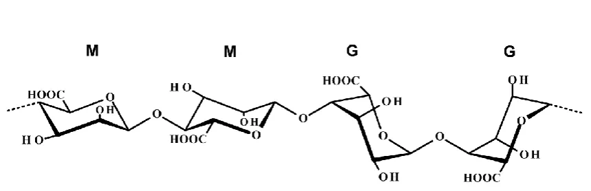
Alginic acid (ALG) is a linear copolymer comprising blocks of (1,4)-linked-D-mannuronate (M) and
C-5 epimer α-L-guluronate (G) residues, connected in different sequences or blocks (Fig. 1) (Tønnesen and
Karlsen 2002). The wide availability of alginate and its desirable chemical and physical properties makes it an
excellent material for formation of micro- and nanoparticles. Using various cross-linking methods, alginate
forms an inert, biodegradable hydrogel matrix (Lee and Mooney 2012). Porosity of the gel enables high drug
diffusion rates, which additionally can be controlled with polymer coatings (Wee and Gombotz 1998).
Alginates are extensively utilised in biomedical applications as mucoadhesive, biodegradable and
biocompatible polymers (Sinha and Kurmia 2001). Furthermore, alginate has the ability to ionically cross-link
with multivalent cations, such as calcium, forming a network gel, which is stable at low pH and dissolves in
neutral or high pH environments (Augst et al. 2006; George et al. 2006). The pH sensitivity of alginate-based
hydrogels makes them particularly attractive for oral delivery, as such formulations can contract in the
stomach protecting the drug. Passing through the gastrointestinal tract, they subsequently swell and release the
drug when pH increases.
The polyelectrolyte complexation (PEC) method of NP preparation has received an increasing
attention, as NPs formed by this method have several advantages for cellular uptake and colloidal stability,
including suitable diameters and surface charge, spherical morphology and low polydispersity indices (Pdl)
(Avadi et al. 2011). Furthermore, the preparation of NPs by PEC methods can be carried out in completely
aqueous conditions and at ambient temperature, without the use of organic solvents and/or surfactants, thus the
stability and biological activity of loaded peptides is expected not to be affected (Hu et al. 2012). Additionally,
NPs formed by the PEC method have been to show to improve encapsulation efficiency of drugs and to control
the rate of drug release due to the nature of the bonds formed between the drug and polymer (Cheow and
4
2001). The PEC method can also be applied to the preparation of membranes, coating on films and fibres,
isolation and fractionation of proteins as well as isolation of nucleic acids (Lankallapali and Kolapalli 2009).
Silva et al. developed alginate microspheres coated with chitosan (CHIT) prepared by an
emulsification/internal gelation method with a mean diameter that ranged from 65 to 106 μm (Silva et al. 2006).
An ionic pre-gelation method of alginate with calcium chloride, followed by complexation between alginate and
CHIT was used to prepare NPs loaded with insulin. The particle size of the formed NPs increased from 764 to
2209 nm when the CHIT to alginate mass ratio was decreased from 6:1 to 3.1:1 (Sarmento et al. 2006). The PEC
formation using alginate and CHIT as oppositely charged polymers has been described by Saether et al. (2008).
As reported, the net charge ratio between CHIT and alginate and the molecular weights (MWs) of both the
alginate and CHIT components were the most significant parameters that influenced the particle size and zeta
potential (ZP) of the formed PECs (Saether et al. 2008). A similar study was conducted by Sarmento’s group,
where colloidal carriers prepared by complexation of two oppositely charged polymers: dextran sulphate (as an
anionic polymer) and CHIT (as a cationic polymer) were used for the delivery of insulin (Sarmento et al. 2007).
The use of cell-penetrating peptides for drug delivery has been extensively studied (Temsamani and
Vidal 2004). Protamine (PROT) is an example of a cell-penetrating substance, which was selected herein to
form PECs with alginate. PROT has been reported to have in vitro membrane-translocating ability, believed to be
associated with its positively charged polyarginine chains (Reynolds et al. 2005). The studies performed by
Umerska et al. (2014) showed that PROT/hyaluronate can form PECs at the nanoscale, however depending on
the ratio of constituents in such PECs their colloidal stability varied considerably. The association efficiency of
the drug (salmon calcitonin, sCT) that was loaded into those NPs was up to 100% with notably high drug
loading (9.6-39% w/w). The release of the cargo from those NPs was ∼70–80% after 24h (Umerska et al. 2014).
The release of sCT from chondroitin sulphate/PROT NPs, on the other hand, was susceptible to the pH and ionic
strength of release medium (Umerska et al. 2015). Wernig et al. (2008) studied encapsulation of vasoactive
intestinal peptide into biodegradable PROT 18mer nonsense oligonucleotides formed by self-assembly of the
components. The encapsulation efficiency of the payload was up to 80% with 77-87% of the oligonucleotide
released of over 24h. A recent study published by Yu et al. (2016) described chitosan/PROT NPs loaded with
fluorouracil with high encapsulation efficiency (82.4%) and sustained release (more than 60% of the drug
released into medium over 48h) of the drug.
Our previous study demonstrated that PROT is able to complex to carrageenans (CARs) forming PECs
5
formation between CAR and PROT was corroborated by infrared analysis and, in the case of iota carrageenan,
the polymer chains underwent structural changes when forming NPs.
The aim of current work was to investigate the process of preparation of covalent cross-linker free and
stable NPs composed of only alginate and PROT. To achieve this goal, the alginate/PROT PEC manufacturing
process was comprehensively studied including examination of the molecular weight of alginate, the component
mixing ratios as well as the sequence of addition and speed of mixing of the constituents guided by the
outcomes of previous studies (Dul et al. 2015). As one of the most important parameters determining
pharmaceutical suitability of a nanoparticle is its colloidal stability, the physical stability of the nanoparticulate
dispersions on storage at room temperature and upon the exposure to environments with varying pH was also
examined.
2 Materials and methods 2.1 Materials
Alginic acid (as a sodium salt), low (AAL) and medium (AAM) viscosity grades as well as protamine
(PROT) (as a sulphate salt, from salmon) were obtained from Sigma-Aldrich (Arklow, Ireland). All other
reagents and chemicals used were of analytical grade.
2.2 Preparation and characterisation of polymer and PROT solutions
Aliquots of 100 ml of 0.1% (1 mg/ml) dispersions or solutions of AAM, AAL, PROT, PROT with pH2
(PROT_pH2) and PROT with pH3 (PROT_pH3) were prepared in deionised water. Polymers as powders were
dispersed at 25 ºC and stirred at 500 rpm for 30 min on a magnetic stirrer until clear dispersions developed. pH
of PROT solutions was adjusted to pH2 or pH3 using 1M HCl.
Viscosity of the liquids containing either alginate or PROT starting/native and NP dispersions was
determined using a low frequency vibration viscometer (SV-10 Vibro Viscometer, A&D Company Limited,
Japan). Deionised water was employed to calibrate the instrument before use. Triplicate measurements at 25±0.2
ºC were carried out for each sample and temperature of each of the aliquots was equilibrated prior to the
measurement in a water bath (Reciprocal Shaking Bath Model 25, Precision Scientific, UK). The results are
presented as an average value ± standard deviation (SD).
pH measurements at 25 ºC were carried out with a Thermo Electron Orion 420A+ Basic pH/mV/ORP
pH meter connected to an Orion RoseTM 8103SC pH semi-micro electrode. Standard buffer solutions at pH 4, 7
and 10 were employed to calibrate the pH meter on regular basis. The results of measurements are given as an
6
Gel Permeation Chromatography (GPC) measurements of the molecular weight of alginates were
performed using a HPLC system as described previously (Dul et al. 2015). The separative column used was a
Plaquagel–OH mixed 8 µm, 300 × 7.5 mm column (Polymer Laboratories Ltd., UK). The mobile phase was
composed of 0.2M NaCl and 0.01M NaH2PO4 adjusted to pH 7.4 with NaOH solution and its flow rate
employed was 1 ml/min. The calibration curves were made using Pullulan standards (PL Polymer Laboratoires,
Germany). Solutions of standards and samples at a concentration of 1 mg/ml were injected onto the column in
triplicate (at a volume of 100 μl).
Fourier transform infrared spectroscopy (FTIR) of alginate and PROT was carried out as described
previously (Umerska et al. 2012; Dul et al. 2015).
2.3 Synthesis and characterisation of NPs composed of alginate and PROT
Aliquots of 0.1% w/v solutions of AAL, AAM and PROT, PROT_pH2 and PROT_pH3 were prepared
according to the method described in Section 2.2. The polymer and PROT dispersions/solutions were combined
together in various v/v ratios at room temperature (RT) and mixed under magnetic stirring at 500 rpm for around
10 min to allow stabilisation of the system. Since the concentration of both solid components was the same
(0.1% w/v), those v/v ratios were equivalent to alginate/PROT weight mixing ratios (WMRs) and WMRs are
used throughout this manuscript.
The impact of the rate of addition of PROT solution to the dispersion of AAL and the sequence of
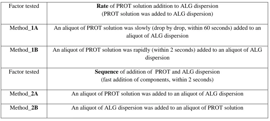
alginate/PROT mixing have been tested. Variations in the assembly method tested are summarised in Table 1. In
all cases after combining both solutions the stirring was continued for 10 minutes.
2.4 Physicochemical characterisation of NPs
The mean particle size (hydrodynamic particle diameter), polydispersity index (PdI) and zeta potential
(ZP) values of the polyelectrolyte complexes were determined at 25 ºC by dynamic light scattering (DLS) and
laser Doppler velocimetry using a Zetasizer Nano ZS instrumnt (Malvern Instruments Ltd., UK) as described
previously (Dul et al., 2015). The results are given as average values of triplicate measurements ± SD. Viscosity
and pH measurements were performed as described in Section 2.2. FTIR analysis was performed on
AAL/PROT systems produced by combining 0.1% w/v ALG and PROT solutions at a WMR of 1 as described
previously (Umerska et al. 2012). Colloidal stability of NP formulations (in native dispersions) was visually
examined instantly after the preparation and prior to conducting any measurements. The presence or absence of
instability (for instance aggregation) was noted. Changes in the hydrodynamic particle size, PdI and ZP
7
observations were performed up to 3 days (72 h). The following solutions/buffers: 0.01M HCl, 0.1M acetate
buffer pH4.5, 0.1M 4-(2-hydroxyethyl)-1-piperazineethanesulfonic acid (HEPES) buffer pH6.5 and 0.015M
phosphate buffered saline (PBS) pH7.4 were also employed as media to determine the colloidal behaviour and
susceptibility of the NPs to changes in pH/ionic strength. The native dispersions containing the nanocomplexes
were diluted with each of the above liquid media in the 1:1 v/v ratio. The physical stability of such dispersions
was estimated by measuring changes in the mean hydrodynamic particle size over time, for up to 3 days (72 h).
2.5 Statistical analysis
The statistical significance of the differences between samples was determined by one-way analysis of
variance (ANOVA) using Origin software version 7.5.
3 Results and discussion
As reported by Boddohi et al. (2008) and Umerska et al. (2012) the molecular weight of the polymer
and its concentration in solution are the crucial parameters determining the successful polyelectrolyte NP
process formation. The study performed by Carneiro-da-Cunha et al. (2011) showed that the polymer
concentration affected the mean diameter and ZP of multilayer NPs formed by sodium alginate, carrageenan and
CHIT solutions. The mean hydrodynamic diameter of NPs made from a 0.6% w/v solution of alginate was
approximately a 2-fold greater than those of NPs formed by 0.2% w/v alginate solution (an increase from 1180
to 2155 nm) (Carneiro-da-Cunha et al. 2011). However, it is important to emphasise that lowering the polymer
concentration reduces the production yield of NPs, which can lead to a decrease in the final loading capacity of
particles. Therefore, in the present work, to balance the desirable NP characteristics and production capacity of
the process, 0.1% w/v solutions of ALG and PROT were used.
3.1 Impact of the rate and sequence of components addition on properties of NPs
As previously reported, the manner in which solutions of polyions in a PEC method are combined
influences the properties of formed complexes (Chen et al. 2003; Dragan et al. 2006; Birch & Schiffman 2014;
Dul et al. 2015). First, to examine the impact of the rate at which the PROT solution was introduced to an
aliquot of ALG dispersion, NPs were prepared by slow (Method_1A) or rapid (Method_1B) addition of
components, as described in Table 1. Two formulations of NPs with an ALG/PROT WMR of 4.5 and 5 were
selected for these trials (Table 2).
A significant difference in the particle size was observed. Smaller in size NPs were formed when the
PROT solution was rapidly added to the aliquot of ALG dispersion (Method_1B) in comparison to the slow
8
an ALG/PROT WMR=5, but not NPs with an ALG/PROT WMR=4.5 (Table 2). Therefore, to obtain NPs with
small sizes, a rapid addition of components was used for further studies.
The sequence of mixing of the NP component solutions/dispersions was also investigated. These
methods were called Method_2A (PROT solution was added to ALG dispersion) and Method_2B (ALG
dispersion was added to PROT solution) as explained in Table 1. Interestingly, Method_2A yielded smaller in
size, in comparison to Method_2B, NPs for WMRs of 1, 1.5, 3 and 5 (with negative ZP values), while
Method_2B gave smaller particles, in comparison to Method_2A, for WMRs of 0.2, 0.33 and 0.67 (positively
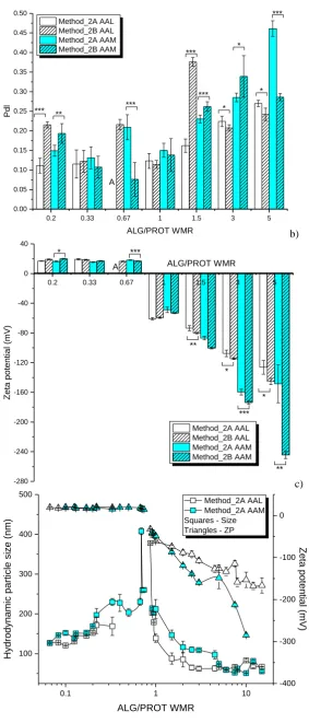
charged NPs), as presented in Fig. 2a. However, comparing the Pdl values, it was noticed that NPs with higher
Pdl indices (less homogenous) were chiefly generated using Method_2B (Fig. 2b). The order of mixing was
seen to impact less the ZP values (Fig. 2c).
The impact of the order of component mixing on the properties of NPs, when preparing PECs, was
investigated previously. Birch and Schiffman (2014) showed that that the order of solution addition resulted in
differences in particle sizes and ZP values of CHIT/pectin NPs, however the rationale for this behaviour was not
provided. Dul et al. (2015) investigated this phenomenon in relation to formation of carrageenan/PROT NPs.
Generally, smaller in size, with lower Pdl values and negatively charged NPs were formed when PROT solution
was added to the dispersion of carrageenan (Method_2A), similar to what is observed for the ALG/PROT
systems here. It was concluded that PROT can either stabilise or destabilise the formed NPs, depending on the
mixing method and also on the type of polymer (carrageenan in this case) used (Dul et al. 2015).
3.2 Impact of molecular weight of alginate on the properties of NPs
The molecular weight of AAL was measured to be ~100 kDa, however the molecular range of AAM
ranged from 42 to 537 kDa (Table 3). The high polydispersity of AAM may have an impact on the formation
and properties of polyelectrolyte complexes. Polydispersity of the polycation has been identified as an issue in
studies on DNA complexation and was shown to lead to structural heterogeneity and different biological activity
of NPs (Mustafaev 1995; Polexe and Delair 2013), while high viscosity of polymer dispersions may negatively
impact on the mixing process leading to the formation of aggregates (Mackay et al. 2006, Umerska et al. 2012).
Viscosity of 0.1% w/v dispersions (used to produce NPs) were in line with the average molecular weights of
alginates, with ~2.7 mPa·s for AAL and ~4.2 mPa·s for AAM.
Results presented in Section 3.1 show that both polymers (AAL and AAM) have the ability to form
polyelectrolyte complexes with PROT. Furthermore, it was observed that the rate and sequence of components
9
alginate with various viscosity/molecular weights (AAL and AAM), a wide range of WMR was tested. The
nanocomplexes were prepared using Method_2A (an aliquot of PROT solution was added to an aliquot of ALG
dispersion quickly), as this method of preparation was selected to be the most favourable for ALG/PROT NPs
self-assembly (Section 3.1).
In was difficult to discern a trend for the particle size when comparing NPs made by AAL and AAM
(Fig. 2a and d). The particle size ranged from 51 to 319 nm for the WMR tested (Fig. 2d). An increase in WMR
resulted in the formation of NPs, which were smaller in size and for WMRs of 4.5 and above NPs with sizes
below 100 nm were formed for both, AAL and AAM. Both combinations tested, AAL/PROT and AAM/PROT,
showed the formation of large entities, most likely agglomerates/flocs, occurring for 0.40-0.83 AAL/PROT and
0.77-0.87 AAM/PROT WMRs (Fig. 2d), consistent with charge neutralisation (Boddohi et al. 2008; Umerska et
al. 2012, Dul et al. 2015). The intrinsic pKa of ALG is around 3 and at pH of the ALG solution (pH around 6,
Table 3) complete ionisation of carboxyl groups is expected, thus notionally all the acidic groups of ALG should
electrostatically bind to the basic groups of PROT (Andriamanantoanina and Rinaudob 2010). Pdl values ranged
from 0.11 to 0.38 for AAL/PROT NPs and from 0.08 to 0.46 for AAM/PROT NPs and were seen to increase
with an increasing ALG/PROT WMR (Fig. 2b). ZP measurements showed that ALG/PROT NPs can be either
positively or negatively charged (Fig. 2c). The cationic NPs had similar ZP values of approximately 20 mV for
both types of ALG tested, which could be due to the PROT presence on their surface. The anionic NPs
composed of AAL/PROT had ZP values of around -110 mV and around -160 mV for AAM/PROT NPs at
WMR of 3. The comparison of NP properties containing the different grades of ALG suggested that NPs based
on AAL offered more promising characteristics: smaller particles with narrower size distributions. Those NP
characteristics have been shown to have an impact on the stability, biodistribution, cellular uptake and
bioavailability of those NPs (Elsabahy and Wooley 2012).
Several reports show the use of alginates in drug delivery, however in most of the cases this polymer is
combined with CHIT (a different polycation than PROT). Similar to the current study, NPs of alginate and
CHIT were formed by addition of 0.1% w/v alginate solution (pH6.5) under high shear conditions to 0.1% w/v
CHIT solution (pH4) (Saether et al. 2008). Those NPs were formed in a one step process, where one polymer
solution was added in a dropwise manner into the other solution. Also, homogenisers operating at various
speeds were facilitating the mixing process. The size of the obtained particles was 500 nm and greater, reaching
values of 2 μm. The component charge ratio and molecular weight were shown to be parameters that affected
10
however without influencing their ZP and pH. At the charge ratios close to 1, the sizes of the largest particles
were more variable, however, by using polymers with low molecular weights and employing a process whereby
an addition of the polymer solution to an excess volume of the other polymer solution, small in size particles
resulted (Saether et al. 2008). Li et al. (2008) also explored alginate/CHIT NPs prepared by the PEC method as
a drug delivery system for nifedipine. Described by this group NPs had very small sizes, 20-50 nm, and were
prepared by ionic pre-gelation of alginate core (dropwise addition of calcium chloride to aqueous solution of
sodium alginate under stirring) followed by CHIT polyelectrolyte complexation (Li et al., 2008).
The importance of the molecular weight ratio of the polycationic and polyanionic components and its
charge density was highlighted by Umerska et al. (2014). It was shown that, while it was possible to form NPs
between hyaluronic acid (HA, molecular weight ~260 kDa) and PROT (molecular weight ~5 kDa), they were
not stable due to the considerable disparity in the molecular weight of HA in comparison to that of PROT. The
ratio of molecular weights for AAM/PROT is ~50 (for AAL/PROT ~20), yet this combination yields physically
stable complexes in the nanoscale. It could be related to the strength of ionic interactions between then
components as HA is regarded as a relatively weak acid with low charge density (Polexe and Delair 2013).
3.3 Impact of pH adjustment on the formation and properties of NPs
Comparing the various strategies that have been used to improve drug solubility, changing pH of
solution is one of the common approaches for ionisable drug molecules. Therefore, changing pH of one of the
polyion solutions prior to mixing with the other polyion solution can facilitate dissolution of an active
compound to be loaded into the NPs, however it will also alter binding of components in the nanoparticle via
modification of electrostatic interactions. The results presented in Sections 3.1 and 3.2 were generated when pH
of the solutions/dispersions was unadjusted (pH values are given in Table 3).
At first, pH of AAL dispersion was adjusted to 2 maintaining pH of PROT solution at the native value
(pH5.8). However, only large in size and sedimenting entities (aggregates, floccules and/or microparticles)
formed immediately after mixing both solutions, regardless of the WMR. Analysis of physicochemical
properties of alginate shows the pH dependent behaviour of this polymer, consistent with this substance being a
polyanion. It has been shown that viscosity of sodium alginate dispersion is almost constant in the pH range of
6-8. However, at moderate concentrations and following a pH decrease, viscosity increases and reaches a
maximum at pH of 3-3.5 (intrinsic pKa of alginic acid is around 3), as carboxylate groups in the alginate
11
2015). Dispersions of alginic acid at higher concentrations form physical gels at these pH values, where
hydrogen bond attractions predominate over electrostatic repulsions.
As no NPs were formed when pH of AAL dispersion was adjusted to 2, further manipulations with
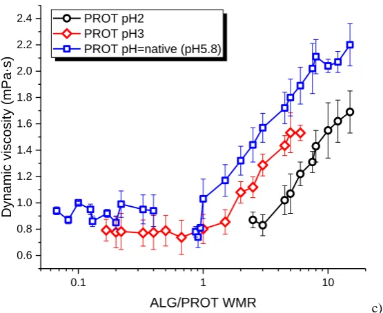
solution pH were performed for PROT only. Lowering pH of PROT solution to 2 (PROT_pH2) resulted in the
formation of particles larger in size in comparison to AAL/PROT (pH unadjusted) NPs (Fig. 3a) and with only
negatively charge, as ZP values ranged from -25 to -113 mV (Fig. 3b).
Aggregates/microparticles were formed at an AAL/PROT_pH2 WMR of 2 and lower. The particle size
of anionic NPs ranged from 142 to 262 nm (Fig. 3a), while Pdl values ranged from 0.21 to 0.28. Comparison of
AAL/PROT and AAL/PROT_pH2 formulations showed that lowering pH of PROT solution resulted in the
formation of NP dispersions with lower dynamic viscosity and higher ZP values for AAL/PROT_pH2 NPs (Fig.
3b and c). As an example, at a WMR of 5 the NPs had ZP of -126 mV for AAL/PROT NPs and -44 mV for
AAL/PROT_pH2 NPs. The decrease in viscosity may be due to the different degree of AAL/PROT interactions.
Additionally, pH of PROT solution was adjusted using a HCl solution, which resulted in an excess of negatively
charged ions (Cl-) in the system, which may have an impact on the surface charge of formed NPs as well as the
WMR at which the neutralisation of charge occurs. The addition of PROT solution at such a low pH could have
resulted in changes in the nature of interactions between the polymer and PROT driven by partial/local
protonation of AAL and/or agglomeration via hydrogen bond interactions (Lee and Mooney 2012).
As decreasing pH of PROT solution to 2 resulted in the formation of anionic NPs only, to enhance the
possibility of formation of NPs that would bear a positive charge, pH of PROT solution was adjusted to 3
(PROT_pH3). This time both, cationic and anionic NPs resulted. The particle size of negatively charged
AAL/PROT_pH3 NPs ranged from 71 to 137 nm, while in the case of positively charged NPs they ranged from
172 to 305 nm (Fig. 3a). Aggregation and an increase in the particle size was observed at an AAL/PROT_pH3
WMR of 1 (Fig. 3a). Considering homogeneity (Pdl) of the formed NPs, negatively charged NPs at
AAL/PROT_pH3 WMRs of 1.5 to 6 had greater Pdl values, which ranged from 0.17 to 0.37 respectively,
compared to cationic NPs (at AAL/PROT_pH3 WMRs of 0.17 to 0.67), ranging from 0.11 to 0.20. ZP values of
positively charged AAL/PROT_pH3 NPs were 14-17.9 mV, while in the case of negatively charged
AAL/PROT_pH3 NPs the ZP values varied from -49 to -107 mV (Fig. 3b). Generally, lowering pH of PROT
solution to 3 changed the WMR at which the agglomeration/flocculation of the formed NPs occurred (Fig. 3a).
pH of the NP dispersions ranged from 6.0 for AAL/PROT_pH3 with a WMR of 15 to 3.3 for AAL/PROT_pH3
12
formulation was 3.8, which can be regarded as an isoelectric point. For the formulations where pH of the PROT
solution was not adjusted, physical instability was observed at AAL/PROT WMRs of 0.50 – 0.83 with pH of
those formulations ranging from 5.7 to 6.0.
There were no major differences within the particle size between AAL/PROT_pH3 and AAL/PROT
NPs. An increase in the ZP value of anionic AAL/PROT_pH3 NPs was observed, in comparison to AAL/PROT
NPs. The differences in the particle size, ZP and dynamic viscosity values for AAL/PROT_pH2,
AAL/PROT_pH3 and AAL/PROT NPs are presented in Fig. 3.
3.4 Interactions between alginate and PROT
FTIR spectra of AAL, PROT and NPs are presented in Fig. 4. The spectrum of AAL showed
absorption bands associated with hydroxyl, ether and carboxylic functional groups. Bands observed at 1649 and
1460 cm-1 were attributed to asymmetric and symmetric stretching vibrations of the carboxylate ion,
respectively. The weak bands at 1301, 1125 and 1094 cm-1 might be assigned to C-C-H and O-C-H deformation,
C-O stretching and C-O and C-C stretching vibrations of pyranose rings, respectively. The band at 1035 cm-1 is
due to C-O stretching vibrations. The fingerprint region of 950-750 cm-1 is typically the most discussed for
carbohydrates. The band observed at 948 cm-1 can be assigned to the C-O stretching vibration of uronic acid
residues, while the peak at 888 cm-1 was assigned to the C
1-H deformation vibration of β-mannuronic acid
residues. The band at 820 cm-1 is characteristic for mannuronic acid residues. In case of PROT the most
prominent peaks appeared between 1400 and 1700 cm-1, which are characteristic for amide I and II vibrations.
Analysis of NPs spectra showed that no new covalent bonds were formed, however shifts of some absorption
bands were observed (Table 4). It was noticed, that the peaks associated with carboxyl groups of AAL and
amine groups of PROT moved slightly (Table 4). Additionally, the band characteristic of mannuronic acid (at
820 cm-1) was less intense following AAL complexation with PROT.
Similar changes were observed for carrageenan complexed to PROT, where the peaks associated with
the sulphate and amine groups of carrageenans and PROT moved significantly for the polyelectrolyte complexes
containing kappa and lambda carrageenan, while for the iota carrageenan/PROT system only the amide I band,
but not sulphate peaks, shifted. It was suggested that, while all the carrageenans studied are able to form
polyelectrolyte complexes with PROT, the mechanism of complexation was different for iota carrageenan (Dul
et al. 2015). Additionally, another reports show a large shift in the amide I band of chitosan, observed after its
13
complexation of chitosan with kappa carrageenan (Li et al. 2013). Therefore, it appears that AAL is another
polyanion that has an ability to bind to PROT via electrostatic interactions.
3.5 Colloidal stability of NPs
A challenging, but very important, aspect of NPs characterisation is the stability studies of formulations
under conditions that resemble in vitro and in vivo environment. Stability and the degree of NP
agglomeration/aggregation in physiological conditions or different media for biotechnological applications are
important parameters to be known. Properties of NPs such as the particle size and/or surface charge determine
the quality and applicability of given NPs. A range of formulations with various AAL/PROT WMRs and
properties were chosen for colloidal stability testing, which were carried out for up to 72h at RT.
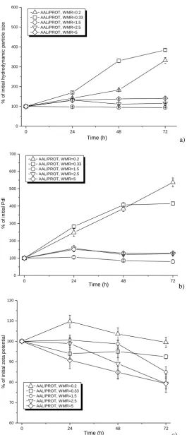
Generally, anionic NPs in their native dispersions were more stable compared to cationic NPs (Fig. 5).
A large increase in the particle size was observed only for the cationic NPs (Fig. 5a). A very rapid increase in
particle size was observed after 24 h for AAL/PROT NPs with a WMR of 0.5. The AAL/PROT formulation
with a WMR of 0.2 showed the best stability comparing the positively charged NPs, as the increase in the
particle size was observed after 72 h (Fig. 5a). An increase in the Pdl values was observed only for the
positively charged NPs (Fig. 5b). A gradual, but relatively small decrease in the ZP values was observed for the
anionic NPs (Fig. 5c), which might be caused by depolymerisation of polymer due to hydrolysis (Holme et al.
2003 and 2008). The best colloidal stability of NPs in the native dispersions was seen for AAL/PROT NPs with
WMRs of 2.5 and 5, while the least stable formulation was that with a WMR of 0.5, perhaps as its ZP is closest
to the neutralisation point.
Colloidal stability studies of selected AAL/PROT formulations in buffers at various pH were also
conducted. Instability and formation of larger particles occurred only in media with very low pH (in HCl) (Fig.
6). Interestingly, better stability in HCl was observed for positively charged NPs (Fig. 6a-c). The cationic NPs
(AAL/PROT_pH3 at WMR of 0.2, 0.33 and 0.5) were physically unstable in the other media tested (acetate
buffer, HEPES buffer and PBS). Immediate aggregation of cationic NPs occurred in acetate buffer at pH 4.5 and
PBS, pH 7.4. Better stability was observed for these NPs in HEPES buffer, pH 6.5. The anionic NPs were stable
in most of the media tested (Fig. 6d-f). No considerable difference in the colloidal stability of anionic NPs was
observed in the acetate, HEPES and diluted PBS media, even though the concentration of the PBS solution was
over a 6-fold lower (0.1M versus 0.015M) than the other two solutions. This might be contributed to the more
kosmotropic (order inducing) character of the acetate and 4-(2-hydroxyethyl)-1-piperazineethanesulphonate
14
2015). Thus lower concentrations of the chloride anions are needed to induce physical instability of NP
dispersions.
As the component, which is used in excess is responsible for charge stabilisation of PEC complex
(PROT for cationic formulations and ALG for anionic NPs), its ability to remain unchanged on the surface of
particle in a varied range of conditions will define the colloidal stability of NPs. In the case of negatively
charged NPs, its poor stability at low pH can be explained by the properties of ALG. Similarly to ionically
cross-linked hydrogels, PECs exhibit pH-sensitive swelling under acidic conditions (Berger et al. 2004). As the
pH value changes, the charge balance between the components inside the formed PECs is altered, resulting in
weakening interactions between the two polymers and possibly leading to swelling of particles due to
dissociation of the complex. In an acidic medium, the carboxylic groups of the polyacid are protonated, thus
forming hydrogen and weaker than ionic bonds with PROT. The study performed by Sankalia et al. (2007)
showed that at pH2, the ionic interactions between CHIT and alginate was greatly reduced and folding of
alginate was observed with an increased micropore size, which allowed better penetration of particles with the
dissolution medium. A similar behaviour of ALG was observed in our study, however an increase in pH of the
liquid medium caused instability of cationic formulations. As PROT comprises small, approximately 5 kDa
fragments, it can be easily removed from the NP surface. Ionic interactions, which occur between positively
charged groups of PROT and ionic groups present in the media often lead to neutralisation of the charge and
formation of larger particles/aggregates.
4 Conclusions
We have demonstrated that alginates can be used as suitable polymers for the formation of novel NPs
by polyelectrolyte complexation with PROT. The conditions for the formation of nanoparticulate carriers were
determined and the self-assembly process was optimised by testing the impact of a range of process and
formulation variables, such the grade of polymer (alginate), the mixing ratio of the polymers as well as the
speed and order of addition of the components during mixing on the properties of NPs. It was shown that the
smallest in size and with lowest Pdl nanocarriers were formed when 0.1% w/v dispersion of AAL and 0.1% w/v
solution of PROT were mixed by Method_2A (when an aliquot of PROT solution was introduced into a stirred
aliquot of ALG dispersion in a fast way).
To facilitate incorporation of drugs which are soluble at low pH, pH of AAL solution was lowered to 2
(from native 6.4), however no NPs were obtained. Therefore further studies were carried out by adjusting pH of
15
decreased to 2, while the adjustment of pH of PROT solution to 3 resulted in the formation of both, anionic and
cationic NPs, depending on the WMR. Physical stability of selected AAL/PROT NPs formulations was tested
upon storage at room temperature and in liquid media at various pH. Good colloidal stability was observed for
anionic NPs stored as native dispersions and with media with a range of pH values.
In summary, a simple, organic solvent and surfactant-free production method by mixing aqueous
solutions of polyelectrolytes at room temperature was successfully employed to formulate novel, cross-linker
free and non-sedimenting NPs comprising alginate and protamine. It was possible to obtain NPs with good
physical properties (i.e. small and homogenously dispersed), which could be used for loading a variety of active
molecules including proteins and peptides. It is proposed that those NPs (positively or negatively charged) can
be used for parenteral and non-parenteral (such as oral, nasal or pulmonary) applications, the route of
administration also dependent of the loaded cargo.
Funding
This study was funded by Merrion Pharmaceuticals Ireland. This work was also supported by the Synthesis and
Solid State Pharmaceutical Centre funded by Science Foundation Ireland under grant number 12/RC/2275.
Conflict of interest
The authors declare that they have no conflict of interest.
References
Andriamanantoanina H, Rinaudo M (2010) Relationship between the molecular structure of alginates and their
gelation in acidic conditions. Polym Int 59:1531-1541.
Augst AD, Kong HJ, Mooney, DJ (2006) Alginate hydrogels as biomaterials. Macromol Biosc 6:623-633.
Avadi MR, Sadeghi AMM, Dounighi NM, Dinarvand R, Atyabi F, Rafiee-Tehrani M (2011) Ex vivo evaluation
of insulin nanoparticles using chitosan and arabic gum. ISRN Pharmaceutics article ID 860109.
Awotwe-Otoo D, Agarabi C, Keire D, Lee S, Raw A, Yu L, Habib MJ, Khan MA, Shah RB (2012)
Physicochemical characterization of complex drug substances: evaluation of structural similarities and
differences of protamine sulfate from various sources. The AAPS Journal 14:619-626.
Berger J, Reist M, Mayer JM, Felt O, Gurny R (2004) Structure and interactions in chitosan hydrogels formed
by complexation or aggregation for biomedical applications. Eur J Pharm Biopharm 57:35-52.
Bertoluzza A, Bonora S, Fini G, Morelli MA, Simoni R (1983) Phospholipid–protein molecular interactions in
16
Birch NP, Schiffman JD (2014) Characterization of self-assembled polyelectrolyte complex nanoparticles
formed from chitosan and pectin. Langmuir 30:3441-3447.
Boddohi S, Killingsworth CE, Kipper MJ (2008) Polyelectrolyte multilayer assembly as a function of pH and
ionic strength using the polysaccharides chitosan and heparin. Biomacromolecules 9:2021-2028.
Carneiro-da-Cunha MG, Cerqueira MA, Souza BWS, Teixeira JA, Vicente AA (2011) Influence of
concentration, ionic strength and pH on zeta potential and mean hydrodynamic diameter of edible
polysaccharide solutions envisaged for multinanolayered films production. Carbohydr Polym 85:522–528.
Chen JH, Heitmann JA, Hubbe MA (2003) Dependency of polyelectrolyte complex stoichiometry on the order
of addition. 1. Effect of salt concentration during streaming current titrations with strong poly-acid and
polybase. Colloids Surf A 223:215-230.
Cheow WS, Hadinoto K (2012) Self-assembled amorphous drug-polyelectrolyte nanoparticle complex with
enhanced dissolution rate and saturation solubility. J Colloid Interface Sci 367:518-526.
Daemi H, Barikani M (2012) Synthesis and characterization of calcium alginate nanoparticles, sodium
homopolymannuronate salt and its calcium nanoparticles. Scientia Iranica F 19:2023-2028.
Delie F, Blanco-Prieto M (2005) Polymeric particulates to improve oral bioavailability of peptide drugs.
Molecules 10:65-80.
Dragan ES, Mihai M, Schwarz S (2006) Polyelectrolyte complex dispersions with a high colloidal stability
controlled by the polyion structure and titrant addition rate. Colloids Surf A 290:213-221.
Dul M, Paluch KJ, Kelly H, Healy AM, Sasse A, Tajber L (2015) Self-assembled carrageenan/protamine
polyelectrolyte nanoplexes-Investigation of critical parameters governing their formation and characteristics.
Carbohydr Polym 123:339-349.
Elsabahy M, Wooley KL (2012) Design of polymeric nanoparticles for biomedical delivery applications. Chem
Soc Rev 41(7):2545-2561.
George M, Abraham TE (2006) Polyionic hydrocolloids for the intestinal delivery of protein drugs: Alginate
17
Guarino V, Caputo T, Altobelli R, Ambrosio L (2015) Degradation properties and metabolic activities of
alginate and chitosan polyelectrolytes for drug delivery and tissue engineering applications. AIMS Mater Sci
2:497-502.
Holme HK, Lindmo K, Kristiansen A, Smidsrød O (2003) Thermal depolymerisation of alginate in the solid
state. Carbohydr Polym 54:431-438.
Holme HK, Davidsen L, Kristiansen A, Smidsrød O (2008) Kinetics and mechanisms of depolymerisation of
alginate and chitosan in aqueous solution. Carbohydr Polym 73:656-664.
Hu Y, Yang T, Hu X (2012) Novel polysaccharide-based nanoparticle carriers prepared by polyelectrolyte
complexation for protein delivery. Polym Bull 68:1183-1199.
Lankalapalli S, Kolapalli VRM (2009) Polyelectrolyte complexes: a review of their applicability in drug
delivery technology. Indian J Pharm Sci 71(5):481-487.
Le-Tien C, Milette M, Mateescu M-A, Lacroix M (2004) Modified alginate and chitosan for lactic acid bacteria
immobilization, Biotechnol Appl Biochem 39:347-354.
Leal D, Matsuhiro B, Rossi M, Caruso F (2008) FT-IR spectra of alginic acid block fractions in three species of
brown seaweeds. Carbohydr Res 343:308-316.
Lee KQ, Mooney DJ (2012) Alginate: Properties and biomedical applications. Prog Polym Sci 37:106-126.
Li P, Dai Y-N, Wei Q (2008) Chitosan-alginate nanoparticles as a novel drug delivery system for nifedipine. Int
J Biomed Sci 4:221-228.
Li C, Hein S, Wang K (2013) Chitosan-carrageenan polyelectrolyte complex for the delivery of protein drugs.
ISRN Biomaterials, article ID 629807.
Liechty WB, Kryscio, DR, Slaughter BV, Peppas NA (2010) Polymers for Drug Delivery Systems. Annu Rev
Chem Biomol Eng 1:149-173.
Mackay ME, Tuteja A, Duxbury PM, Hawker CJ, van Horn B, Guan Z, Chen G, Krishnan RS (2006) General
strategies for nanoparticle dispersion. Science 311:1740-1743.
Mustafaev MI (1996) Polyelectrolyes in immunology. Turk J Chem 20:126-138.
Ngwuluka NC, Ochekpe NA, Aruoma OI (2014) Naturapolyceutics: the science of utilizing natural polymers for
18
Polexe RC, Delair T (2013) Elaboration of stable and antibody functionalized positively charged colloids by
polyelectrolyte complexation between chitosan and hyaluronic acid. Molecules 18:8563-8578.
Reynolds F, Weissleder R, Josephson L (2005) Protamine as an efficient membrane-translocating peptide.
Bioconjugate Chem 16:1240-1245.
Saether HV, Holme HK, Maurstad G, Smidsrod O, Stokke BT (2008) Polyelectrolyte complex formation using
alginate and chitosan. Carbohydr Polym 74:813-821.
Sankalia MG, Mashru RC, Sankalia JM, Sutariya VB (2007) Reversed chitosan–alginate polyelectrolyte
complex for stability improvement of alpha-amylase: Optimisation and physicochemical characterisation. Eur J
Pharm Biopharm 65:215–232.
Sarmento B, Ferreira D, Veiga F, Ribeiro A (2006) Characterisation of insulin-loaded alginate nanoparticles
produced by ionotropic pre-gelation through DSC and FTIR studies. Carbohydr Polym 66:1–7.
Sarmento B, Bibeiro A, Veiga F, Ferreira D, Neufeld R (2007) Oral Bioavailability of Insulin Contained in
Polysaccharide Nanoparticles. Biomacromolecules 8:3054-3060.
Silva CM, Ribeiro AJ, Ferreira D, Veiga F (2006) Insulin encapsulation in reinforced alginate microspheres
prepared by internal gelation. Eur J Pharm Sci 29:148-159.
Sinha VR, Kumria R (2001) Polysaccharides in colon-specific drug delivery. Int J Pharm 224:19–38.
Soppimath KS, Aminabhavi TM, Kulkarni AR, Rudzinski WE (2001) Biodegradable polymeric nanoparticles as
delivery devices. J Control Release 70:1-20.
Srivastava A, Yadav T, Sharma S, Nayak A, Kumari A, Mishra N (2016) Polymers in Drug Delivery. J Biosci
Med 4:69-84.
Temsamani J, Vidal P (2004) The use of cell-penetrating peptides for drug delivery. Drug Discov Ther
9:1012-1019.
Tønnesen HH, Karlsen J (2002) Alginate in Drug Delivery Systems. Drug Dev Ind Pharm 28:621–630.
Umerska A, Paluch KJ, Inkielewicz-Stepniak I, Santoz-Martinez MJ, Corrigan OI, Medina C, Tajber L (2012)
Exploring the assembly process and properties of novel cross-linker free hyaluronate-based polyelectrolyte
19
Umerska A, Paluch KJ, Santos-Martinez MJ, Corrigan OI, Medina C, Tajber L (2014) Self-assembled
hyaluronate/protamine polyelectrolyte nanoplexes: synthesis, stability, biocompatibility and potential use as
peptide carriers. J Biomed Nanotechnol 10:3658-3673.
Umerska A, Paluch KJ, Santos-Martinez MJ, Corrigan OI, Medina C, Tajber L (2015) Chondroitin-based
nanoplexes as peptide delivery systems – Investigations into the self-assembly process, solid-state and extended
release characteristics. Eur J Pharm Biopharm 93:242–253.
Wee S, Gombotz WR (2012) Protein release from alginate matrices. Adv Drug Del Rev 31:267-285.
Wernig K, Griesbacher M, Andreae F, Hajos F, Wagner J, Mosgoeller W, Zimmer A (2008) Depot formulation
of vasoactive intestinal peptide by protamine-based biodegradable nanoparticles. J Control Release
130(2):192-198.
Yu X, Hou J, Shi Y, Su Ch, Zhao L (2016) Preparation and characterization of novel chitosan-protamine
20
FiguresFig. 1 Structural units of alginate. M = β-D-mannuronic acid, G = α-L-guluronic acid
0.2 0.33 0.67 1 1.5 3 5
0 50 100 150 200 250 300 350
***
***
*** *** *
**
*** ***
***
H
yd
ro
d
yn
a
mi
c
p
a
rt
icl
e
si
ze
(n
m)
ALG/PROT WMR
Method_2A AAL Method_2B AAL Method_2A AAM Method_2B AAM
***
A
21
0.2 0.33 0.67 1 1.5 3 5
0.00 0.05 0.10 0.15 0.20 0.25 0.30 0.35 0.40 0.45 0.50 A ** * * * *** *** *** *** Pd l ALG/PROT WMR Method_2A AAL Method_2B AAL Method_2A AAM Method_2B AAM *** b)
0.2 0.33 0.67 1 1.5 3 5
-280 -240 -200 -160 -120 -80 -40 0 40 A * *** * ** * *** Z e ta p o te n ti a l (mV) ALG/PROT WMR Method_2A AAL Method_2B AAL Method_2A AAM Method_2B AAM ** c)
0.1 1 10
100 200 300 400 500 Hydro dyn am ic pa rticle size ( nm ) ALG/PROT WMR Method_2A AAL Method_2A AAM Squares - Size Triangles - ZP
-400 -300 -200 -100 0 Ze ta po ten tial ( mV) d)
Fig. 2 Comparison of: a) hydrodynamic particle size, b) polydispersity index (Pdl), c) zeta potential and d)
[image:21.595.71.355.64.727.2]22
composed of alginate low viscosity/protamine (AAL/PROT) and alginate medium viscosity/protamine
(AAM/PROT) formed by the following variation in method preparation: Method_2A: an aliquot of PROT
solution was added to an aliquot of ALG dispersion under magnetic stirring and Method_2B: an aliquot of ALG
dispersion was introduced to an aliquot of PROT solution under magnetic stirring. A – instantaneous
aggregation/flocculation; statistical analysis: *p<0.05, **p<0.01, ***p<0.001
0.1 1 10
50 100 150 200 250 300 350 400
Hydro
dyn
am
ic par
ticle size (
nm
)
ALG/PROT WMR
PROT pH2 PROT pH3
PROT pH=native (pH5.8)
a)
0.1 1 10
-200 -150 -100 -50 0 50
Ze
ta
po
ten
tial (
mV)
ALG/PROT WMR PROT pH2
PROT pH3
PROT pH=native (pH5.8)
23
0.1 1 10
0.6 0.8 1.0 1.2 1.4 1.6 1.8 2.0 2.2 2.4
Dynam
ic viscosity (
mPa·
s)
ALG/PROT WMR
PROT pH2 PROT pH3
PROT pH=native (pH5.8)
c)
Fig. 3 Comparison of: a) hydrodynamic particle size, b) zeta potential (ZP) and c) dynamic viscosity for NPs
made by combining 0.1% w/v dispersion of alginate low viscosity (AAL) and 0.1% w/v solution of PROT with
pH2, pH3 and native pH (pH5.8). NPs were prepared by Method_2A: an aliquot of PROT solution was added to
an aliquot of AAL dispersion under magnetic stirring
800 900
1000 1100
1200 1300
1400 1500
1600 1700
(C-C-H,O-C-H)
s(COO-)
a(COO-)
a(COC)
s(C-O,C-OH)
(C-O,C-OH)
a(SO)
(CH
2,NH)
(NH3+)
amide II
Wavenumber (cm
-1)
PROT
AAL
AAL/PROT
amide I
Arg
M
Fig. 4 FTIR analysis of PROT, AAL and AAL/PROT NPs. Band assignments was done based on studies of
Bertoluzza et al. (1983), Le-Tien et al. (2004), Leal et al. (2008), Awotwe-Otoo et al. (2012) and Daemi and
Barikani (2012). v – stretching, vS – symmetric stretching, vA – asymmetric stretching and δ - bending
[image:23.595.74.352.68.295.2] [image:23.595.89.502.406.692.2]24
0 24 48 72
0 100 200 300 400 500 600 % o f in it ia l h yd ro d yn a mi c p a rt icl e si ze Time (h) AAL/PROT, WMR=0.2 AAL/PROT, WMR=0.33 AAL/PROT, WMR=1.5 AAL/PROT, WMR=2.5 AAL/PROT, WMR=5 a)
0 24 48 72
0 100 200 300 400 500 600 700 % o f in it ia l Pd l Time (h) AAL/PROT, WMR=0.2 AAL/PROT, WMR=0.33 AAL/PROT, WMR=1.5 AAL/PROT, WMR=2.5 AAL/PROT, WMR=5 b)
0 24 48 72
[image:24.595.72.338.88.706.2]60 70 80 90 100 110 120 % o f in it ia l ze ta p o te n ti a l Time (h) AAL/PROT, WMR=0.2 AAL/PROT, WMR=0.33 AAL/PROT, WMR=1.5 AAL/PROT, WMR=2.5 AAL/PROT, WMR=5 c)
Fig. 5 Colloidal stability of AAL/PROT native dispersions at room temperature: a) hydrodynamic particle size,
c) polydispersity index (Pdl) and c) zeta potential. WMR – weight mixing ratio, AAL – alginate low viscosity,
25
HCl ACETATE HEPES PBS0 100 200 300 400 500 600 700 800 900 A
A A A
% o f i ni ti al h y dr od y na m ic p ar ti c le s iz e AAL/PROT, WMR=0.2 0h - medum 0h + medium 72h + medium
A
a) HCl ACETATE HEPES PBS 0 100 200 300 400 500 600 700 800 900 A A A A A % o f i ni ta il h y dr on am ic p ar ti c le s iz e AAL/PROT, WMR=0.33 0h - mediun 0h + medium 72h + medium
b)
HCl ACETATE HEPES PBS 0 100 200 300 400 500 600 700 800 900 A A A % o f i ni ti al h y dr od y na m ic p ar ti c le s iz e AAL/PROT, WMR=0.5 0h - mediun 0h + medium 72h + medium
c) HCl ACETATE HEPES PBS 0 50 100 150 200 250 300 350 400 450 A A A A % o f i n iti a l h y d ro d y n a m ic p a rti c le s iz e AAL/PROT, WMR=1.5 0h - medium 0h + medium 24h + medium 48h + medium 72h + medium
d)
HCl ACETATE HEPES PBS 0 50 100 150 200 250 300 350 400 450 % o f i ni ti al h y dr od y na m ic p ar ti c le s iz e AAL/PROT, WMR=2.5 0h - medium 0h + medium 24h + medium 48h + medium 72h + medium
A A A A
e) HCl ACETATE HEPES PBS 0 50 100 150 200 250 300 350 400 450 % of i ni ti al h y dro dy na m ic p art ic le s iz e AAL/PROT, WMR=5 0h - medium 0h + medium 24h + medium 48h + medium 72h + medium
A A A A
[image:25.595.74.513.67.592.2]f)
Fig. 6 Colloidal stability studies based on monitoring the change in hydrodynamic particle size of AAL/PROT
nanoparticle dispersions in media with various pH. The weight mixing ratios (WMRs) of AAL/PROT were: a)
0.2, b) 0.33, c) 0.5, d) 1.5, e) 2.5 and f) 5. A – aggregation, AAL – alginate low viscosity, PROT– protamine,
26
TablesTable 1 Summary of the methods evaluated for NPs preparation. ALG – alginate, PROT – protamine
Factor tested Rate of PROT solution addition to ALG dispersion
(PROT solution was added to ALG dispersion)
Method_1A An aliquot of PROT solution was slowly (drop by drop, within 60 seconds) added to an
aliquot of ALG dispersion
Method_1B An aliquot of PROT solution was rapidly (within 2 seconds) added to an aliquot of ALG
dispersion
Factor tested Sequence of addition of PROT and ALG dispersion
(fast addition of components, within 2 seconds)
Method_2A An aliquot of PROT solution was added to an aliquot of ALG dispersion
Method_2B An aliquot of ALG dispersion was added to an aliquot of PROT solution
Table 2 Comparison of properties of AAL/PROT NPs prepared using two various speeds of PROT solution
addition to polymer solution. Method_1A: an aliquot of PROT solution was slowly (within 30 seconds) added to
an aliquot of polymer dispersion, Method_1B: an aliquot of PROT solution was quickly (within 2 seconds)
added to an aliquot of polymer dispersion. Statistical analysis: *p<0.05, **p<0.01, ***p<0.001
AAL/PROT weight mixing
ratio
Hydrodynamic particle size (nm)
Polydispersity index Zeta potential (mV)
Method of preparation:
1A 1B 1A 1B 1A 1B
4.5
131±1 57±1 0.239±0.010 0.233±0.012 -132.8±2.0 -129.4±6.4
*** not significant not significant
5
133±3 72±0 0.312±0.032 0.380±0.018 -145.6±2.7 -131.6±8.4
*** ** *
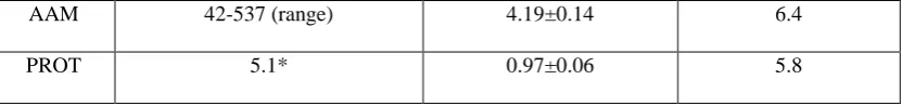
Table 3 Physicochemical properties of alginates and protamine solutions/dispersions studied in present work.
AAL – alginate low viscosity, AAM – alginate medium viscosity and PROT – protamine (* manufacturer data)
Polymer Molecular weight (kDa) Viscosity (mPa·s) of 0.1%
w/v solution/dispersion
pH of 0.1% w/v, native (pH non-adjusted) aqueous solution/dispersion
[image:26.595.69.528.415.601.2] [image:26.595.71.487.666.754.2]27
AAM 42-537 (range) 4.19±0.14 6.4
[image:27.595.70.486.72.121.2]PROT 5.1* 0.97±0.06 5.8
Table 4 FTIR band positions (in cm-1) in alginate (AAL), protamine (PROT) and AAL/PROT nanoparticle (NP)
formulations. The number in parenthesis indicates the magnitude of the shift in cm-1. AAL – alginate low
viscosity, PROT – protamine, v – stretching, vS – symmetric stretching, vA – asymmetric stretching.
Band AAL PROT AAL/PROT NPs
vS (C-O, C-OH) 947 - 949 (2)
v (C-O, C-OH) 1027 - 1028 (1)
vS (COO-) 1408 - 1403 (5)
vA (COO-) 1597 - 1592 (5)