Case Report
Primary primitive neuroectodermal tumor of urinary
bladder in a child: a case report and literature review
Yuedong Chen, Xuegang Wang, Fei Liu, Jinchun Xing
Department of Urology and Center of Urology, The First Affiliated Hospital of Xiamen University, Xiamen, P. R. China
Received February 26, 2016; Accepted June 4, 2016; Epub October 15, 2016; Published October 30, 2016
Abstract: Objectives: The present study is to report the clinical and pathological features of the primary primitive neuroectodermal tumor, Ewing’s sarcoma (PNET/EWS) of urinary bladder. Methods: A case of PNET/EWS in urinary bladder was reported. The literatures of 14 cases of PNET/EWS were reviewed according to clinical manifesta-tion and pathological characteristics. Results: A 2-year-old boy presented with dysuria was referred to our institu-tion for further management in December 2014. Pelvic computed tomography showed a large intravesical tumor. Cystoscopy showed a large intravesical mass arising from the neck and left side of the bladder, and then biopsy was made. Histopathological examination revealed sheets of uniform, small, round, and oval cells, which presented scarce cytoplasm, hyperchromatic nuclei, inconspicuous nucleoli, and abundant atypical mitotic figures. From the further immunohistochemical characterization, the tumor cells demonstrated strong reactivity to CD99 and Vim. A definitive diagnosis of PNET of the bladder was established. Catheterization was performed, but chemotherapy was refused by the parents, and the patient was discharged according to his parents’ will. The patient died 4 months later. Conclusions: Bladder PNET is an extremely rare malignant tumor. The diagnosis is based on histological, im-munohistochemical and molecular pathologic findings. It is a kind of highly aggressive tumor and has very poor prognosis. Radical excision combined with adjuvant chemotherapy and radiotherapy appears to be the best treat-ment.
Keywords: Primary primitive neuroectodermal tumor/Ewing’s sarcoma of the urinary bladder, pathology, therapy, prognosis
Introduction
Ewing’s sarcoma (EWS) is a kind of rarely seen malignant tumor originated from neural ecto-derm. Clinically, EWS is diagnosed as primitive neuroectodermal tumor (PNET) [1]. PNET/EWS is mainly composed of primitive neuroectoder-mal cells, and possesses the potential of multi-directional differentiation. It can be classified into central type and peripheral type [2]. Peripheral PNET/EWS usually occurs in chil-dren and adolescents, with strong invasiveness and poor prognosis. It is commonly observed in bone and cartilage tissues, especially in trunk, limbs, spine, and chest wall [3, 4]. The main clinical manifestations of PNET/EWS include gradually enlarged local masses and mass compression-related symptoms. On computed tomography, PNET/EWS is mainly presented as low density images of circular or irregularly
Case report
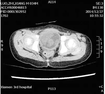
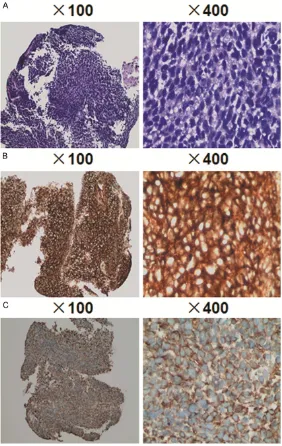
A child (boy; 2 years and 7 months old) was ad- mitted at our hospital in December 2014 due to dysuria for the past one week. Past medical history was unremarkable. Computed tomogra-phy showed that the child had developed blad-der occupation 12 days ago. The patient had no fever, emaciation, hematuria, or family genetic history. Body examination showed that the patient was conscious, with normal develop-ment both physically and develop-mentally. The sizes of superficial lymph nodes were not large, and the patient had normal movements of trunk and limbs. No weight loss was registered. Routine blood parameters were normal. No distant metastasis was detected by chest computed tomography. Computed tomography revealed a large tumor measuring 7 cm in diameter at the base of the bladder. Enhanced scanning by computed tomography showed that arterial phase was significantly enhanced, and venous phase and muscle equilibrium phase were con-tinuously enhanced (Figure 1). Cystoscopy showed a piece of mass on bladder neck and left wall blocked the bladder cavity, and the mass had mucous and smooth surface with grey color (Figure 2). Pathological examinations of the mass showed that the tumor cells had diffuse lamellar arrangement, with variable sizes. The cytoplasm was scarce and dyed pink. The nucleus showed round or oval shapes, with dust-like chromatin. Nuclear deviation was
observed, the cytoplasm was eosinophilic, and no obvious nucleolus was visualized. In addi-tion, pathological mitotic figures were observed (Figure 3A).
Immunohistochemistry showed positive expres-sion of CD99 (++) and Vim (Figure 3B and 3C). The pathological diagnosis was small round cell malignant tumor. Morphology observation under light microscope, immunophenotyping and specific staining supported the diagnosis of classical EWS. Catheterization was perfor- med on the patient to relieve dysuria. Che- motherapy was suggested but rejected by the parents of the patient after the condition of the patient was informed to them. Four months later, the patient died.
Discussion
[image:2.612.323.523.72.189.2]PNET is a kind of malignant tumor in soft tis-sues and bones that are composed of small round cells. As the development of immunohis-tochemistry, more and more cases are report-ed. PNET is also found to occur in internal organs. By December 2014, a total of 14 cases of bladder EWS have been reported. Together with the case reported here, the ages of the 15 bladder EWS patients range from 2 years and 7 months to 81 years (average, 39.1 years). Although PNET/EWS is common in children, 13 out of the 15 cases are older than 14 years. Among the 15 patients, 9 are males and 6 are females. The clinical manifestations of bladder EWS include hematuria (8 cases; 57.1%), dys-uria (7 cases; 42.9%), lower urinary tract irrita-tion (2 cases), bilateral hydronephrosis (2 ca- ses), lymphatic edema (1 case), lower abdomi-nal mass (1 case), and lower abdomiabdomi-nal pain (1
[image:2.612.89.290.73.253.2]Figure 1. Computed tomography with contrast en-hancement for the patient with EWS. The scan showed a large abdominal mass localized in the bladder.
case). These phenomena are local irritation and oppressive symptoms caused by rapid growth of the mass. At the time of diagnosis, 5 cases already have metastasis into external tis-sues of the bladder, such as prostate, seminal vesicle gland, uterus, and rectum. Two cases have ureteral obstruction induced by compres-sion. Only one case has shown distant metasta-sis in the lungs. Four cases have radical total resection of bladder, three cases have partial
shows positive CD99, vim and CD177 expres-sion. Sometimes, PNET tissues are accompa-nied by local focus positive expression of cyto-keratin AE1 or AE3. However, negative expres-sion of smooth muscle actin, chromogranin, epithelial membrane-like antigen, desmin, and leukocyte antigen antibody [14]. These mani-festations can help distinguish PNET cells from neuroendocrine carcinoma, lymphoma and melanoma [21]. Although the positive
immuno-Figure 3. Histopathological examination of tumor tissue biopsy. (A) Hema-toxylin and eosin staining showing the presence of sheets of small round blue cells that were separated by minimal desmoplastic fibrous stroma. (B) CD99 and (C) Vim immunostaining showing positive cytoplasmic staining in tumor cells.
resection of bladder, four cases have received transurethral re- section of bladder tumor and 9 cases have undergone chemo-therapy (vincristine + doxorubi-cin + cyclophosphamide and ifo- sfamide + etoposide). Among the reported 14 cases, 8 cases were followed up. The follow-up results show that one case has survived for 3 years and is still alive [14], and two cases have survived for 18 months. Among the remaining 5 patients, two cases died 2-3 weeks after dia- gnosis due to metastasis [10, 12], one case died 2 weeks after transurethral resection of bladder tumor and bilateral nep- hrostomy [13], one case died 4 months after diagnosis [19], and one case died 22 months after total resection of bladder [16].
[image:3.612.90.372.72.518.2]histochemical expression rate of CD99 in PNET is more than 95% [22], its expression is not specific for PNET. Some lymphoblastic lympho-ma, poorly differentiated synovial sarcoma and rhabdomyosarcoma also have positive expres-sion of CD99 [23].
Cell biology and molecular genetics studies show that the occurrence of PNET/EWS is closely associated with chromosomal translo-cations, with more than 85% showing t [11, 22] (q24;q12) abnormal karyotype, which is the result of the fusion of EWS gene (located at 22q12) with FLI-1 gene (located at 11q24). In addition, about 10% NET/EWS cases show t [21, 22] (q22;q11) karyotype, which is the result of the fusion of EWS gene with Ets-related gene [24]. Fusion genes produced by chromosome translocations have stable struc-tures, and tumor specificity. Therefore, EWS translocation gene test combined with FLSH is of great value in the clinical diagnosis of PNET/ EWS [25].
Currently, surgical resection is the primary method for the treatment of PNET/EWS. Patients with tumor foci that can be completely resected should receive the surgery. Early and complete resection of the tumor can reduce local recurrence and metastasis, eliminate drug-resistant tumor cells, and help to enhance the effectiveness of postoperative chemother-apy. Chemotherapy is a supplementary treat-ment in addition to surgery for adults, but is debatable for child patients. The efficacy of chemotherapy for PNET/EWS is already proven, and novel adjuvant chemotherapy can increase resection rate. In a case report, the size of blad-der mass on a ten-year-old patient was decreased from 14 cm × 13 cm × 13 cm to 4.7 cm × 3.4 cm × 3.3 cm after three courses of novel adjuvant chemotherapy (vincristine + doxorubicin + cyclophosphamide and ifos-famide + etoposide), after which the mass in bladder was resected [20]. Recent studies show that EWS/FLI-1 can cause abnormal acti-vation of cyclin dependent kinase and the deactivation of its inhibitors, which may be a key reason for the uncontrolled proliferation of EWS cells. It is already proven that inhibition of cyclin dependent kinase can induce the apop-tosis of tumor cells and thus, cyclin dependent kinase may become a potential target in the treatment of EWS [26]. In conclusion, bladder EWS is a kind of rare malignant soft tissue
tumor that occurs in neural ectoderm of blad-der. Preoperative diagnosis is dependent on histological, pathological and immunohisto-chemical examinations, especially the determi-nation of EWS translocation gene. Complete resection, accompanied with chemotherapy and radiotherapy, may prolong the survival duration of patients with EWS.
Acknowledgements
This study was funded by the First Affiliated Hospital of Xiamen University. Informed con-sent was obtained from the family of the patient included in the study.
Disclosure of conflict of interest
None.
Address correspondence to: Jinchun Xing, Depart- ment of Urology and Center of Urology, The First Hospital Affiliated to Xiamen University, 55 Zhenhai Road, Xiamen 361003, Fujian Province, P. R. China. E-mail: xmurology@126.com
References
[1] Parharm DM, Roloson GJ, Feely M, Green DM, Bridge JA and Beckwith JB. Primary malignant neuroepithelial tumors of the kidney: A clinico-pathologic analysis of 146 adult and pediatric cases from the National Wilms’ Tumor Study Group Pathology Center. Am J Surg Pathol 2001; 25: 133-146.
[2] Burkhardt JK, Kockro RA, Dohmen-Scheufler H, Woernle CM, Bellut D, Kollias S and Bertalanffy H. Small supratentorial, extraaxial primitive neuroectodermal tumor causing large intracerebral hematoma. Neurol Med Chir (Tokyo) 2011; 51: 441-444.
[3] Ellinger J, Bastian PJ, Hauser S, Biermann K and Müller SC. Primitive neuroectodermal tu-mor: rare, highly aggressive differential diag-nosis in urologic malignancies. Urology 2006; 68: 257-262.
[4] Banerjee SS, Eyden BP, McVey RJ, Bryden AA and Clarke NW. Primary peripheral primitive neuroectodermal tumor of urinary bladder. Histopathology 1997; 30: 486-490.
[5] Duan XH, Ban XH, Liu B, Zhong XM, Guo RM, Zhang F, Liang BL and Shen J. Intraspinal prim-itive neuroectodermal tumor: imaging findings in six cases. Eur J Radiol 2011; 80: 426-431. [6] Kamphues CH, Rocken C, Neuhaus P and
[7] Ho DM, Hsu CY, Wong TT, Ting LT and Chiang H. Atypical teratoid/rhabdoid tumor of the central nervous system: A comparative study with primitive neurocetodermal tumor/medullo-blastoma. Acta Neuropathol 2000; 99: 482-488.
[8] Li WY and Zhou JL. Research progress of prim-itive neuroectodermal tumor. Int J Med Radiol 2014; 37: 221-224.
[9] Gousse AE, Roth DR, Popek EJ, Cooley LD and Horowitz ME. Primary Ewing’s sarcoma of the bladder associated with an elevated antinucle-ar antibody titer. J Urol 1997; 158: 2265-2266. [10] Mentzel T, Flaschka J, Mentzel HJ, Eschholz G
and Katenkamp D. Primary primitive neuroec-todermal tumor of the urinary bladder. Clinicopathologic case report and differential small cell tumor diagnosis of this site. Pathologe 1998; 19: 154-158.
[11] Desai S. Primary primitive neuroectodermal tumor of the urinary bladder. Histopathology 1998; 32: 477-478.
[12] Colecchia M, Dagrada GP, Poliani PL and Pilotti S. Immunophenotypic and genotypic analysis of a case of primary peripheral primitive neuro-ectodermal tumor (pPNET) of the urinary blad-der. Histopathology 2002; 40: 108-109. [13] Krüger S, Schmidt H, Kausch I, Böhle A,
Holzhausen HJ, Johannisson R and Feller AC. Primitive neuroectodermal tumor (PNET) of the urinary bladder. Pathol Res Pract 2003; 199: 751-754.
[14] Lopez-Beltran A, Perez-seoane C, Montironi R, Hernández-Iglesias T, Mackintosh C and de Alava E. Primary primitive neuroectodermal tu-mor of the urinary bladder: a clinico-pathologi-cal study emphasising immunohistochemiclinico-pathologi-cal, ultrastructural and molecular analyses. J Clin Pathol 2006; 59: 775-778.
[15] Osone S, Hosoi H, Tanaka K, Tsuchiya K, Iehara T, Morimoto A, Hashida T, Yamashita M, Kawabata K, Nishijo K, Toguchida J, Hata J and Sugimoto T. A case of a Ewing sarcoma family tumor in the urinary bladder after treatment for acute lymphoblastic leukemia. J Pediatr Hematol Oncol 2007; 29: 841-844.
[16] Okada Y, Kamata S, Akashi T, Kurata M, Nakamura T and Kihara K. Primitive neuroec-todermal tumor/Ewing’s sarcoma of the uri-nary bladder: a case report and its molecular diagnosis. Int J Clin Oncol 2011; 16: 435-438.
[17] Rao RN, Sinha S, Babu S and Mehrotra R. Fine-needle aspiration cytology of primitive neuro-ectodermal tumor of the urinary bladder: a case report. Diagn Cytopathol 2011; 39: 924-926.
[18] Busato WF Jr, Almeida GL and Ogata DC. Primary primitive neuroectodermal tumor of the bladder: histogic and clinical features of 9 cases. Clin Genitourin Cancer 2011; 9: 63-67. [19] Zheng Y, Tan F, Wang L, Xu N and Mou H.
Primary primitive neurocetodemal tumor of the urinary bladder: a case report and literature review. Med Oncol 2011; 28: 388-391. [20] Sueyoshi R, Okawada M, Fujimura J, Saito M,
Koga H, Lane GJ and Yamataka A. Successful complete resection of Ewing sarcoma arising from the bladder in a 10-year-old boy after che-motherapy. Pediatr Surg Int 2014; 30: 965-969.
[21] Lott S, Lopez-Beltran A, Montironi R, MacLennan GT and Cheng L. Soft tissue tu-mors of the urinary bladder. Part II: malignant neoplasms. Hum Pathol 2007; 38: 963-977. [22] Huang KH, Shun CT, Huang SY, Yu HJ, Chueh
SC and Chen J. Primary primitive neuroecto-dermal tumor of the urinary tract. J Formos Med Assoc 2006; 105: 1008-1012.
[23] Busato Jr WFS, Bettega LBP and Pereira E. Paratesticular pleomorphic rhabdomyosarco-ma in an elderly rhabdomyosarco-man. Chirurgia 2009; 22: 49-50.
[24] Skubitz KM and D’Adamo DR. Sarcoma. Mayo Clin Proc 2007; 82: 1409-1432.
[25] Hua W, Jie Z and Yu-ping W. Molecular detec-tion of EWS-Ets fusion transcripts and their clinicopathologic significance in Ewing’s sar-coma/peripheral primitive neuroectodermal tumor. Chin Med J 2005; 118: 1323-1329. [26] Kauer M, Ban J, Kofler R, Walker B, Davis S,