Original Article
Clinical significance of NGAL and MMP-9 protein
expression in epithelial ovarian cancers
Jian Wu1, An-Quan Shang2, Wen-Ying Lu2
1Department of Laboratory Medicine, Yancheng People’s First Hospital, Yancheng 224005, Jiangsu Province, China; 2Pathological Science Laboratory, The Sixth People’s Hospital of Yancheng, Yancheng 224000, Jiangsu, China
Received March 13, 2015; Accepted January 7, 2016; Epub February 15, 2016; Published February 29, 2016
Abstract: Objective: To investigate the significance of neutrophil gelatinase-associated lipocalin (NGAL) and matrix metalloproteinase-9 (MMP-9) protein expression in epithelial ovarian cancers. Methods: NGAL and MMP-9 serum
and ovarian tissue levels were detected in 150 ovarian epithelial cancers. 42 benign ovarian tumors and 30 healthy women as controls by immunohistochemistry technique and reverse transcriptase-polymerase chain reaction
(RT-PCR). Results: NGAL and MMP-9 expression in tissue and serum in ovarian cancer group were significantly higher
than that in benign ovarian tumor group and normal control group (both P<0.05). Two high expression and clinical
staging, lymph node metastasis positive correlation (both P<0.05). Moreover, in the serum and ovarian tissue of pa
-tients with epithelial ovarian cancer, the expression of NGAL had positive correlation with histological differentiation (both P<0.05) and the expression level of MMP-9 had negative correlation with histological differentiation (P<0.05). In patients with ovarian carcinoma tissue or serum, NGAL expression were positive expression of MMP-9 (r=0.740, r=0.676, both P<0.05). Conclusions: The high expression of NGAL and MMP-9 in epithelial ovarian cancer may be associated with the initiation and progression of epithelial ovarian cancer.
Keywords: Epithelial ovarian cancers, neutrophil gelatinase-associated lipocalin, matrix metalloproteinase-9
Introduction
The molecular biological characteristics of the latest to epithelial ovarian cancer (EOC) is a highly heterogeneous disease, its clinical char-acteristics is the disease starts without symp-toms until advanced disease [1, 2], generally diagnosed. In the past few decades, mortality statistics has almost no change in patients with EOC. According to the latest cancer statistics, EOC (including the fallopian tube and primary peritoneal carcinoma) become the cancer of the female in western countries the fifth most common cause of death, the top four is lung cancer, breast cancer, colon cancer, pancreatic cancer.
As a new protein [3, 4], neutrophil gelatinase-associated lipocalin (NGAL) is found when Kjeldsen et al. study matrix metalloprotein-ase-9 in neutrophil in 1993. High expression of NGAL in inflammatory diseases and various tumor is cancer research field in recent years.
As a secreted protein, relative molecular mass of NGAL was about 25000 bp. In the late stage of young and immature little synthesis in bone marrow neutrophil cells under physiological conditions. Matrix metalloproteinase-9 (MMP-9) is the Zn2+ dependent activation of proteo -lytic enzyme catalysis [5, 6].
The main degradation of type IV collagen and gelatin, which can promote the formation of malignant tumor growth and angiogenesis. MMP-9 can reduce the extracellular matrix and basement membrane and the influence of tis -sue remodeling, thereby promoting tumor inva-sion and metastasis.
Patients and controls
From recently five years, samples of epithelial ovarian cancers were collected from 150 patients (93 cases of serous carcinoma and 57 cases of mucinous carcinoma) who received surgical resection in the First People’s Hospital of Yancheng City (Yancheng, China) and had been diagnosed by pathological confirmation. There are 42 cases of benign ovarian epithelial tumors (30 patients with serous cystadenocar-cinoma and 12 cases of mucinous cystadeno -carcinoma). Each case had detailed clinical and pathological data and none received preopera-tive chemotherapy or radiotherapy. Cancer patients age 36 to 73 years (mean age 53.9±11.6 years). Select 18 cases in our hospi-tal as control group with normal ovarian tissue resection of uterine fibroids surgery and blood specimens. Ages 35 to 69 years (mean age 49.5±10.4 years).
According to the International Federation of gynecology and obstetrics (FIGO) clinical stag -ing criteria in 2000:51 cases of stage I-II, 99 cases of stage III-IV.
No statistically significant difference was detected in age between the two groups. All specimens were obtained under informed con -sent with approval by the Ethics Committee of our hospital (Identification No. HMU (Ethics) 20131103).
Immunohistochemical staining techniques
Immunohistochemical method to EnVision staining was used to detect the distribution of NGAL and MMP-9. Immunohistochemical pro -cedures were performed strictly with the kit manual operation. The EnVision and DAB chro -mogenic reagent kit (Antibody Diagnostic Inc., USA) was used to immunohistochemical stain -ing. All slice staining were operated under the same conditions, the tissue was sliced to 4 μm, dehydration, dewaxing and antigen repaired by using PH 6.0, 0.01 mol/L citric acid. Normal goat serum was dropped on the slice by incu-bating for 10 min at room temperature, then corresponding specific antibodies were drop-ped on the slice by incubating for 1.5 h at room temperature. It was washed with PBS for 3 min by 3 times. The second antibody was dropped on the slice by incubating for 30 min at room temperature. It was colored by DAB, nucleus was stained by hematoxylin, dehydrated by gra-dient ethanol, cleared by xylene, sealed by nat-ural gum. Each batch dyeing all has positive control (with the known positive section reagent which was offered by reagent company) and negative control (the corresponding specific antibody was replaced by PBS).
[image:2.612.93.521.84.140.2]The nucleus in yellow or tan reactant particles is positive. Four independent random were detected by optical microscope of Olympus (BH-2) at high magnification (×200). According to the positive staining degree and the percent-age of tumor cells, the criteria for judgment was as follows. There is no express (-): Or a small amount of cell shading. Expression is less than
Table 1. Primer sequences for RT-PCR analysis
Primers Sense sequences Antisense sequences Product size
NGAL 5’-TCACCTCCCTCCTGTTTA-3’ 5’-CTCCTTGGTTCTCCCGTA-3’ 233 bp
MMP-9 5’-GCTCTTCCCTGGAGACCTGA-3’ 5’-CTGCCTAACCCTGGACCT-3’ 203 bp
β-action 5’-CTGGGACGACTGGAGAAAA-3’ 5’-AAGGAAGGCTGGAAGAGTGC-3’ 564 bp
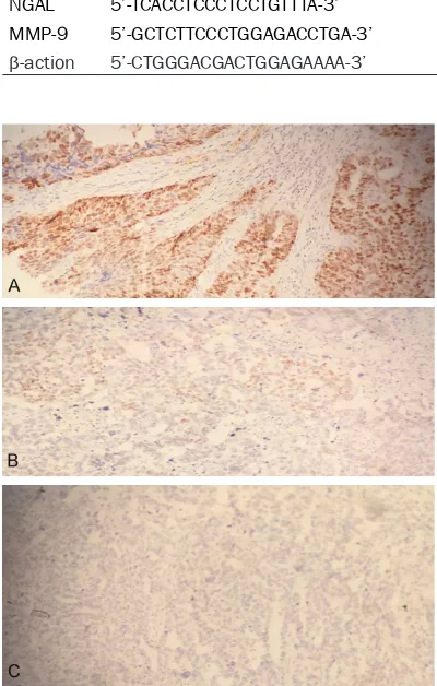
Figure 1. The staining result of EnVision immunohis
-tochemistry for NGAL in ovary tissues and normal
tissues. A: Staining strong positive result in ovary
tis-sues; B: Staining positive result in ovary tistis-sues; C:
[image:2.612.89.289.103.417.2]5%. Lower expression (+): Pale yellow or posi-tive cells was 5% to 29%. Moderate expression (2+): Yellow or positive cells was 30% to 59%. High expression (3+): A tan or positive cells was more than 60%.
ELISA detection of serum NGAL and MMP-9 Extract of patient fasting venous blood 3 ml in the 3-4 days before operation. In a test tube without anticoagulant, standing for 30 minutes
noassay instrument. Draw standard curve, and calculate the content of NGAL and MMP-9 in the sample.
Reverse transcriptase-polymerase chain reac-tion
[image:3.612.92.365.98.189.2]Reverse transcriptase-polymerase chain reac-tion (RT-PCR) was used to detect NGAL and MMP-9 expression. Total RNA was extracted from frozen tissue using Trizol reagent (KeyGEN). The total mRNA was reverse tran -scribed into cDNA by reverse transcriptase 8 mL. β-action as a quantitative control. The cDNA products were amplified by PCR. Taking the 5 mL PCR amplification product, the experi -ment was carried out in 2% agarose gel electro-phoresis. Gel imaging system will gel bands imaged, and semi quantitative analysis was performed by software of quantity one. RT-PCR kit was purchased from Beijing Zhong Shan Jinqiao Company. NGAL and MMP-9 primers were purchased from Nanjing ginster. NGAL, MMP-9 and control primer sequences see
Table 1.
Statistical methods
SPSS13.0 statistical software was used for sta -tistical analysis. The χ2 test was used to com-pare distribution of NGAL and MMP-9 between normal and cancer tissues, and spearman cor-relation was used to analyze the cor-relationship of distribution among NGAL and MMP-9. P<0.05 was considered to be statistically significant.
Results
Cell nucleus dyeing distribution of NGAL and MMP-9 in ovary tissue and normal tissue The positive rate of NGAL staining in ovary tis -sues 86.7% (130/150) is significantly higher than normal skin tissue and benign ovarian
Table 2. The staining positive result of EnVision immunohisto -chemistry for NGAL in ovary, benign ovarian and normal tissue
Group n NGAL positive Negative (-) Rate (%)Positive tive Rate (%)Strong Posi-+ 2Posi-+ 3Posi-+
Normal 30 0 0 0 30 0 0
benign ovarian 42 6 4 2 30 28.6 11.9 ovary 150 30 45 55 20 86.7 36.7
P* value 0.000
P▲ value 0.021
Note: *The χ2 test was used to compare staining positive result between normal
and ovary tissues, P<0.05; ▲The χ2 test was used to compare staining positive
result between benign ovarian and ovary tissues, P<0.05.
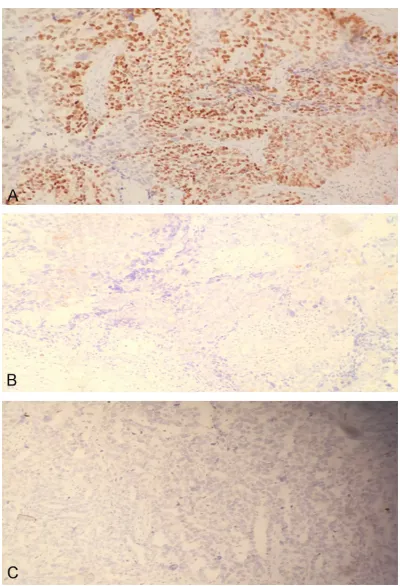
Figure 2.The staining result of EnVision immunohis
-tochemistry for MMP-9 in ovary tissues and normal
tissues. A: Staining strong positive result in ovary
tis-sues; B: Staining positive result in ovary tistis-sues; C:
Staining negative result in normal tissues.
at room temperature. 4°C 3000 r/min rotating centrifugal for 15 minutes. Serum placed -80°C refrigerator. The intraoperative and cut a part of ovarian tissue into 8 degrees refrigerator rap -idly within 0.5 hours, for the extraction of mRNA.
[image:3.612.89.289.241.534.2]-tumors (P<0.05; Figure 1 and Table 2). And positive rate MMP-9 in ovary tissues was 66.7% (100/150), and there was significant difference in statistics than normal skin tissue and benign ovarian tumors (P<0.05; Figure 2 and Table 3). The expression of NGAL mRNA and MMP-9 mRNA in ovary tissue
NGAL and MMP-9 mRNA expression in epithe -lial ovarian cancer group were significantly higher than that in the normal group and ovari-an benign tumor group (both P<0.05; Figure 3;
Table 4). In ovarian cancer tissues, the expres-sion of NGAL and MMP-9 mRNA increased with elevated clinical staging (both P<0.05). Lymph node metastasis group was significantly higher in no lymph node metastasis group (both P<0.05). The expression of NGAL mRNA in high, medium, low differentiation cancer reduce falls in turn (both P<0.05), while the expression of NGAL mRNA in high, medium, low differentiation cancer were enhanced in turn (both P<0.05; Table 5).
The expression of NGAL and MMP-9 in serum
NGAL and MMP-9 in the serum of patients with ovarian cancer were higher than those of nor -mal group and benign ovarian tumors (both P<0.05; Table 6). In ovarian cancer patient
The Pearson Correlation Analysis showed that the level of serum NGAL was positively corre -lated with the expression of NGAL mRNA in epi -thelial ovarian cancer tissues (r=0.762, P<0.05). The level of serum MMP-9 was posi -tively correlated with the expression of MMP-9 mRNA in epithelial ovarian cancer tissues (r=0.601, P<0.05). Expression of NGAL mRNA correlated positively with the expression of MMP-9 mRNA in epithelial ovarian cancer (r=0.740, P<0.05). Level of serum NGAL corre -lated positively with the level of serum MMP-9 in epithelial ovarian cancer (r=0.676, P<0.05).
Discussion
NGAL is the one member of lipocalin family [7, 8]. Also the molecular weight of 25 KD. NGAL contains 178 amino acid residues and is locat-ed in human chromosome of 9q34. With the length for 5869 bp, NGAL 135 KD NGAL can not only through the two disulfide bonds and neutrophil MMP-9 is formed by the combina -tion of molecular weight 135 KD heterologous dimerization of two, but also exist as mono -mers, dimers and other forms of homologous two. The high expression of NGAL is in a variety of tumors, such as breast cancer, rectal cancer, colon cancer, gastric cancer [9-11]. It was sug-gested that NGAL may be a novel human oncogene.
Table 3. The staining positive result of EnVision immunohistochem -istry for MMP-9 in ovary, benign ovarian and normal tissue
Group n MMP-9 positive Negative (-) Positive rate (%) tive Rate (%)Strong
posi-+ 2+ 3+
Normal 30 0 0 0 30 0 0
Benign ovarian 42 3 2 1 36 14.3 2.3
Ovary 150 25 35 40 50 66.7 26.7
P* value 0.219
P▲ value 0.198
Note: *The χ2 test was used to compare staining positive result between normal and
ovary tissues, P<0.05; ▲The χ2 test was used to compare staining positive result
between benign ovarian and ovary tissues, P<0.05.
Figure 3. Electrophoresis results of expression of NGAL mRNA and MMP-9 mRNA in different ovarian tissues. M: Marker; 1: Normal group; 2: Ovarian
benign tumor group; 3: Epithelial ovarian cancer group.
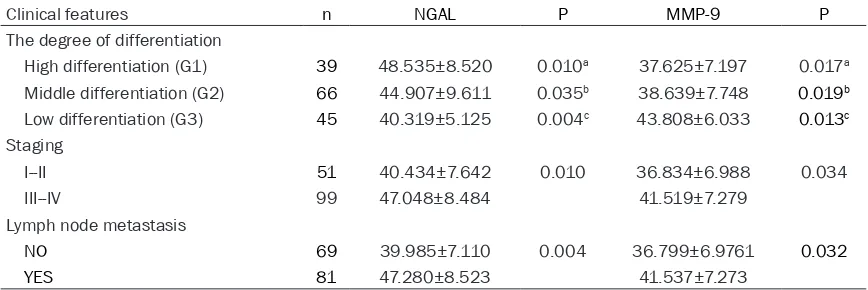
serum, the NGAL and MMP-9 levels in stage III--IV were sig -nificantly higher than in stage I--II (both P<0.05). NGAL and MMP-9 in lymph node metas-tasis group was significantly higher than that in the group without lymph node metasta-sis (both P<0.05). The degree of differentiation with ovarian cancer. Tissue differentiation was positively correlated with the level of NGAL. While the degree of differentiation with ovarian cancer. Tissue differ -entiation was negatively cor-related with the level of MMP-9. Both difference have statistical significance (both P<0.05; Table 7).
Proved by experiments, using NGAL gene as a target, the design of small interfering RNA, downregulation of NGAL gene expression by gene silencing, observed cell apoptosis signifi -cantly increased tumor cell proliferation signifi -cantly reduce. Suggesting that NGAL could be blocked by specific apoptosis inducing tumor cell apoptosis factor induced, thus protecting the survival of tumor cells [12, 13]. In vitro, NGAL can activate ERalpha/Slug pathway, induced by mesenchymal-epithelial transition. You can also recombine cancer cell skeleton by ectopic E-cadherin, Rac1 and catenins, so that the adhesion decrease between cells and matrix, so as to improve the invasive ability of tumor cells [14, 15].
In this study, the expression of NGAL mRNA in ovarian cancer tissue was significantly higher than that in normal ovarian tissue and ovarian benign tumors. NGAL mRNA expression in ovar -ian cancer tissue increased with elevated clini-cal staging. Lymph node metastasis group was significantly higher than that in the group with -out lymph node metastasis. These differences indicate that NGAL play some role in the forma -tion, invasion and metastasis of the ovarian cancer. In addition, the expression of NGAL
MMP-9 is the Zn2+ dependent activation of proteolytic enzyme catalysis. MMP-9 main deg-radation of type IV collagen and gelatin, and thus promote the malignant tumor growth and angiogenesis. The study found that the expres -sion of MMP-9 mRNA in ovarian cancer tissues is increased, which suggesting that MMP-9 may play a certain role in the carcinogenesis of ovarian tissue. Expression of MMP-9 mRNA was markedly increased in ovarian carcinoma. The expression levels during III-IV period in epi -thelial ovarian carcinoma was significantly high -er than that I-II p-eriod, which suggesting that MMP-9 may play a certain role in the invasion, metastasis and evolution of ovarian tissue. The level of serum MMP-9 in ovarian cancer has higher degree with clinical staging, pathological grading and lymph node metastasis in the data organization. Therefore, MMP-9 has important significance in judging the malignant degree of ovarian cancer invasion and metastasis, and prognosis estimation.
[image:5.612.88.524.85.139.2]The study by Nuntago [16] et al. found that NGAL through the formation of complexes with MMP-9, stable MMP-9 activity, reduce the deg-radation, and promote bile duct cancer cell invasion. NGAL/MMP-9 complex in the serum
Table 4. The expression of NGAL and MMP-9 mRNA in different ovarian tissue
Group n NGAL P MMP-9 P
Normal group 30 0.121±0.037 0.017a 0.111±0.042 0.021a
Ovarian benign tumor group 42 0.442±0.155 0.019b 0.312±0.064 0.015b
Epithelial ovarian cancer group 150 0.747±0.146 0.008c 0.625±0.144 0.002c
a: Normal group vs ovarian benign tumor group; b: Ovarian benign tumor group vs epithelial ovarian cancer group; c: Normal group vs epithelial ovarian cancer group.
Table 5. The relationship between NGAL and MMP-9 mRNA expression and clinical pathological characteristics of ovarian cancer
Clinical features n NGAL P MMP-9 P
The degree of differentiation
High differentiation (G1) 39 0.824±0.065 0.012a 0.505±0.142 0.014a Middle differentiation (G2) 66 0.770±0.064 0.007b 0.635±0.111 0.022b Low differentiation (G3) 45 0.620±0.221 0.004c 0.715±0.120 0.000c
Staging
I--II 51 0.684±0.179 0.009 0.528±0.136 0.000
III--IV 99 0.779±0.116 0.676±0.124
Lymph node metastasis
NO 69 0.678±0.179 0.032 0.539±0.132 0.002 YES 81 0.806±0.073 0.699±0.109
a: High differentiation vs Middle differentiation; b: Middle differentiation vs low differen -tiation; c: High differentiation vs low differentiation.
[image:5.612.91.389.206.353.2]of clear cell renal cell carcinoma has high expression, and also the patients has poor prognosis. Compared with low expression group, the total survival rate was decreased obviously [17]. In this experiment, the expres-sion of NGAL and MMP-9 in ovarian cancer tis -sue and serum has the positive correlation. It can be inferred that both of them may have syn -ergistic effects in the occurrence and develop -ment of ovarian cancer. However, NGAL in serum and tissue has positive correlation to the degree of differentiation in ovarian cancer, while MMP-9 was negatively correlated with the extent of tissue differentiation. Therefore, both the mechanism of action are still needing fur -ther research [18, 19].
Levels of NGAL and MMP-9 in the ovarian can -cer tissues and serum were significantly higher than that of other research group. Between the NGAL and MMP-9 relationship with clinical pathological features and RT-PCR findings are consistent. The level of NGAL in serum is relat -ed to NGAL mRNA in ovarian cancer tissue, and the level of MMP-9 in serum is related to MMP-9 mRNA in ovarian cancer tissue. It can be inferred that, MMP-9 and NGAL elevated in
serum may partly derived from cancer tissue. Therefore, monitoring of NGAL and MMP-9 can serve as an important index to judge the prog-nosis of patients with ovarian cancer before operation [20-22]. To explore the combined detection of NGAL and MMP-9 serum levels, the aim is to make it clear to be used as a diag -nostic index used for play a certain clinical value in early diagnosis of ovarian cancer, which is needed to further research.
Disclosure of conflict of interest
None.
Address correspondence to: Dr. Wen-Ying Lu,
Path-ological Science Laboratory, The Sixth People’s
Hospital of Yancheng, Yancheng 224500, Jiangsu,
China. Tel: +86-515-88508709; E-mail: piaox-ue1982717@sina.com
References
[1] Mezzanzanica D. Ovarian cancer: a molecular -ly insidious disease. Chin J Cancer 2015; 34: 1-3.
[2] Kim Y, Guntupalli SR, Lee SJ, Behbakht K, The
-odorescu D, Lee JK, Diamond JR. Retrospec
-Table 6. The level of NGAL and MMP-9 in different serum of the patients
Group n NGAL P MMP-9 P
Normal group 30 17.787±2.655 0.024a 24.176±3.405 0.017a
Ovarian benign tumor group 42 31.944±4.190 0.031b 35.321±8.759 0.019b
Epithelial ovarian cancer group 150 44.799±8.723 0.003c 39.925±7.455 0.008c
[image:6.612.89.523.84.138.2]a: Normal group vs ovarian benign tumor group; b: Ovarian benign tumor group vs epithelial ovarian cancer group; c: Normal group vs epithelial ovarian cancer group.
Table 7. The relationship between NGAL and MMP-9 level and clinical pathological characteristics of ovarian cancer (ng/ml)
Clinical features n NGAL P MMP-9 P
The degree of differentiation
High differentiation (G1) 39 48.535±8.520 0.010a 37.625±7.197 0.017a Middle differentiation (G2) 66 44.907±9.611 0.035b 38.639±7.748 0.019b Low differentiation (G3) 45 40.319±5.125 0.004c 43.808±6.033 0.013c
Staging
I--II 51 40.434±7.642 0.010 36.834±6.988 0.034
III--IV 99 47.048±8.484 41.519±7.279
Lymph node metastasis
NO 69 39.985±7.110 0.004 36.799±6.9761 0.032 YES 81 47.280±8.523 41.537±7.273
[image:6.612.89.523.209.354.2]tive analysis of survival improvement by mo
-lecular biomarker-based personalized chemo-therapy for recurrent ovarian cancer. PLoS One
2014; 9: e86532.
[3] Bolignano D, Donato V, Lacquaniti A, Fazio MR, Bono C, Coppolino G, Buemi M. Neutrophil ge
-latinase-associated lipocalin (NGAL) in human
neoplasias: a new protein enters the scene. Cancer Lett 2010; 288: 10-6.
[4] Chae H, Ryu H, Cha K, Kim M, Kim Y, Min CK. Neutrophil gelatinase-associated lipocalin as a
biomarker of renal impairment in patients with
multiple myeloma. Clin Lymphoma Myeloma
Leuk 2015; 15: 35-40.
[5] Akter H, Park M, Kwon OS, Song EJ, Park WS, Kang MJ. Activation of matrix metalloprotein -ase-9 (MMP-9) by neurotensin promotes cell invasion and migration through ERK pathway
in gastric cancer. Tumour Biol 2015; 36:
6053-62.
[6] Tang D, Piao Y, Zhao S, Mu X, Li S, Ma W, Song
Y, Wang J, Zhao W, Zhang Q. Expression and
correlation of matrix metalloproteinase-9 and
heparanase in patients with breast cancer. Med Oncol 2014; 31: 26.
[7] Wu BL, Li CQ, Du ZP. Functional analysis of the mRNA profile of neutrophil gelatinase-associ -ated lipocalin overexpression in esophageal squamous cell carcinoma using multiple
bioin-formatic tools. Mol Med Rep 2014; 10:
1800-12.
[8] Kos FT, Sendur MA, Aksoy S. Evaluation of re
-nal function using the level of neutrophil gelati
-nase-associated lipocalin is not predictive of
nephrotoxicity associated with cisplatin-based chemotherapy. Asian Pac J Cancer Prev 2013; 14: 1111-4.
[9] Shimura T, Dagher A, Sachdev M, Ebi M, Ya
-mada T, Ya-mada T, Joh T, Moses MA. Urinary ADAM12 and MMP-9/NGAL Complex Detect the Presence of Gastric Cancer. Cancer Prev
Res (Phila) 2015; 8: 240-8.
[10] Peres LA, da Cunha AD Jr, Assumpção RA. Eval
-uation of the cisplatin nephrotoxicity using the
urinary neutrophil gelatinase-associated
lipo-calin (NGAL) in patients with head and neck cancer. J Bras Nefrol 2014; 36: 280-8.
[11] Duvillard L, Ortega-Deballon P, Bourredjem A. A case-control study of pre-operative levels of
serum neutrophil gelatinase-associated
lipo-calin and other potential inflammatory mark
-ers in colorectal cancer. BMC Cancer 2014;
14: 912.
[12] Ding L, Zhang X, Zhang Y. Clinical significance of the detection of serum neutrophil gelatin -ase-associated lipocalin in human colorectal cancer. Zhonghua Wei Chang Wai Ke Za Zhi 2014; 17: 589-93.
[13] Lippi G, Meschi T, Nouvenne A, Mattiuzzi C, Borghi L. Neutrophil gelatinase-associated li -pocalin in cancer. Adv Clin Chem 2014; 64: 179-219.
[14] Volpe V, Raia Z, Sanguigno L, Somma D, Mas -trovito P, Moscato F, Mellone S, Leonardi A,
Pacifico F. NGAL controls the metastatic poten
-tial of anaplastic thyroid carcinoma cells. J Clin
Endocrinol Metab 2013; 98: 228-35.
[15] Barresi V, Leni A, Tuccari G. Neutrophil gelatin
-ase-associated lipocalin (NGAL) immunohisto
-chemical expression in follicular cell-derived
thyroid tumors: a novel diagnostic tool? Histol Histopathol 2012; 27: 329-36.
[16] Nuntagowat C, Leelawat K, Tohtong R. NGAL knockdown by siRNA in humancholangiocarci -noma cells suppressed invasion by reducing
NGAL/MMP-9 complex formation. Clin Exp Me -tastasis 2010; 27: 295-305.
[17] Perrin C, Patard JJ, Jouan F, Collet N, Théoleyre
S, Edeline J, Zerrouki S, Laguerre B, Bellaud-Roturaud MA, Rioux-Leclercq N, Vigneau C.
The neutrophil gelatinase-associated lipocalin,
or LCN 2, marker of aggressiveness in clear cell renal cell carcinoma. Prog Urol 2011; 21:
851-858.
[18] Shimura T, Dagher A, Sachdev M, Ebi M, Ya
-mada T, Ya-mada T, Joh T, Moses MA. Urinary ADAM12 and MMP-9/NGAL Complex Detect the Presence of Gastric Cancer. Cancer Prev
Res (Phila) 2015; 8: 240-8.
[19] Ruiz-Morales JM, Dorantes-Heredia R, Arrieta O, Chávez-Tapia NC, Motola-Kuba D. Neutro
-phil gelatinase-associated lipocalin (NGAL)
and matrix metalloproteinase-9 (MMP-9) prog-nostic value in lung adenocarcinoma. Tumour
Biol 2015; 36: 3601-10.
[20] Odabasi M, Yesil A, Ozkara S, Paker N, Ozkan S, Eris C, Yildiz MK, Abuoglu HH, Gunay E, Tekeşin K. Role of human neutrophil gelatin
-ase associated lipocalin (NGAL) and Matrix
Metalloproteinase-9 (MMP-9) overexpression in neoplastic colon polyps. Int J Clin Exp Med 2014; 7: 2804-11.
[21] Gilet A, Zou F, Boumenir M, Frippiat JP, Thorn -ton SN, Lacolley P, Ropars A. Aldosterone
up-regulates MMP-9 and MMP-9/NGAL expres -sion in human neutrophils through p38, ERK1/2 and PI3K pathways. Exp Cell Res 2015; 331: 152-63.
[22] Surlin P, Silosi I, Rauten AM, Cojocaru M, Foia
L. Involvement of TSP1 and MMP9/NGAL in
angiogenesis during orthodontic periodontal
remodeling. ScientificWorldJournal 2014;