Original Article
Resection marginal width and positive margin of
transurethral resection of bladder tumor are
associated with bladder cancer early recurrence
Xiao-Song Wei1,2, Fan Li1,3, Khurram Mutahir Siddiqui3, Qian-Yuan Zhuang1, Zhi-Quan Hu1, Xiao-Dong Song1,
Zhong Chen1, Wei-Min Yang1, Shao-Gang Wang1
1Department of Urology, Tongji Hospital, Tongji Medical College, Huazhong University of Science and Technology, Wuhan 430030, Hubei, P. R. China; 2Department of Urology, The First Affiliated Hospital of Zhengzhou University, Zhengzhou 450052, Henan, P. R. China; 3Departments of Surgery (Urology), Western University, London Health Sciences Centre, London N6A 5W9, Ontario, Canada
Received July 11, 2016; Accepted September 29, 2016; Epub November 15, 2016; Published November 30, 2016
Abstract: Objective: To investigate the relationship between the marginal width of transurethral resection of blad-der tumor (TURBT) and bladblad-der cancer recurrence to define the ideal resection marginal width. Methods: Medical records of 143 patients with non-muscle invasive bladder cancer were retrospectively reviewed. The patients were divided into three groups according to the width of resection margin. All patients were followed for a minimum of 24 months to investigate the timing of tumor recurrence and recurrence in situ. Log-rank test, Cox regression, and Kaplan-Meier estimator were performed to compare the three groups, respectively. Results: Tumor size, primary/ recurrent tumor, pathological grade, resection marginal width, and margin status affected the tumor recurrence. The same factors also affected the tumor recurrence in situ. The recurrence and the recurrence in situ rate of 10 mm marginal width group were significantly higher than 15 mm and 20 mm marginal width groups (P = 0.005); similarly, the recurrence and the recurrence in situ rate in patients with positive margin were higher than those with negative margin (P < 0.001). The postoperative complication rate in 20 mm marginal width group was significantly higher compared with the 10 mm and 15 mm groups (P = 0.041). Conclusions: The positive margin of TURBT is associated with increased risk of both tumor recurrence and recurrence in situ. We recommend a marginal width of 15 mm as the preferable standard, based on reduced the rates of recurrence, without any significant increase in the postoperative complications found in this group.
Keywords: Non-muscle invasive bladder cancer, transurethral resection of bladder tumor, tumor margin, tumor recurrence, tumor recurrence in situ
Introduction
Bladder cancer (BC) is 9th most common
malig-nant tumors in the world. The majority (70%) of the cases of BC are diagnosed with non-muscle invasive bladder cancer (NMIBC) [1]. Presently,
the standard first-line treatment for NMIBC
is transurethral resection of bladder tumor (TURBT), followed by instillation of intravesical chemotherapy [2]. The key to a high-quality and successful TURBT is the complete removal of bladder tumor without leaving any residual tumor. However, TURBT is not able to prevent recurrence due to either presence of residual disease or instability of the urothelium, making
it a clinically important problem that needs urgent attention. Although urothelial carcinoma
is regarded as a field change disease, early
recurrence is mostly a result of residual cancer,
infinite proliferation or re-growth of the tumor or
progression. These nomograms are based on tumor number, size, grade, T stage, concomi-tant CIS and prior recurrence rate [7, 8].
TURBT is the cornerstone of management of NIMBC. A complete TURBT is the key to reduc-ing early postoperative recurrence of BC. Presently, no reliable clinical determinant can
confirm the complete removal at TURBT. The
overall accuracy of CT scan for local staging is only 50% [9]. The presence of bladder detrusor muscle in the surgical specimen helps to
evalu-ate the efficacy of the TURBT surgery and guide
repeat-TURBT [10-12]. Adjunctive aids used during transurethral resection, i.e. narrow-band
imaging and fluorescence cystoscopy, have helped in better identification of local disease,
but their use is limited due to cost constrains [13]. For high-grade bladder tumor, repeat-TURBT has been shown to decrease tumor recurrence and to upstage the disease in up to 40% [14]. However, even after TURBT, where the resection depth reached the visible bladder detrusor muscle and with a negative muscular pathological report, some patients later
pro-gressed to the muscle-invasive stage and required radical cystectomy. Specimens from these patients reported a residual tumor rate of up to 41.2% [6, 15, 16]. Thus, it is logical to not only completely remove the tumor but also resect beyond the visible margin [17]. Presently, the ideal bladder tumor resection marginal width to reduce the tumor recurrence is not
clearly specified in the guidelines. Kolozsy et al.
found that 20 mm marginal width could reduce the tumor residual rate to less than 35% [18]. Herr et al. found that 20 mm marginal width led to more complications such as post-operation
bleeding, ureter orifices injury, and bladder per -foration or rupture [19]. Therefore, presently, the tumor marginal width in TURBT does not
have a unified standard. This ambiguity moti -vated us to interrogate a more reasonable resection margin.
[image:2.612.89.524.71.364.2]In this study, patients with NIMBC who under-went TURBT were reviewed to investigate the relationship of resection marginal width and tumor recurrence (entire bladder) and recur-rence in situ (local recurrence at the site of the
previous resection). We also looked at the com-plication and morbidity rate to determine the ideal resection margin for TURBT.
Material and methods
Clinical information
A retrospective chart review was performed for 161 patients undergoing TURBT for non-muscle invasive (pTa or pT1) urothelial carcinoma at the Department of Urology, Tongji Hospital (China), from January 2011 to December 2013. Eigh- teen patients were excluded. One hundred and forty-three patients undergoing TURBT (only
once) were finally included in this study. The
exclusion criteria were as followed: (1) the pa- tients with follow-up of fewer than 24 months, (2) those who did not receive the standard of care, i.e. intravesical chemotherapy, (3) associ-ated diagnosis of carcinoma in situ (Tis), (4)
abnormal blood coagulation profile or blood
glucose, raised creatinine and poor cardiopul-monary function. This study was approved by the local independent ethics committee of the Tongji Hospital (No.TJ-C20141216).
Surgical procedure
Patients under general anesthesia were placed in dorsal lithotomic position. The Karl-Storz white light electrocautery resection system was used. During the TURBT, we estimated the dis-tance of the margin by using the width of the loop as a reference, and calculated the dis-tance from the gross tumor by multiplying this distance (5 mm). The margin of resection was marked circumferentially around the tumor by fulguration. The diathermy was used to dissect the visible tumor tissue, followed by resection
of deep detrusor muscular tissues, and finally,
the areas around the tumor lateral to the excised tumors were resected. Based on the
finding noted on the surgical report, we divided
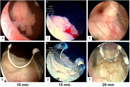
the patients into three groups with the margin of resection 10 mm, 15 mm and 20 mm away from the edge of resected tumors (Figure 1). Tumors with diameters less than 1 cm were resected en-bloc, and larger lesions were frac-tionated. In case of multiplicity, the same strat-egy or resection margin was applied for each tumor. For pathological reporting of positive surgical margin, the last circumferential TURBT chip furthest away from the tumor was sepa-rately submitted for histology. All pathology
specimens were initially evaluated by three junior pathologists and later reviewed by a sin-gle senior pathologist.
One hundred and thirty-four (94%) patients underwent single instillation of chemotherapy (50 mg epirubicin hydrochloride/40 ml) within 24 h after TURBT. Nine patients (6%) with sus-pected bladder perforation underwent delayed instillation of intravesical chemotherapy on day
7 after TURBT. The patients were stratified and
underwent auxiliary treatment according to the postoperative risk (2012 European Association
of Urology Guidelines for risk stratification).
Low-risk patients no longer underwent chemo-therapy. Moderate or high-risk patients under-went intravesical instillation chemotherapy,
once a week for the first 8 weeks, then once a
month for 10 months. All the patients were
fol-lowed up for 24 months [20]. The first surveil -lance cystoscopy was performed in the third month after surgery and then repeated once every 3 months. The follow-up was terminated if the tumor recurrence happened. Local
recur-rence single or multiple were defined as recur -rence in situ, if they occurred at the same loca-tion as the primary tumor after initial resecloca-tion, and recurrence if it occurred elsewhere in the bladder. For multiple lesions, recurrence was regarded as local recurrence if tumor appeared at the same location as one of the sites of pri-mary tumors [21]. The variables studies includ-ed the following: (1) postoperative pathological reports, (2) intra-operative bladder perforation or rupture and postoperative bleeding
evaluat-ed by using the Clavien-Dindo classification
system.
Statistical analysis
The parametric data are presented as the mean ± standard deviation. The categorical data is compared by Chi-square test. The Log-rank test was used to compare tumor recur-rence and recurrecur-rence in situ among the three groups. With tumor recurrence as a termination event, the tumor size, primary/recurrent, patho-logical stage, resection scope, positive/nega-tive margin, tumor recurrence, and recurrence
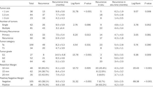
Table 1. Difference in tumor recurrence and recurrence in situ by Log-rank test in NMIBC patients
Total Recurrence Recurrence time (months) Log-Rank P value Recurrence in situ situ time (months)Recurrence in Log-Rank P value Tumor size
< 1 cm 36 13 9.9 ± 5.6 31.78 < 0.001 5 6.2 ± 1.9 9.57 0.008
1-3 cm 84 37 7.7 ± 4.9 18 5.4 ± 2.8
> 3 cm 23 19 4.2 ± 4.3 8 1.4 ± 0.5
Number of tumors
Single 62 26 9.0 ± 5.9 2.76 0.096 9 4.8 ± 1.3 3.78 0.052
Multiple 81 43 6.1 ±4.4 22 4.4 ± 3.4
Primary/Recurrence
Primary 83 33 7.5 ± 5.4 6.20 0.013 14 4.7 ± 4.0 3.05 0.081
Recurrence 60 36 6.9 ± 5.0 17 4.3 ± 1.8
Tumor category
Ta 109 49 8.2 ± 5.3 4.54 0.331 23 5.4 ± 2.8 0.74 0.390
T1 34 20 4.7 ± 3.9 8 1.9 ± 1.1
Tumor grade
G1 27 8 14.0 ± 6.8 15.78 < 0.001 3 9.0 ± 3.5 9.38 0.009
G2 52 21 8.5 ± 4.8 8 5.6 ± 3.0
G3 64 40 5.1 ± 3.5 20 3.4 ± 2.0
Resection Margin
10 mm 46 30 (65.2%) 6.1 ± 4.9 10.72 0.005 20 (43.4%) 4.3 ± 3.0 20.43 < 0.001
15 mm 62 24 (38.7%) 8.4 ± 5.4 8 (12.9%) 5.4 ± 3.2
20 mm 35 15 (42.9%) 7.4 ± 5.3 3 (8.6%) 3.7 ± 1.5
Positive/Negative Margin
Negative 105 40 (38.1%) 9.0 ± 5.3 31.32 < 0.001 7 (6.7%) 5.6 ± 2.5 68.38 < 0.001
Armonk, NY, USA). Statistical significance was
at P < 0.05. Results
Clinical characteristics
One hundred and forty-three patients with blad-der cancer were enrolled, comprising 123 males and 20 females, aged 31 to 82 years, with an average age of 57.13 years. They under-went TURBT and were followed up postopera-tively for 24 months, with the tumor recurrence as a termination event. Among the recruited patients, 83 were primary patients (58.0%) and 60 were recurrent patients (42.0%), including 62 cases with single tumor (43.4%) and 81 with multiple tumors (56.6%). Of these, 46 (32.2%), 62 (43.4%), and 35 (24.5%) cases had a mar-ginal width of 10 mm, 15 mm, and 20 mm, respectively; 109 cases (76.2%) were pTa, and
Cox multiple regression analysis of NMIBC recurrence and recurrence in situ
Cox regression analysis was performed, taking tumor recurrence as a termination event, with tumor size, primary/recurrent tumor, pathologi-cal stage, marginal width, and positive/nega-tive margin as covariates. The results showed that if the tumor was recurrent and tumor diam-eter > 3 cm, the positive postoperative margin was the independent risk factor, and the increase in marginal width was the indepen-dent protective factor affecting NMIBC recur-rence (Table 2). With tumor recurrecur-rence in situ
as a termination event, the tumor size, patho-logical stage, marginal width, and positive/ negative margin were analyzed as covariates. The results showed that positive postoperative margin was the independent risk factor, and an increase in marginal width was the indepen-Table 2. Multivariate Cox analysis on tumor recurrence
P value HR 95% CI
Tumor size < 0.001
1-3 cm vs. < 1 cm 0.900 0.954 0.456-1.996 > 3 cm vs. < 1 cm < 0.001 7.802 2.980-20.428 Primary vs. Recurrence 0.002 2.665 1.437-4.945
Tumor Grade 0.184
G2 vs. G1 0.805 1.125 0.411-2.873
G3 vs. G1 0.203 1.845 0.719-4.733
Resection Margin < 0.001
15 mm vs. 10 mm 0.001 0.346 0.186-0.646 20 mm vs. 10 mm < 0.001 0.267 0.128-0.556 Positive margin vs. Negative margin 0.049 1.781 1.002-3.168 HR: Hazard ratio, CI: Confidence interval.
34 cases (23.8%) were pT1 stage. Moreover, 27 (18.9%), 52 (36.4%), and 64 (44.8%) cases were patho-logical grade G1, G2, and G3, re- spectively; and 38 cases (26.6%) had a positive margin. The rates of positive marginal in the three groups (10 mm, 15 mm, and 20 mm) were 43.4% (20/46), 19.4% (12/62), and 17.1% (6/35), res- pectively. Furthermore, 69 cases (48.3%) developed recurrence and 31 cases (21.7%) had in situ
recurrence.
Difference in tumor recurrence and recurrence in situ among groups
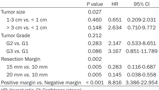
[image:5.612.90.358.84.252.2]The Log-rank test of tumor recur-rence and recurrecur-rence in situ am- ong the tested groups was per-formed (Table 1). The results showed that tumor size, primary/ recurrent tumor, pathological sta- ge, resection scope, and positive/ negative margin affected the blad-der tumor recurrence (P < 0.05), whereas tumor size, pathological stage, resection scope, and posi-tive margin affected the tumor recurrence in situ (P < 0.05). Table 3. Multivariate Cox analysis on tumor recurrence in situ
P value HR 95% CI
Tumor size 0.027
1-3 cm vs. < 1 cm 0.460 0.651 0.209-2.031 > 3 cm vs. < 1 cm 0.148 2.634 0.710-9.772
Tumor Grade 0.212
G2 vs. G1 0.283 2.147 0.533-8.651
G3 vs. G1 0.086 3.167 0.851-11.789
Resection Margin 0.002
[image:5.612.91.358.291.442.2]dent protective factor affecting NMIBC recur-rence in situ (P < 0.05) (Table 3).
Survival curve analysis of NMIBC patients by using the Kaplan-Meier estimator
With tumor recurrence as a termination event, the survival curve of patients with NMIBC hav-ing different resection marginal width and the positive/negative margin was plotted using the Kaplan-Meier estimator. The tumor recurrence
rate was significantly higher for patients with
10 mm marginal width compared with those with 15 mm and 20 mm marginal width (P =
0.005). The recurrence rate was also signifi -cantly higher for patients with positive margin compared with those with negative margin (P <
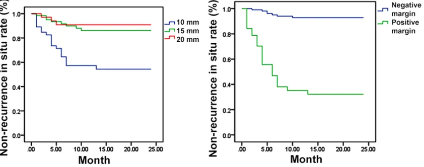
With tumor recurrence in situ as a termination event, the survival curve of patients with NMIBC having different resection marginal width and the positive/negative margin was plotted. The rate of tumor recurrence in situ was significant -ly higher in patients with 10 mm marginal width compared with those with 15 mm and 20 mm marginal width (P < 0.001). The rate of tumor recurrence in situ was similarly more in patients with positive margin compared with those with negative margin (P < 0.000) (Figure 3).
Patterns for peri-operative factors to margin positive rate and width of resection margin
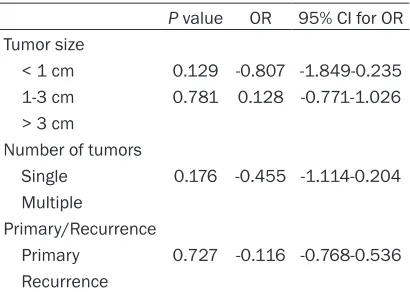
[image:6.612.100.520.74.241.2]No statistic correlation had been found bet-
Figure 2. Kaplan-Meier estimator survival curve estimates of 24-month recurrence-free survival for three different resection marginal width groups (Left), and for positive or negative resection margin groups (Right) (tumor recur-rence as the termination event).
[image:6.612.97.518.307.473.2]ry/recurrence to the width of resection margin in a multivariate logistic regression model (Table 4). And for Ta and T1 groups, resection margin positive rate and width of resection margin were not statistically different (P = 0.668, P = 0.968). However, the margin
posi-tive rate was influenced by grade of the tumor
(P < 0.001), which meant high-grade bladder tumor was likely to have a higher margin posi-tive rate (Table 5).
Incidence of complications in patients with dif-ferent marginal width
The Clavien-Dindo postoperative complication grading system was employed to evaluate dif-ferent groups. No grade IV or V case was found in this study. The results showed that the surgi-cal bleeding rate increased with the growth of marginal width. The bladder perforation or
rup-ture rate was significantly (P = 0.041) higher for patients with 20 mm margin compared with those with 10 mm and 15 mm margins. The incidence of complications increased in patients with 20 mm margin who underwent TURBT (Table 6).
Discussion
TURBT is a mini-invasive and highly repeatable standard surgical procedure for treatment of NMIBC. A high-quality TURBT should strictly comply with the principle of achieving negative
resection margin, which is of great significance
to improve the outcome. However, due to fac-tors like tumor size, single/multiple tumors, pri-mary/recurrent tumor, and surgeon’s surgical
skills, about 70% patients are found to have recurrent tumor after initial resection. Previous clinical research mostly focused on the resec-tion depth of TURBT to ensure the detrusor muscular tissues were obtained in the speci-mens. We acknowledge the importance of the resection depth. However, we also consider that the width of margin of resection surround-ing the tumor has long been overlooked. There is a recent interest in the width of peripheral tumor margin surrounding a resection area as an important factor to prevent local recurrence [20]. Jancke et al. conducted a 3 years follow-up study of TURBT oncological outcomes and demonstrated that the positive margin rate was 26%, and the recurrence rate of patients with positive margin was 83%. On the contrary, the recurrence rate of patients with negative margin was 57% [21]. The rate of recurrence in situ was 58% for patients with positive margin, while it was only 19% for those with negative margin. Ectopic recurrence was found in 39%
vs. 25% cases with positive and negative
mar-gin, respectively. According to the findings pre -sented in this work, they provided novel insight into the role of positive margin as a risk factor causing the tumor recurrence and recurrence
in situ in patients with NMIBC. Another study conducted by Jancke et al. found that the tumor maximum diameter > 3 cm was related to ecto-pic tumor recurrence and recurrence in situ
[22]. In the current study, we noted that posi-tive margin, primary tumor, and tumor diameter > 3 cm were independent predictive risk fac-tors for NMIBC tumor recurrence. Furthermore, the tumor diameter > 3 cm was also an inde-pendent risk factor predicting NMIBC recur-rence in situ. In our study, the rates of tumor recurrence and recurrence in situ were signifi -cantly higher in patients with positive margin than those with negative margin (recurrence rate 76.3% vs. 38.1%, P < 0.001; the rate of re- currence in situ was 63.2% vs. 6.7%, P < 0.001).
These are highly consistent with the findings of
the previous studies which suggested that the positive margin was an important risk factor contributing to the higher recurrence and recur-rence in situ of bladder cancer. Thus, in short, positive lesions in the margin may be a proxy variable. Richterstetter et al. expanded the width and depth of margin (expanded TUR) and noted that the total tumor margin residual rate was 22%, and about 94% of the residues were found in the margin rather than in the base-Table 4. Multivariate logistic regression model
for factors on different width of resection margin
P value OR 95% CI for OR Tumor size
< 1 cm 0.129 -0.807 -1.849-0.235 1-3 cm 0.781 0.128 -0.771-1.026 > 3 cm
Number of tumors
Single 0.176 -0.455 -1.114-0.204 Multiple
Primary/Recurrence
Primary 0.727 -0.116 -0.768-0.536 Recurrence
[image:7.612.91.296.107.253.2]ment [17]. Furthermore, expanded TUR could pronouncedly reduce the tumor margin positive rate, as well as the rate of tumor recurrence in situ to 5.1%. These results had unambiguously demonstrated that adequate width, as well as depth during TURBT, was necessary to reduce the tumor recurrence. In our study, we also
noted a significant difference in the postopera -tive recurrence rate among 10 mm, 15 mm and 20 mm groups (38.7% vs. 65.2%, P = 0.001; 42.9% vs. 65.2%, P < 0.001); hazard ratios (HR) were 0.346 and 0.267, respectively. Multivari- ate relating to the recurrence in situ was
ana-lyzed by Cox regression. Significant differences
were found in the postoperative recurrence in situ among 10 mm, 15 mm and 20 mm groups (12.9% vs. 43.4%, P = 0.005; 8.5% vs. 43.4%, P
= 0.005). Similar patterns were found in HR as 0.283 and 0.145, respectively. This strongly suggests that the increase in marginal width plays as an independent protective role in pre-venting both the recurrence and recurrence in situ of NMIBC.
The tumor recurrence rates and recurrence in situ rates in patients with 15 mm and 20 mm margins were compared in current study, and we noted that the tumor recurrence rates for the two groups were 38.7% vs. 42.8%, respec-tively (P > 0.05), while the rates of tumor
recur-vs. 30.6% and 14.3% vs. 3.2%, respectively; P
= 0.041). Overall, these findings reveal that the
20 mm marginal resection would increase the risk of bladder perforation or rupture and post-operation bleeding rates without promising fur-ther advantage of reduction in tumor recur-rence rate, whereas the 15 mm margin rese-
ction might be sufficient to reduce the rates of
tumor recurrence and recurrence in situ
with-out leading a significant increase in the occur -rence of postoperative complications. Hence, we propose that the 15 mm margin resection should be considered as a potential standard for TURBT. In particular, fulguration around the margin as mentioned before may reduce tumor dissemination and recurrence by sealing the blood vessels and achieving further tumor necrosis, without increasing the risk of postop-erative complications.
In the last several years, the re-TURBT, per-formed in 2-6 weeks after TURBT, has come to the forefront of attention for NMIBC. The
poten-tial benefits reported included ensuring com -plete tumor removal, re-staging as well as pre-venting postoperative tumor recurrences [5, 23, 24]. Ali et al. reported that patients with multiple tumors and those with maximum
diam-eter > 3 cm benefited from re-TURBT [25]. Our
study proposes that patients with tumor residu-Table 5. Patterns for T stage and grade to margin positive rate and
width of resection margin
Margin Resection Margin
Negative Positive P value 10 mm 15 mm 20 mm P value
T stage 0.668* 0.968**
Ta 81 28 35 48 26
T1 24 10 11 14 9
Grade < 0.001** 0.061**
G1 24 3 15 9 3
G2 45 7 13 26 13
G3 36 28 18 27 19
[image:8.612.90.379.95.214.2]*Chi-square test, **Fisher exact test, statistical significance is at P < 0.05.
Table 6. Comparison of surgical complications in different resec-tion margins
Resection Margin Total Bleeding Bladder perforation I II III Total Rate% I II III Total Rate%
10 mm 46 7 2 1 10 21.7 0 2 0 2 4.3
15 mm 62 17 1 1 19 30.6 0 1 1 2 3.2
20 mm 35 8 1 5 14 40.0 0 2 3 5 14.3
rence in situ were 12.9% vs. 8.5%, respectively. Nonethe-
less, no significant difference
was found between the two groups (P > 0.05).
[image:8.612.91.376.275.343.2]al on margins might also benefit from re-TURBT.
However, this was not the objective of the cur-rent study.
The limitations of this study should also be noted. Firstly, being a retrospective study, selection bias was possible. The patients enrolled in this study were not randomly divid-ed, but according to the surgical methods of different urologists. Secondly, in the multiple linear regression analysis, the effect of multi-collinearity among variables between tumor size, single/multiple tumors, primary/recurrent tumor, clinical stage, marginal width, pathologi-cal type, surgipathologi-cal skills and learning curve of surgeons cannot be eliminated and may inter-fere within prediction of recurrence rate. Thirdly, this study was a single-center study; hence, the lack of multicenter data, to a certain extent,
reduced the significance of the present find -ings. In addition, the follow-up time in this study
was limited. Thus, the results may only reflect
the outcome of the relationship between the marginal width and the short-term prognosis for patients who underwent TURBT.
Acknowledgements
This study was supported by National Natural Sciences Foundation of China (No. 81302219).
Disclosure of conflict of interest
None.
Address correspondence to: Dr. Fan Li, Department of Urology, Tongji Hospital, Tongji Medical College, Huazhong University of Science and Technology, No. 1095 Jiefang Ave., Wuhan 430030, Hubei, P. R. China. Tel: +86-1388-616-9740; Fax: +86-027-83- 662851; E-mail: [email protected]
References
[1] Siegel R, Naishadham D and Jemal A. Cancer statistics, 2013. CA Cancer J Clin 2013; 63: 11-30.
[2] Okamura T, Ando R, Akita H, Kawai N, Tozawa K, Kohri K and Arano H. Prophylactic effects of Bacillus Calmette-Guerin intravesical instilla-tion therapy: time period-related comparison between Japan and Western Countries. Curr Urol Rep 2014; 15: 374.
[3] Legrand G, Soliman H, Dubosq F, Verine J, Des-grandchamps F, de The H, Mongiat-Artus P and Ploussard G. Prevalence and spectrum of mic-rosatellite alterations in non-muscle invasive
bladder cancers. Am J Cancer Res 2011; 1: 595-603.
[4] Kurth KH, Denis L, Bouffioux C, Sylvester R, De-bruyne FM, Pavone-Macaluso M and Ooster-linck W. Factors affecting recurrence and pro-gression in superficial bladder tumors. Eur J Cancer 1995; 31A: 1840-1846.
[5] Adiyat KT, Katkoori D, Soloway CT, De los San-tos R, Manoharan M and Soloway MS. “Com-plete transurethral resection of bladder tu-mor”: are the guidelines being followed? Urology 2010; 75: 365-357.
[6] Zurkirchen MA, Sulser T, Gaspert A and Hauri D. Second transurethral resection of superfi-cial transitional cell carcinoma of the bladder: a must even for experienced urologists. Urol Int 2004; 72: 97-102.
[7] Cho KS, Hwang TK, Kim BW, Yoon DK, Chang SG, Kim SJ, Park JY, Cheon J, Sung GT and Hong SJ. Differences in tumor characteristics and prognosis in newly diagnosed Ta, T1 uro-thelial carcinoma of bladder according to pa-tient age. Urology 2009; 73: 828-832. [8] Hu Z, Mudaliar K, Quek ML, Paner GP and
Bar-kan GA. Measuring the dimension of invasive component in pT1 urothelial carcinoma in transurethral resection specimens can predict time to recurrence. Ann Diagn Pathol 2014; 18: 49-52.
[9] Hoosein MM and Rajesh A. MR imaging of the urinary bladder. Magn Reson Imaging Clin N Am 2014; 22: 129-134.
[10] Mariappan P, Zachou A and Grigor KM. Detru-sor muscle in the first, apparently complete transurethral resection of bladder tumor speci-men is a surrogate marker of resection quality, predicts risk of early recurrence, and is depen-dent on operator experience. Eur Urol 2010; 57: 843-849.
[11] Jurewicz M and Soloway MS. Approaching the optimal transurethral resection of a bladder tumor. Turk J Urol 2014; 40: 73-77.
[12] Dalbagni G, Herr HW and Reuter VE. Impact of a second transurethral resection on the stag-ing of T1 bladder cancer. Urology 2002; 60: 822-824.
[13] Maruniak NA, Takezawa K and Murphy WM. Ac-curate pathological staging of urothelial neo-plasms requires better cystoscopic sampling. J Urol 2002; 167: 2404-2407.
[14] Dobruch J, Borówka A and Herr HW. Clinical value of transurethral second resection of bladder tumor: systematic review. Urology 2014; 84: 881-885.
[16] Cao M, Yang G, Pan J, Sun J, Chen Q, Chen Y, Chen H and Xue W. Repeated transurethral re-section for non-muscle invasive bladder can-cer. Int J Clin Exp Med 2015; 8: 1416-1419. [17] Richterstetter M, Wullich B, Amann K,
Haeber-le L, Engehausen DG, Goebell PJ and Krause FS. The value of extended transurethral resec-tion of bladder tumour (TURBT) in the treat-ment of bladder cancer. BJU Int 2012; 110: E76-79.
[18] Kolozsy Z. Histopathological “self control” in transurethral resection of bladder tumors. Br J Urol 1991; 67: 162-164.
[19] Herr HW and Donat SM. Quality control in transurethral resection of bladder tumours. BJU Int 2008; 102: 1242-1246.
[20] Chen SY, Du LD and Zhang YH. Pilot study of intravesical instillation of two new generation anthracycline antibiotics in prevention of su-perficial bladder cancer recurrence. Chin Med J (Engl) 2010; 123: 3422-3426.
[21] Jancke G, Rosell J and Jahnson S. Residual tu-mour in the marginal resection after a com-plete transurethral resection is associated with local recurrence in Ta/T1 urinary bladder cancer. Scand J Urol Nephrol 2012; 46: 343-347.
[22] Jancke G, Damm O, Rosell J and Jahnson S. Risk factors for local recurrence with pTa/pT1 urinary bladder cancer. Scand J Urol Nephrol 2008; 42: 417-421.
[23] Divrik RT, Sahin AF, Yildirim U, Altok M and Zorlu F. Impact of routine second transurethral resection on the long-term outcome of patients with newly diagnosed pT1 urothelial carcinoma with respect to recurrence, progression rate, and disease specific survival: a prospective randomized clinical trial. Eur Urol 2010; 58: 185.
[24] Herr HW and Donat SM. A re-staging transure-thral resection predicts early progression of superficial bladder cancer. BJU Int 2006; 97: 1194-1198.