HYBRID IMAGING: A COMPLEMENTARY EFFECT ON HEAD
*Dr. Nikhat Mukhtar Gazge, Dr. Balaji, P., Dr. Sowbhagya, M
Department of Oral Medicine and Radiology, Rajarajeswari Dental College and Hospital, #14 Ramohalli Cross,
Mysore Road, Bangalore
ARTICLE INFO ABSTRACT
It is well known that cross
tomography (CT), magnetic resonance imaging (MRI)
diagnosis and in tumour treatment strategies. Likewise, nuclear med
emission tomography (PET) and single photon emission computerized tomography (SPECT) are unparalleled in their ability to assess information about metabolic function. Despite their advantages over conventional imaging, the
can be overcome by combining anatomical and functional imaging techniques. These techniques have different working principles and consequently complement each other with respect to the information obtained. The combination (fusion) of two
defining the so
techniques (ultrasound with CT or MR imaging), as well as associations between anatomical (CT or MR imaging) and molecular (SPECT or PET) imaging modalities are currently being used in clinical practice.
arena of head and neck cancers.
Copyright © 2015 Dr. Nikhat Mukhtar Gazge et al. This
unrestricted use, distribution, and reproduction in any medium, provided the original work is properly cited.
INTRODUCTION
From advances in X-ray films and cassettes to introduction of computers and digital images, diagnostic imaging has never stopped reinventing its technology to improve patient care. Today, diagnostic imaging is on the cusp of explosive growth in an arena known as “Fusion imaging”. This technology merges two independent imaging modalities
demonstrates an organ’s anatomy and the other that demonstrates the organ’s function- to produce a diagnostically and clinically superior diagnostic study. Curren
imaging has found its application in many areas of medicine. However, this review paper highlights the clinical applications of fusion imaging in head and neck cancers.
Methods of image fusion
Image fusion can be performed at 3 different levels:
1. Visual fusion
2. Software fusion
3. Hardware fusion
In traditional visual image fusion, the physician compares 2 separate imaging modalities viewed next to each other and the
*Corresponding author: Dr. Nikhat Mukhtar Gazge
Department of Oral Medicine and Radiology, Rajarajeswari Dental College and Hospital, #14 Ramohalli Cross, Mysore Road, Bangalore – 560074, Karnataka, India.
ISSN: 0975-833X
Article History:
Received 17th December, 2014
Received in revised form 26th January, 2015
Accepted 23rd February, 2015
Published online 17th March, 2015
Key words: Fusion imaging, Hybrid imaging, PET/CT.
RESEARCH ARTICLE
HYBRID IMAGING: A COMPLEMENTARY EFFECT ON HEAD AND NECK ONCOLOGY
*Dr. Nikhat Mukhtar Gazge, Dr. Balaji, P., Dr. Sowbhagya, M. B. and Dr. Yogesh Pawar
Department of Oral Medicine and Radiology, Rajarajeswari Dental College and Hospital, #14 Ramohalli Cross,
Mysore Road, Bangalore – 560074, Karnataka, India
ABSTRACT
It is well known that cross-sectional anatomical imaging techniques such as ultrasound, computed tomography (CT), magnetic resonance imaging (MRI) etc. have important roles in non
diagnosis and in tumour treatment strategies. Likewise, nuclear med
emission tomography (PET) and single photon emission computerized tomography (SPECT) are unparalleled in their ability to assess information about metabolic function. Despite their advantages over conventional imaging, they have their own limitations in oncologic imaging.
can be overcome by combining anatomical and functional imaging techniques. These techniques have different working principles and consequently complement each other with respect to the information obtained. The combination (fusion) of two imaging techniques has been developed in recent years, defining the so-called “hybrid techniques’’ or “fusion imaging”. Combinations of anatomical imaging techniques (ultrasound with CT or MR imaging), as well as associations between anatomical (CT or imaging) and molecular (SPECT or PET) imaging modalities are currently being used in clinical practice. This paper highlights the fusion of various imaging modalities and their applications in the arena of head and neck cancers.
This is an open access article distributed under the Creative Commons Att use, distribution, and reproduction in any medium, provided the original work is properly cited.
ray films and cassettes to introduction of computers and digital images, diagnostic imaging has never stopped reinventing its technology to improve patient care. Today, diagnostic imaging is on the cusp of explosive growth own as “Fusion imaging”. This technology merges two independent imaging modalities- one, that demonstrates an organ’s anatomy and the other that to produce a diagnostically and clinically superior diagnostic study. Currently, fusion imaging has found its application in many areas of medicine. However, this review paper highlights the clinical applications
Image fusion can be performed at 3 different levels:
In traditional visual image fusion, the physician compares 2 separate imaging modalities viewed next to each other and the
Mukhtar Gazge,
Department of Oral Medicine and Radiology, Rajarajeswari Dental College and Hospital, #14 Ramohalli Cross, Mysore Road, Bangalore
fusion takes place in his or her mind. Interpreting images obtained on two different modalities side by side is time consuming and logistically demanding. It also results in diagnostic inaccuracy because of imperfect anatomical matching (Becker and Zaidi, 2014)
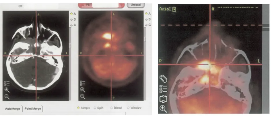
uses anatomic landmarks to coregister images from separate CT or MRI scanners to the images acquired from PET or SPECT. Softwares (Figure 1) for image fusion have been developed by various vendors and is
all sorts of image sets. It provides evaluation of 2 modalities in 1 integrated image set. Currently,
more common because it is less expensive and more readily available.
Until recently radiologists had to obtain physiological and anatomical information on separate machines and use special registration softwares to digitally superimpose the two images. Today, hybrid equipment are available that are capable of performing both types of examination simultaneously, automatically merging the data to form a composite image
(Clarke et al., 2002).The scanners for the different imaging
modalities are combined within the same equipment. This makes it possible to sequentially acquire SPECT/PET and CT/MRI in a single imaging session. By uniting
function with anatomic form, fusion imaging depicts the human body with a level of precision never achievable before with fusion of clinically significant anatomic and metabolic data.
International Journal of Current Research
Vol. 7, Issue, 03, pp.13309-13315, March, 2015
INTERNATIONAL
NECK ONCOLOGY
B. and Dr. Yogesh Pawar
Department of Oral Medicine and Radiology, Rajarajeswari Dental College and Hospital, #14 Ramohalli Cross,
sectional anatomical imaging techniques such as ultrasound, computed . have important roles in non-invasive diagnosis and in tumour treatment strategies. Likewise, nuclear medicine procedures such as positron emission tomography (PET) and single photon emission computerized tomography (SPECT) are unparalleled in their ability to assess information about metabolic function. Despite their advantages y have their own limitations in oncologic imaging. These limitations can be overcome by combining anatomical and functional imaging techniques. These techniques have different working principles and consequently complement each other with respect to the information imaging techniques has been developed in recent years, called “hybrid techniques’’ or “fusion imaging”. Combinations of anatomical imaging techniques (ultrasound with CT or MR imaging), as well as associations between anatomical (CT or imaging) and molecular (SPECT or PET) imaging modalities are currently being used in clinical This paper highlights the fusion of various imaging modalities and their applications in the
is an open access article distributed under the Creative Commons Attribution License, which permits
takes place in his or her mind. Interpreting images obtained on two different modalities side by side is time consuming and logistically demanding. It also results in diagnostic inaccuracy because of imperfect anatomical , 2014). The software technique uses anatomic landmarks to coregister images from separate to the images acquired from PET or ) for image fusion have been developed by various vendors and is universally applicable to l sorts of image sets. It provides evaluation of 2 modalities in 1 integrated image set. Currently, the software technique is common because it is less expensive and more readily
The first commercial system to combine functional and anatomic imaging capabilities was a SPECT-CT unit introduced commercially in 1998. In 1999, manufacturers began working on Hybrid PET-CT system and the first
commercial PET-CT unit was introduced in 2000 (Clarke
et al., 2002). Several ultrasound systems for fusion imaging of real-time ultrasound with CT or MRI or PET/CT are commercially available. All systems are based on an electromagnetic tracking system—a transmitter and a small sensor mounted on the ultrasound probe— that provides the position and orientation of the transducer in the transmitter’s spatial volume. The previously recorded CT or MRI or PET/CT or MRI dataset is transferred to the ultrasound system, and a coregistration from external or internal markers is performed. Afterward, the CT, MRI, or PET/CT or MRI dataset is reformatted in a projection to fit the real-time ultrasound image. The images may be shown side by side or
superimposed (Ewertsen et al., 2013).
Application of fusion imaging in Head and Neck cancer
Head and neck cancer is the sixth most common cancer in the world with approximately 640,000 cases per year with
approximately 350,000 deaths per year (Ghosh, 2012).With
such a significant prevalence, oncologic imaging plays an important role in head and neck cancers. The imaging findings can aid significantly in detection, staging, restaging, and therapy response assessment of these tumours. Accurate staging at the time of diagnosis is critical for selection of the appropriate treatment strategy. After therapy, early detection of recurrence is also critical to achieve an optimal outcome. Imaging modalities such as Computerized tomography (CT), Magnetic resonance imaging (MRI) are superior at depicting normal anatomy and anatomic changes. They are the standard conventional imaging modalities for evaluation of patients
with head and neck cancer (El-Khodary et al., 2011). These
anatomic imaging modalities, however, are based on morphologic diagnostic criteria, such as nodal size and contrast enhancement patterns that do not always accurately reflect the presence of active malignancy. Moreover, they may be unable to differentiate between normal and pathologic tissues with
similar densities. They provide relatively little information about the viability or metabolic activity of organs and lesions, thus lacking sufficient sensitivity and specificity to answer a number of important clinical questions. Well-known examples are the differentiation of viable tumour from scar tissue after external beam radiation or chemotherapy and the detection of metastases in normal-size lymph nodes. The regional anatomy is distorted by surgery and/or radiation making the distinction between post-treatment changes and recurrence or residual
tumour difficult with imaging tests (El-Khodary et al., 2011).
Hence, anatomical imaging modalities have a limited accuracy in response assessment after treatment of head and neck cancers as well as early diagnosis of recurrence.
On the other hand, nuclear medicine procedures such as positron emission tomography (PET) and single photon emission computerized tomography (SPECT) are unparalleled in their ability to assess information about metabolic function. Fluorine-18 fluorodeoxyglucose (FDG) positron emission tomography (PET), a functional imaging modality, is based on its capability to assess the metabolic status of tumours. It permits the differentiation of viable malignant tissue or active infection from normal tissue and from nonviable remnants by direct visualization of metabolic activity in vivo. Other tracers currently under development may prove useful in visualizing other important parameters, such as DNA synthesis, mitotic activity, protein synthesis, local ischemia, and expression of tumour-specific receptors. Thallium-201 (201Tl) SPECT has unique features that enable it to reveal metabolically active tissue by virtue of its cellular uptake by malignant cells. PET and SPECT are superior to CT and MRI in diagnosing and differentiating recurrence from post radiation effects and surgical scars in sites of tumours of the head and neck and also in the detection of cervical lymph node status in cases of head and neck cancer. However, they are limited by the lack of anatomic landmarks, and the precise localization of suspicious findings is difficult due to the low background tracer
uptake.5Despite high contrast resolution, their major
[image:2.595.68.536.58.258.2]structures. This may result in uncertainty or even failure in correctly localizing detected abnormalities. In addition, variable degrees of physiologic and inflammatory non cancer-related uptake of FDG in the region of the head and neck mainly after treatment can confound interpretation of
suspicious foci (El-Khodary et al., 2011). As rightly said by
Sir Stephen Wainwright, “Structure without function is a corpse… and function without structure is a ghost”. The limitations in separate anatomic and functional imaging may be compensated for when the two modalities are used in a complementary way. High-resolution anatomic information produced by CT adds significant information to tissue characterization delivered by PET. In addition, fusion of high resolution MRI anatomic and functional information with PET will provide an extra dimension. When applying the integration of different imaging modalities, adequate anatomic alignment of both image sets is required. This permits convenient visualization of all information in one study.
The benefits of fusion imaging include: (Săftoiu and Vilmann,
2011 and Ghom et al., 2011)
Improved lesion characterization thereby increasing the
diagnostic accuracy. This is recognized as a beneficial diagnostic effect
Direct comparison of the lesions using different imaging
modalities
Better identification of small recurrent tumours obscured
by scar tissue at site of incipient radiation or postoperative necrosis
Detection of large tumours in clinically inaccessible areas
such as hypopharynx or maxilla
Improved lesion localization may lead to better results in
other successive diagnostic procedures (e.g., easier CT-guided biopsy)
Moreprecise monitoring of interventional procedures
Reduced radiation exposure
Hence, fusion imaging is an imaging modality with high diagnostic performance in assessment of head and neck cancer. Clinical applications of fusion imaging in the head and neck cancer include:
Detection of unknown primary tumour (Ghom et al., 2011
and Subramaniam et al., 2010)
Detection of synchronous second primaries (Subramaniam
et al., 2010)
Tumourstaging (Ghom et al., 2011 and Subramaniam
et al., 2010)
Diagnosis of distant metastases (Subramaniam et al., 2010)
Detection of residual or recurrent disease (Subramaniam
et al., 2010)
Emerging applications are:
Precise delineation of the tumour volume for radiation
therapyplanning(Ghom et al., 2011)
Evaluating treatment and providing prognostic information
(Ghom et al., 2011)
Reference tool for surgical planning (Al-Ibraheem et al.,
2009)
DISCUSSION
Staging of cancer
A literature survey on the use of 18F-FDG PET in head and neck cancer (HNC) compared to CT indicates that PET has a higher sensitivity (87% versus 62%) and specificity (89% versus 73%) for staging cancer. Addition of PET/CT to initial staging of patients with HNC has also been shown to have a measurable impact on the treatment selection. Table 1 shows various studies that compare the accuracy of FDG PET and PET/CT with CT and MRI for detection of lymph node metastases in head and neck squamous cell carcinoma
(HNSCC)(Al-Ibraheem et al., 2009). Table 2 shows various
studies evaluation the performance of FDG PET for the
detection of distant metastases and synchronous 2nd tumour in
HNC. These studies showed that PET detected distant metastases or 2nd primaries in up to 15.6% of the patients. The true positive findings were 82%. Moreover, PET showed a better accuracy once it was compared to conventional imaging
as demonstrated by Ng et al., 2009; Chua et al., 2009 and Liu
et al., 2007.
A study was conducted by Loeffelbein et al. (2014) to assess
the diagnostic value of retrospective PET-MRI fusion and to compare the results with side-by-side analysis and single modality use of PET and of MRI alone for locoregional tumour and nodal staging of head-and-neck cancer. The overall sensitivity/specificity for tumour staging for MRI, PET, side-by-side analysis and retrospective PET-MRI fusion was
79%/66%, 82%/100%, 86%/100% and 89%/100%,
respectively. The overall sensitivity/specificity for nodal staging on a patient basis for MRI, PET, side-by-side analysis and PET-MRI fusion was 94%/64%, 94%/91%, 94%/82% and 94%/82%, respectively. MRI, PET, side-by-side analysis and retrospective image fusion were associated with correct
diagnosis/over-staging/under-staging of N-staging in
70.4%/18.5%/11.1%, 81.5%/7.4%/11.1%, 81.5%/11.1%/7.4%
and 81.5%/11.1%/7.4%, respectively (Feichtinger et al., 2007).
Unknown primary
Cervical lymph node metastases from an unknown primary tumour account for approximately 1-2% of newly diagnosed head and neck cancers. In 5% to 80%, depending on the patient selection, the primary tumour are not be identified by physical examination and conventional imaging, including CT and/or MRI. Treatment of these patients often includes extensive fields of irradiation to include the entire pharyngeal mucosa, larynx, and bilateral neck. The wide-field irradiation reduces the risk of tumour recurrence. However, it also causes significant morbidity, particularly in terms of xerostomia. Therefore, the accurate identification of occult primary sites is important because the therapy can then be focused to the known site of origin, decreasing treatment-related morbidity, and improving therapeutic efficacy. Table 3 shows studies evaluating performance of FDG PET or PET/CT in detecting carcinoma of unknown primary in patients who presented with cervical lymph node metastases and negative or inconclusive standard workup. For this group, 18F-FDG PET and PET/CT detected the primary tumour in 51 of 180 patients (28%)
(Al-Ibraheem et al., 2009).
Table 1.
Author year Number of patients Tumour Subtypes Results Notes
Beak et al.(2009) 15 Periorbital PET/CT accuracy (98%) > CT 84% CT: 16 slice PET modified Tx in 39%
Roh et al.(2007) 167 HNSCC PET or PET/CT accuracy (92%-93%)
> CT/MR 85%-86%
PET/CT significantly better for detection of primary tumour
Gordin et al. (2007) 35 Nasopharyngeal PET/CT accuracy 91% > PET 80% >
CT 60%
Retrospective
PET/CT modified Tx in 57% Kim et al.(2007) 32 Oropharyngeal PET sensitivity 21% higher than
CT/MR (p< .05)
PET/CT significantly better for detection of primary tumour
Dammann et al. (2005) 79 Oral cavity and oropharynx PET accuracy 96% > MRI 94% > CT
92% Nonhybrid PET/CT used
Ng et al.(2006) 124 Oral cavity SCC PET accuracy 98.4% > CT/MR 87.1% Prospective
Table 2.
Author year Number of
patients
Positive PET
True positive (distant mets +
2nd primary) False positive Notes
Ng et al.(2009) 111 16 13/16 3/16 CT/MR detect 4/16
Chua et al. (2009) 68 6 5/6 1/6 CT + BS detect 4/6
Liu et al. (2007) 300 61 50/61 11/61
Yen et al.(2005) 118 32 24/32 8/32
Goerres et al. (2003) 34 8 7/8 1/8 PET modified Treatment in 15%
Sigg et al. (2003) 58 8 7/8 1/8 PET modified Treatment in 5%
Schwartz et al. (2003) 33 7 7/7 0/7
Total 722 138 113/138 25/138
Table 3.
Author year Number of patients All positive True positive False positive Percent detected by PET
Padovani et al. (2009) 13 9 7 2 54%
Silva et al. (2007) 25 9 3 6 12%
Fakhry et al. (2006) 20 10 7 3 35%
Wong and Saunders (2003) 17 8 5 3 29%
Fogarty et al. (2003) 21 6 1 5 5%
Johansen et al. (2002) 42 20 10 10 24%
Kresnik et al. (2001) 15 12 11 1 73%
Jungehulsing et al.(2000) 27 7 7 0 26%
Total 180 81 51 30 28%
Table 4.
Authors year Number of patients Sensitivity Specificity Accuracy Notes
Abgral et al. (2009) 91 100% 85% 90% FDG PET/CT
Wang et al. (2009) 44 100% 98% 98% Prospective PET performance > CT
Cermik et al. (2007) 50 83% 93%
Alvarez perez et al. (2007) 60 98% 90% Prospective
Salaun et al. (2007) 30 100% 95% 97%
Goerres et al. (2004) 26 91% 93% Prospective
Kubota et al. (2004) 36 90% 78% Prospective
[image:4.595.54.549.182.740.2]Accuracy significantly higher than CT/MR
Table 5.
Author year Number of patients Study type Results Notes
Soto et al. (2008) 61 (9 LRF) Retrospective 8/9 LRF within BTV-PET
Rothschild et al. (2007) 45 Case-control analysis PET/CT with IMRT improved cure rates Advanced pharyngeal
carcinoma
Wang et al. (2006) 28 Prospective PET/CT-based GTV significantly different from
CT scans alone in 50% of cases
PET/CT upgraded T and N stage in 18 cases
Breen et al. (2007) 10 No significant differences in the GTVs between
PET/CT and CT alone
CT volumes were larger than PET/CT
El-Bassiouni et al. (2007) 25 PET/CT based volume significantly smaller
than CT
Koshy et al. (2005) 36 Retrospective TNM changed in 36%, RT volume and dose
changed in 14%
Heron et al. (2004) 21 Prospective PET/CT improves delineation of normal tissues
from tumour areas
PET/CT improves staging
Ciernik et al. (2003) 12 HNC of 39 Retrospective PET/CT changed GTV in 50% compared to CT
Nishioka et al. (2002) 21 PET improves GTV, normal tissue sparing PET alone
Recurrence
18F-FDG PET and PET/CT have a high sensitivity and moderate specificity for detecting recurrent disease at the primary tumour site, regional lymph node metastases and distant metastases. Several studies on the utility of PET or 18F-FDG PET/CT for the detection of recurrence are
summarized in Table 4 (Al-Ibraheem et al., 2009).
Radiation treatment planning
New high-precision radiotherapy (RT) techniques, such as intensity-modulated radiation therapy (IMRT), 3-dimensional conformal radiotherapy (3D-CRT), and proton beam therapy allow conformal treatment of tumour and to avoid unacceptable damage to normal tissues leading to possible improvement of tumour control and decrease of treatment-related toxicity. These techniques depend on imaging modalities allowing accurate tumour volume delineation and response assessment during treatment. The potential application of 18F-FDG PET/CT forintensity modulated radiation therapy (IMRT) planning is an area of major interest. Table 5 summarizes recent studies on the use of 18F-FDG PET
and PET/CT in radiotherapy planning (Al-Ibraheem et al.,
2009).
Conclusion
The additional information provided by image fusion significantly improves diagnostic accuracy and localization of malignant lesions and is superior to conventional anatomical and functional imaging techniques especially in areas of complex anatomy. There is improved utilization of patient data, thereby improving patient care. Hybrid technology has the potential to revolutionize diagnostics and has emerged as a promising diagnostic tool. Their clinical value for HNC has not been fully defined yet and must be analysed systematically. Further detailed clinical studies and imaging are required to substantiate their performance in oncologic imaging.
REFERENCES
A´ lvarezPe´rez, RM., Borrego Dorado, I., Ruiz Franco- Baux, JV. and V´azquez Albertino, RJ. 2007. Evaluation of efficacy and clinical impact of positron emission tomography with 18F fluoro-deoxyglucose in patients with suspicion of recurrent head and neck cancer or
distant metastases. Revista Espanola deMedicina Nuclear;
26(1): 30–39.
Abgral, R., Querellou, S., Potard, G. et al. 2009. Does
18F-FDG PET/CT improve the detection of posttreatment recurrence of head and neck squamous cell carcinoma in patients negative for disease on clinical follow-up?
Journal of Nuclear Medicine; 50(1): 24–29.
Al-Ibraheem, A., Buck, A., Krause, BJ., Scheidhauer, K. and Schwaiger, M. 2009. Clinical Applications of FDG PET
and PET/CT in Head and Neck Cancer. Journal of
Oncology.
Baek, CH., Chung, MK., Jeong, HS. et al. 2009. The clinical
usefulness of 18F-FDG PET/CT for the evaluation of lymph node metastasis in periorbital malignancies.
Korean Journal of Radiology; 10 (1):1–7.
Becker, M. and Zaidi, H. 2014. Imaging in head and neck squamous cell carcinoma: The potential role of PET/MRI. BJR; 87.
Breen, SL., Publicover, J., De Silva, S. et al. 2007.
Intraobserver and interobserver variability in GTV delineation on FDGPET-CT images of head and neck
cancers. International Journal of Radiation Oncology
Biology Physics; 68(3):763–770.
Cermik, TF., Mavi, A., Acikgoz, G., Houseni, M., Dadparvar, S. and Alavi, A. 2007. FDG PET in detecting primary and
recurrent malignant salivary gland tumors. Clinical
Nuclear Medicine; 32(4):286–291.
Chua, ML., Ong, SC., Wee, JT. et al. 2009. Comparison of 4
modalities for distant metastasis staging in endemic
nasopharyngeal carcinoma. Head and Neck; 31(3):346–
354.
Ciernik, IF., Dizendorf, E., Baumert, BG. et al. 2003.
Radiation treatment planning with an integrated positron emission and computer tomography (PET/CT): A
feasibility study. International Journal of Radiation
Oncology Biology Physics; 57(3):853–863.
Clarke, M. et al. 2002. Fusion Imaging: A new type of
technologist for a new type of technology. J Nuclear Med
Tech; 30 (4): 201-204.
Dammann, F, Horger, M., Mueller-Berg, M. et al. 2005.
Rational diagnosis of squamous cell carcinoma of the head and neck region: comparative evaluation of CT, MRI, and
18FDG PET. American Journal of Roentgenology; 184
(4):1326–1331.
El-Bassiouni, M., Ciernik, IF., Davis, JB. et al. 2007. [18FDG]
PET-CT-based intensity-modulated radiotherapy
treatment planning of head and neck cancer. International
Journal of Radiation Oncology Biology Physics; 69(1): 286–293.
El-Khodary, M., Tabashy, R., Omar, W., Mousa, A. and Mostafa, A. 2011. The role of PET/CT in the management
of head and neck squamous cell carcinoma. The Egyptian
Journal of Radiology and Nuclear Medicine; 42: 157–167.
Ewertsen, C., Săftoiu, A., Gruionu, LG., Karstrup, S. and Nielsen, MB. 2013. Real-Time Image Fusion Involving Diagnostic Ultrasound. AJR; 200: W249-W255.
Fakhry, N., Jacob, T., Paris, J. et al. 2006. Contribution of
18-FFDG PET for detection of head and neck carcinomas
with an unknown primary tumor. Ann Otolaryngol Chir
Cervicofac; 123(1):17–25.
Feichtinger, M., Aigner, RM., Santler, G. and Karcher, H. 2007. Case Report: Fusion of positron emission tomography (PET) and computed tomography (CT) images for image-guided endoscopic navigation in maxillofacial surgery: Clinical application of a new
technique. Journal of Cranio-Maxillofacial Surgery;
35:322–328.
Fogarty, GB., Peters, LJ., Stewart, J., Scott, C., Rischin, D. and Hicks, RJ. 2003. The usefulness of fluorine 18-labelled deoxyglucose positron emission tomography in the investigation of patients with cervical lymphadenopathy
from an unknown primary tumor. Head and Neck;
25(2):138–145.
Ghom, S. et al. 2011. Fusion Imaging: The double impact.
JIAOMR; 23(3): 225-228.
Ghosh, P. 2012. PET/CT in Radiation Therapy Planning for Head and Neck Cancer. Imaging life.
Goerres, GW., Schmid, DT., Bandhauer, F. et al. 2004.
Positron emission tomography in the early follow-up of
advanced head and neck cancer. Archives of
Otolaryngology; 130(1):105–109.
Goerres, GW., Schmid, DT., Gratz, KW., Von Schulthess, GK. and Eyrich, GK. 2003. Impact of whole body positron emission tomography on initial staging and therapy in patients with squamous cell carcinoma of the oral cavity.
Oral Oncology; 39(6):547–551.
Gordin, A., Golz, A., Daitzchman, M. et al. 2007. Fluorine-18
fluorodeoxyglucose positron emission tomography/
computed tomography imaging in patients with carcinoma of the nasopharynx: diagnostic accuracy and impact on
clinical management. International Journal of Radiation
Oncology Biology Physics; 68 (2):370–376.
Heron, DE., Andrade, RS., Flickinger, J. et al. 2004. Hybrid
PETCT simulation for radiation treatment planning in head-and neck cancers: a brief technical report.
International Journal of Radiation Oncology Biology Physics; 60(5):1419–1424.
Johansen, J., Eigtved, A., Buchwald, C., Theilgaard, SA. and Hansen, HS. 2002. Implication of 18F-fluoro-2-deoxy-Dglucose positron emission tomography on management of carcinoma of unknown primary in the head and neck:
ADanish cohort study. Laryngoscope; 112(11):2009–
2014.
Jungehulsing, M., Scheidhauer, K., Damm, M. et al. 2000.
2[F]-fluoro-2-deoxy-D-glucose positron emission
tomography is a sensitive tool for the detection of occult primary cancer (carcinoma of unknown primary syndrome) with head and neck lymph node manifestation.
Otolaryngology; 123(3):294–301.
Kim, MR., Roh, JL., Kim, JS. et al. 2007. Utility of
18Ffluorodeoxyglucose positron emission tomography in the preoperative staging of squamous cell carcinoma of
the oropharynx. European Journal of Surgical Oncology;
33(5):633–638.
Koshy, M., Paulino, AC., Howell, R., Schuster, D., Halkar, R. and Davis, LW. 2005. F-18 FDG PET-CT fusion in radiotherapy treatment planning for head and neck cancer.
Head and Neck; 27(6):494–502.
Kresnik, E., Mikosch, P., Gallowitsch, HJ. et al. 2001.
Evaluation of head and neck cancer with 18F-FDG PET:
A comparison with conventional methods. European
Journal of Nuclear Medicine; 28(7):816–821.
Kubota, K., Yokoyama, J., Yamaguchi, K. et al. 2004.
FDGPET delayed imaging for the detection of head and
neck cancer recurrence after radio-chemotherapy:
comparison with MRI/CT. European Journal of Nuclear
Medicine and Molecular Imaging; 31(4):590–595.
Liu, FY., Lin, CY., Chang, JT. et al. 2007. 18F-FDG PET can
replace conventional work-up in primary M staging of
nonkeratinizing nasopharyngeal carcinoma. Journal of
Nuclear Medicine; 48(10):1614–p1619.
Loeffelbein, DJ. et al. 2014. Diagnostic value of retrospective
PET-MRI fusionin head-and-neck cancer. BMC Cancer
2014; 14 (846).
Ng, SH., Chan, SC., Yen, TC. et al. 2009. Staging of untreated
nasopharyngeal carcinoma with PET/CT: comparison with
conventional imaging work-up. European Journal of
Nuclear Medicine and Molecular Imaging; 36(1):12–22.
Ng, SH., Yen, TC., Liao, CT. et al. 2005. 18F-FDG PET and
CT/MRI in oral cavity squamous cell carcinoma: a prospective study of 124 patients with histologic
correlation. Journal of Nuclear Medicine; 46(7):1136–
1143.
Nishioka, T., Shiga, T., Shirato, H. et al. 2002. Image fusion
between 18FDG-PET and MRI/CT for radiotherapy
planning of oropharyngeal and nasopharyngeal
carcinomas. International Journal of Radiation Oncology
Biology Physics; 53(4):1051–1057.
Padovani, D., Aimoni, C., Zucchetta, P., Paluzzi, A. and Pastore, A. 2009. 18-FDG PET in the diagnosis of
laterocervical metastases from occult carcinoma.
European Archives of Oto-Rhino-Laryngology;
266(2):267–271.
Roh, JL., Yeo, NK., Kim, JS. et al. 2007. Utility of 2-[18F]
fluoro-2-deoxy-d-glucose positron emission tomography and positron emission tomography/computed tomography imaging in the preoperative staging of head and neck
squamous cell carcinoma. Oral Oncology; 43(9):887–893.
Rothschild, S., Studer, G., Seifert, B. et al. 2007. PET/CT
staging followed by intensity-modulated radiotherapy (IMRT) improves treatment outcome of locally advanced pharyngeal carcinoma: a matched-pair comparison.
Radiation Oncology; 2(1).
Săftoiu, A. and Vilmann, P. 2011. Hybrid ultrasound imaging
techniques (fusion imaging). World J Gastroenterol;
17(1): 49-52.
Salaun, PY., Abgral, R., Querellou, S. et al. 2007. Does
18fluorofluorodeoxyglucose positron emission
tomography improve recurrence detection in patients treated for head and neck squamous cell carcinoma with
negative clinical follow-up? Head and Neck;
29(12):1115–1120.
Schwartz, DL., Rajendran, J., Yueh, B. et al. 2003. Staging of
head and neck squamous cell cancer with extended-field
FDGPET. Archives of Otolaryngology; 129(11): 1173–
1178.
Sigg, MB., Steinert, H., Gratz, K., Hugenin, P., Stoeckli, S. and Eyrich, GK. 2003. Staging of head and neck tumors: [18F] fluorodeoxyglucose positron emission tomography compared with physical examination and conventional
imaging modalities. Journal of Nuclear Medicine; 61:
1022–1029.
Silva, P., Hulse, P., Sykes, AJ. et al. 2007. Should FDG-PET
scanning be routinely used for patients with an unknown
head and neck squamous primary? Journal of
Laryngology and Otology; 121(2):149–153.
Soto, DE., Kessler, ML., Piert, M. and Eisbruch, A. 2008. Correlation between pretreatment FDG-PET biological target volume and anatomical location of failure after
radiation therapy for head and neck cancers. Radiotherapy
and Oncology 2008; 89(1):13–18.
Subramaniam, RM., Truong, M., Peller, P., Sakai, O. and Mercier, G. 2010. Fluorodeoxyglucose Positron-Emission Tomography Imaging of Head and Neck Squamous Cell Cancer. AJNR; 31: 598-604.
Wang, D., Schultz, C., Jursinic, PA. et al. 2006. Initial
neck carcinoma. International Journal of Radiation
Oncology Biology Physics 2006; 65(1):143–151.
Wang, YF., Liu, RS., Chu, PY. et al. 2009. Positron emission
tomography in surveillance of head and neck squamous
cell carcinoma after definitive chemoradiotherapy. Head
and Neck; 31(4):442–451.
Wong, RJ. 2008. Current status of FDG-PET for head and
neck cancer. Journal of Surgical Oncology; 97(8):649–
652.
Wong, WL. and Saunders, M. 2003. The impact of FDG PET on the management of occult primary head and neck
tumours. Clinical Oncology; 15(8):461–466.
Yen, TC., Chang, JT., Ng, SH. et al. 2005. The value of
18FFDG PET in the detection of stage M0 carcinoma of
the nasopharynx. Journal of Nuclear Medicine;
46(3):405–410.