RESEARCH ARTICLE
A PLANT -MEDIATED SYNTHESIS OF SILVER & GOLD NANOPARTICLES AND THEIR
CHARACTERIZATION USING AQUEOUS LEAF EXTRACT OF SELECTED MEDICINAL TREES
Sree Gayathri S., and Dr. Racheal Regi Daniel
Department of Botany and Microbiology, Lady Doak College, Madurai - 625002, Tamil Nadu, India
ARTICLE INFO ABSTRACT
The development of reliable, environmentally benign processes for the synthesis of nanoscale materials is an important feature of nanotechnology. Metal nanostructures have usual physiochemical properties and biological actions compared to their bulk parent materials. Gold (Au) and Silver (Ag) nanoparticles have a range of interesting properties which emphasize the electrical ones, optical, catalytic, applications in biomedicine. Biosynthetic processes for nanoparticles would be more constructive if nanoparticles were formed extracellularly using leaf extracts of medicinal trees and in a controlled method according to their size, dispersity and shape. In this study, the production of Ag and Au nanoparticles (as a reducing agent) was carried out from the leaves of selected medicinal trees by the shade dry exposure method. Qualitative comparisons of the synthesized nanoparticles of selected trees were measured. The Quantification of nanoparticles synthesized was done using UV-Vis spectroscopy and characterization was done by X-Ray Diffraction (XRD). Further analysis carried out by Fourier Transform Infra Red spectroscopy (FTIR), provided confirmation for the presence of amino groups, which increased the stability of the synthesized nanoparticles. Scanning Electron Microscopy (SEM) and Energy Dispersive Spectrometry (EDS) revealed the size and dispersity of the nanoparticles. Therefore, eco-friendly, low cost blend and non toxicity are the main features that make it more striking potential option for biomedical field and elsewhere. The most outcome of this work will be the progress of value added products for biomedical and nanotechnology based industries.
Copy Right, IJCR, 2012, Academic Journals. All rights reserved.
INTRODUCTION
Nanotechnology is the most captivating area of research in the field of material science. Nanoparticles, generally considered as particles with the size of up to 100nm, exhibit completely new or enhanced properties as compared to the larger particles of the bulk material that they are composed of based on explicit characteristics such as size, distribution and morphology [Williams and Wildrenbery, 2005]. Nanoparticles of noble metals, such as gold, silver and platinum are broadly applied in products that directly come in contact with the human body, such as shampoos, soaps, detergents, shoes, cosmetic products and toothpastes, besides medical and pharmaceutical applications [Balaprasad, 2010]. Therefore, there is a growing necessitate to develop the environment friendly processes for nanoparticle synthesis without using lethal chemicals. Synthesis using bio-organisms, especially medicinal tree extracts that secrete the functional molecules for the reaction, is compatible with the green chemistry principles: the bio-organism is (i) eco- friendly as are (ii) the reducing agent employed, and (iii) the capping agent in the
reaction [Gardea-Torresday, et al., 2003]. There have been
current information on phytosynthesis of silver and gold
nanoparticles by employing Coriander leaves [Naryanan, et
al., 2008], sun dried Cinnamomum camphora leaves
*Corresponding author: [email protected]
[Huang, et al., 2007], Phyllanthin extract [Kasthuri, et al.,
2009], and purified apiin compound extracted from henna
leaves [Kasthuri, et al., 2009]. Synthesis of gold nanoparticles
have been shown by the reduction of aqueous Aucl4 – ions
using the extracts from Emblica officinalis (Indian
Gooseberry) fruit [Ankamwar, et al., 2005] and Tamarindus
indica [Ankamwar, et al., 2005] leaf. Use of silver and gold
nanoparticle is relatively new because of their high reactivity and large surface area to volume ratio. The basic mechanism in all cases involves the accumulation of nanoparticles after the reduction of metal ions. This reduction process was mediated by some reducing agents or enzymes bound to the
cell wall or proteins [Chandran, et al., 2006].
This paper demonstrates the synthesis and monitoring of silver and gold nanoparticles from medicinally important trees like
Azadirachta indica (Neem), Mangifera indica (Mango), and
Eucalyptus polybrachtea (Eucalyptus), Fiscus benghalensis
(Banyan), Fiscus religiosa (peepal), Phyllanthus emblica
(Amla), Tectona grandis (Teak).the dispersion of silver and
gold nanoparticles display intense colours due to the Plasmon resonance absorption. Morphology and crystalline phase in the NPs were determined by X-Ray Diffraction (XRD), stability confirmed by Fourier Transform Infra Red spectroscopy (FTIR ), size and dispersity of NPs were determined by Scanning Electron Microscopy (SEM). This area enlighten the probable potentialities of taking a new look at old systems that
ISSN: 0975-833X
International Journal of Current Research
Vol. 4, Issue, 12, pp. 509-514, December,2012
INTERNATIONAL JOURNAL OF CURRENT RESEARCH
Article History:
Received 14th September, 2012
Received in revised form
24th October, 2012
Accepted 29th November, 2012
Published online 28th December, 2012
Key words:
maintain some of the most attractive Nature’s secrets. Besides this study afford the opportunity of an environmental friendly
method to remediate mining wastes [Gardea-Torresday, et al.,
2002].
MATERIALS AND METHODS
Material collection and preparation of extracts
Fresh leaves of medicinal trees were collected from Lady Doak College campus, Madurai, Tamil Nadu, India. Primarily the leaves were washed with mercuric chloride and dried with water absorbent paper. Then they were cut into small pieces. Finally dispensed in 100ml of sterile distilled water and boiled
for one hour at 80oC. The extract was collected in separate
conical flasks by typical filtration process [Prabhu, et al.,
2010].
Biosynthesis of silver and gold nanoparticles
1mM aqueous solution of silver nitrate (AgNO3) were
prepared and used for the synthesis of silver nanoparticles. 5ml of extract from each sample were taken and 100ml of
AgNO3solution was added to it. The colour change from pale
green to dark brown was checked frequently. They were incubated at room temperature for 24 hours. The colour change indicate the synthesis of silver nanoparticles.
10-3 M aqueous chloroauric acid (HAuCl4) solutions were
prepared, used for the synthesis of gold nanoparticles. 0.2ml
of leaf extract was added to 50ml of 10-3M HAuCl4 solution.
Within an hour (50 minutes) cherry red colour solution was obtained.
UV-Vis Spectroscopy studies
The reduction of pure silver and gold ions were monitored by measuring the UV-Vis range of the reaction medium at 5
hours for AgNO3 along leaf extract and 1 hour for HAuCl4,
after diluting small aliquots of the sample with distilled water. UV-Vis spectral analysis was done by using UV-Vis
spectrometer (HELIOS λ, Thermo Electron Corporation).
FTIR analysis
Fourier Transform Infra Red spectroscopy dimensions were conceded to make out the biomolecules for synthesis of silver and gold nanoparticles. To remove any unconventional biomass residue or composite that was not the capping ligand of the nanoparticles, the remaining solution of 100ml after reaction were centrifuged at 5000 rpm for 10 min and the ensuing suspension was redispersed in 10ml sterile distilled water. Three times centrifuging and redispersing procedure were repeated. Next, the purified pellets were air dried and analyzed by Schimadzu Japan at a resolution of 1cm/1(1cm prefix la minus 1).
XRD measurements
The silver and gold nanoparticle solution thus obtained were purify by frequent centrifugation at 5000rpm for 20 min followed by redispersion of the pellet of Ag and Au nanoparticles into 10ml of deionized water. After air dried of purified NPs, XRD measurement was carried out on films of the dehydrated pellet powder drop coated onto glass substrates on a Schimadzu XRD 6000 instrument operating at a voltage
of 20 kV and a current of 30mA with Cu k α 1 radiation.
SEM analysis
Scanning Electron Microscopic (SEM) analysis were made using JOEL-JSM 6390 SEM machine. Thin films of the sample were arranged on a carbon coated copper grid by just dipping a very small amount of the sample on the grid, extra solution was detached using blotting paper and then the film on the SEM grid were allowed to dry by putting it under a mercury lamp for 5 min.
EDAX measurements
In order to take out EDAX analysis, the leaf extracts reduced to silver and gold NPs were dried out and drops coated on to carbon film and performed on JOEL-JSF 6000 SEM instrument outfitted with a Thermo EDAX measurement.
Table:1. Periodical colour change from green-brown shown by tree extracts with AgNO3
Time Medicinal trees Azadirachta indica Mangifera indica Tectona grandis Fiscus benghalensis Fiscus religiosa Phyllanthus emblica Eucalyptus polybrachtea
0 minute ++ +++ ++++ ++ +++ ++ ++++
24 hours ++++ +++ +++++ ++ +++ ++++ ++++
48 hours +++++ ++++ ++++++ +++ +++ ++++ +++++
72 hours ++++++ +++++ ++++++ ++++ +++++ +++++ ++++++
Dark Green: ++ Reddish Green: +++ Red: ++++ Reddish Brown:+++++ Tinge Brown:++++++
Table: 2. Periodical colour change from yellow-purple red shown by tree extracts with HAuCl4
Time Medicinal trees
Azadirachta indica Mangifera indica Tectona grandis Fiscus benghalensis Fiscus religiosa Phyllanthus emblica Eucalyptus polybrachtea
0 minute + + + + + + +
1 hours ++ + + + + + ++
12 hours +++ + + + + + +++
24 hours _ _ _ _ _ _ _
RESULTS AND DISCUSSION
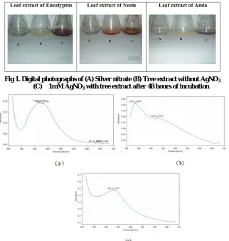
Reduction of silver ion into silver particles in contact to the leaf extracts was identified by colour change from green to brown in aqueous solution owing to Plasmon resonance phenomenon (Fig1). Conical flasks were observed periodically for colour change from green to different shades of brown (Table 1) which indicates the presence of silver nanoparticles during increasing intensity. The results represented in (Fig 2)
showed that Azadirachta indica (Neem) had the absorbance
peak at 335nm and 426nm, Phyllanthus emblica (Amla) at
431nm, and Eucalyptus polybrachtea (Eucalyptus) at 418nm
and 450nm, and broadening of peak indicated that the particles were polydispersed. The reductions of aqueous AuCl4 ions were followed by colour change from yellow-purple red indicating the formation of gold nanoparticles while increasing intensity (Fig 3). Periodical observation of colour change from purple red to dark purple red and than to brown was done (Table 2).
In case of gold ions reduction, the bands corresponds to the
surface Plasmon resonance of Eucalyptus polybrachtea
(Eucalyptus) at 564nm and Azadirachta indica (Neem) at
560nm (Fig 4). The FTIR spectrum (Fig 5.) recorded from the silver nitrate solution after reaction with leaf extracts of
medicinal trees, revealed strong band shifts for Phyllanthus
emblica (Amla) at 1643.24, 1627.18 corresponds to –c-c-
stretching vibration of aromatic amine group and c=o ,
Tectona grandis (Teak) shifts at 1629.74, 1382.87 corresponds
to –c-c- and c=o. X-Ray Diffraction study was used to confirm the crystalline nature of the particle. XRD analysis showed
intense peaks for Phyllanthus emblica (Amla) with AgNO3 at
2θ =32.10, 38.20 and 46.30 which can be indexed at 004, 111
and 100 planes. Eucalyptus polybrachtea (Eucalyptus) with
HAuCl4corresponds peak at 2θ = 38.10 (Fig 6) which can be
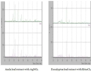
indexed at 111 plane. From EDAX spectrum, it was
understood that Phyllanthus emblica (Amla) leaf extract with
AgNO3 (Fig 7) and Eucalyptus polybrachtea (Eucalyptus)
with HAuCl4 (Fig 8) had recorded weight percent 100.50%
[image:3.595.141.463.291.629.2]and 91.65% of Ag and Au nanoparticle followed.
Fig 1. Digital photographs of (A) Silver nitrate (B) Tree extract without AgNO3
(C) 1mM AgNO3 with tree extract after 48 hours of incubation
Fig 2. UV- Vis absorption spectrum of silver nanoparticles synthesized by treating 1mM aqueous AgNO3 solution with (a) Leaf
[image:3.595.102.490.665.763.2]extract of Eucalyptus, (b) Leaf Extract of Neem, c) Leaf extract of Amla after 24 hours.
Fig 3. Digital photographs of (A) Chloroauric Acid (B) Tree extract without HAuCl4 (C) 10 -3
M HAuCl4 with tree extract after 1
Fig 4. UV- Vis absorption spectrum of gold nanoparticles synthesized by treating 10-3 M HAuCl4 solution with (a) Leaf extract of Neem,
[image:4.595.69.523.265.426.2](b) Leaf Extract of Eucalyptus after 24 hours.
Fig 5. FTIR analysis of silver and gold nanoparticles synthesized with (a) Phyllanthus emblica (Amla) with AgNO3 and Azadirachta
indica (Neem) with HAuCl4
[image:4.595.111.481.470.761.2]The SEM image showed relatively spherical shape
nanoparticle formed for Phyllanthus emblica (Amla) range
from 20-30nm and Eucalyptus polybrachtea (Eucalyptus)
cubiodal shape range from 37-60nm (Fig 9). The bio-reduction of aqueous Ag+ ions and Au+ ions by the leaf extract of the medicinal trees has been demonstrated. In the present study it was found that leaves could be also a good source for production of NPs. This green approach has many compensation such as, ease with which the method can be eco-friendly, scale-up, financial feasibility, etc. With increasing time, the size of the NPs increased and the crystalline nature of NPs changed from polycrystalline to single crystalline. Most importantly, the reaction was simple and convenient to
handle, and it is believed that it was advantageous over other chemical synthesis.
Acknowledgement
Department of Botany and Microbiology, Lady Doak College, Madurai, India for providing facility to carry out the work, Science Instrumentation Center (LDC) for UV -Visible analysis, USIC-Madurai Kamaraj University for FTIR analysis, Nanotech Research Center- Karunya University for XRD, SEM analysis, EDAX measurements.
REFERENCES
[image:5.595.146.450.52.223.2]Ankamwar, B., Chaudhary, M. and Sastry, M. 2005.Gold nanotriangles biologically synthesized using tamarind leaf
[image:5.595.156.438.253.417.2]Fig 7. Energy – Dispersive Absorption Spectroscopy photograph of AgNP with Phyllanthus emblica (Amla)
[image:5.595.144.457.462.612.2]Fig 8. Energy – Dispersive Absorption Spectroscopy photograph of AuNP with Eucalyptus polybrachtea (Eucalyptus)
extract and potential application in vapor sensing. Synth. React. Inorg. Metal-Org. Nanometal. Chem., 35: 19-26. Ankamwar, B., Damle, C., Ahmad, A. and Sastry, M. 2005.
Biosynthesis of gold and silver nanoparticles using Emblica officinalis fruit extract, their phase transfer and transmetallation in an organic solution. J Nanosci Nanotechnol., 5 (10):1665–1671.
Balaprasad Ankamwar. 2010. Biosynthesis of gold
nanoparticles (Green- Gold) using leaf extract of
Terminalia catappa. E-Journal of Chemistry., 7(4):
1334-1339.
Chandran S.P., Chaudhary, M., Pasricha, R., Ahmad, R. and Sastry, M. 2006.Synthesis of gold nanotriangles and silver nanoparticles using Aloe vera plant extract. Biotechnol.Prog., 22: 577.
Elumalai, E. K., Prasad, T. N. V. K. V., Hemachandran, J., Viviyan, T. S., Thirumalai, T. and David, E. 2010. Extracellular synthesis of silver nanoparticles using leaves
of Euphorbia hirta and their antibacterial activities.
Journal of Pharmaceutical Sciences and Research., Vol. 2 (9): 549-554.
Gardea-Torresday, J. L., Gomez, E., Peralta, V. J. R., Parsons, J. G., Troiani, H. and Jose, Y. M. 2003. Alfalfa sprouts: A natural source for the synthesis of silver nanoparticles. Langmuir., 19:1357-1361.
Gardea-Torresday, L. J., Persons, G.J., Gomez, E., Peralta-Videa, J., Troiani, E.H., Santiago, P. and Yacaman, J.M. 2002. Formation and growth of Au nanoparticles inside live Alfalfa plants. Nano Letters., 2 (4): 397-401.
Huang, J., Li, Q., Sun, D., Lu, Y., Su, Y., Yang, X., Wang, H., Wang, Y., Shao, W., He, N., Hong, J. and Chen, C. 2007. Biosynthesis of silver and gold nanoparticles by novel
sundried Cinnamomum camphora leaf. Nanotechnology.,
18:105104-105114.
Kasthuri, J.K., Kathiravan, N. and Rajendiran. 2009. Phyllanthin-assisted biosynthesis of silver and gold nanoparticles: a novel biological approach. J Nanopart Res., 11:1075.
Kasthuri, J.S., Veerapandian, N. Rajendiran . 2009. Biological synthesis of silver and gold nanoparticles using apiin as reducing agent. Colloids Surf B Biointerf., 68:55.
Narayanan, K. B. and Sakthivel. N. 2008.Coriander leaf mediated biosynthesis of gold nanoparticles.Mater Lett., 62:4588.
Prabhu, N., Divya, T. R., Yamuna Gowri, K., Ayisha Siddique, S. and Joseph Puspha Innocent, D. 2010. Synthesis of silver phyto nanoparticles and their antibacterial efficacy. Digest Journal of Nanomaterials and Biostructures., Vol.5. No 1: 185-189.
Shikuo, Li., Yuhua Shen., Anjian Xie., Xuerong Yu., Lingguang Qiu., Li Zhang. and Qingfeng Zhang. 2007. Green synthesis of silver nanoparticles using Capsicum annuum L. extract. Green Chemistry., Vol.1039.
Subramanian Arulkumar. and Muthukumaran Sabesan. 2010. Biosynthesis and characterization of gold nanoparticle
using antiparkinsonian drug Mucuna pruriens plant
extract. Int. Res. Pharm. Sci. Vol.1: Issue. 4. 417- 420. Vedapriya Arya. 2010. Living Systems: Eco-Friendly
Nanofactories. Digest Journal of Nanomaterials and Biostructures. Vol-5: No 1. 9-21.
Willems van den. and Wildenberg. Roadmap report on nanoparticles. 2005 Barcelona, Spain: W&W Espana sl.