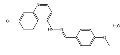
7-Chloro-4-[(
E
)-2-(4-methoxybenzyl-idene)hydrazin-1-yl]quinoline
monohydrate
Marcelle de Lima Ferreira,aMarcus V. N. de Souza,a R. Alan Howie,bEdward R. T. Tiekink,c* James L. Wardelld‡ and Solange M. S. V. Wardelle
aInstituto de Tecnologia em Farmacos, Fundac¸a˜o Oswaldo Cruz (FIOCRUZ), FarManguinhos, Rua Sizenando Nabuco, 100, Manguinhos, 21041-250 Rio de Janeiro, RJ, Brazil,bDepartment of Chemistry, University of Aberdeen, Old Aberdeen AB15 5NY, Scotland,cDepartment of Chemistry, University of Malaya, 50603 Kuala Lumpur, Malaysia,dCentro de Desenvolvimento Tecnolo´gico em Sau´de (CDTS), Fundac¸a˜o Oswaldo Cruz (FIOCRUZ), Casa Amarela, Campus de Manguinhos, Av. Brasil 4365, 21040-900 Rio de Janeiro, RJ, Brazil, andeCHEMSOL, 1 Harcourt Road, Aberdeen AB15 5NY, Scotland
Correspondence e-mail: edward.tiekink@gmail.com
Received 18 February 2010; accepted 19 February 2010
Key indicators: single-crystal X-ray study;T= 120 K; mean(C–C) = 0.005 A˚; Rfactor = 0.069;wRfactor = 0.150; data-to-parameter ratio = 12.6.
The organic molecule in the title hydrate, C17H14ClN3OH2O,
has a small but significant twist from planarity, as seen in the dihedral angle of 12.10 (17) between the quinoline and
benzene rings. The conformation about the C N bond isE. Chains along the baxis are formed in the crystal structure aided by water–quinoline O—H N ( 2) and hydrazone– water N—H O hydrogen bonds. Layers of these chains stack along the a axis via C—H and – interactions [ring centroid–ring centroid distance = 3.674 (2) A˚ ]. C—H O interactions are also present.
Related literature
For background to the pharmacological activity of quinoline derivatives, see: Warshakoon et al.(2006). For recent studies into quinoline-based anti-malarials, see: Andradeet al.(2007); de Souzaet al.(2005). For related structures, see: Kaiseret al.
(2009); de Souzaet al.(2009, 2010).
Experimental
Crystal data
C17H14ClN3OH2O
Mr= 329.78
Triclinic,P1
a= 7.0086 (6) A˚
b= 9.2384 (8) A˚
c= 13.3701 (12) A˚ = 100.026 (4)
= 103.903 (5)
= 107.000 (5)
V= 775.27 (12) A˚3
Z= 2
MoKradiation = 0.26 mm1
T= 120 K
0.120.040.02 mm
Data collection
Nonius KappaCCD area-detector diffractometer
Absorption correction: multi-scan (SADABS; Sheldrick, 2007)
Tmin= 0.686,Tmax= 1.000
10514 measured reflections 2702 independent reflections 2037 reflections withI> 2(I)
Rint= 0.064
Refinement
R[F2> 2(F2)] = 0.069
wR(F2) = 0.150
S= 1.02 2702 reflections 215 parameters
H atoms treated by a mixture of independent and constrained refinement
max= 0.30 e A˚
3 min=0.36 e A˚
[image:1.610.60.271.628.725.2]3
Table 1
Hydrogen-bond geometry (A˚ ,).
Cgis the centroid of the C11–C16 ring.
D—H A D—H H A D A D—H A
O1w—H1w N1i 0.85 (5) 2.02 (5) 2.867 (4) 172 (5)
O1w—H2w N1 0.85 (5) 2.20 (5) 3.047 (5) 175 (5)
N2—H2n O1wii
0.88 2.18 3.007 (4) 157
C5—H5 O1wii
0.95 2.45 3.380 (5) 165
C17—H17a Cgiii
0.98 2.65 3.508 (5) 147
Symmetry codes: (i) xþ1;yþ2;zþ1; (ii) xþ1;yþ1;zþ1; (iii)
x;y;zþ2.
Data collection: COLLECT (Hooft, 1998); cell refinement: DENZO(Otwinowski & Minor, 1997) andCOLLECT; data reduc-tion:DENZO andCOLLECT; program(s) used to solve structure: SHELXS97(Sheldrick, 2008); program(s) used to refine structure: SHELXL97 (Sheldrick, 2008); molecular graphics: ORTEP-3 (Farrugia, 1997) andDIAMOND(Brandenburg, 2006); software used to prepare material for publication:publCIF(Westrip, 2010).
The use of the EPSRC X-ray crystallographic service at the University of Southampton, England, and the valuable assis-tance of the staff there is gratefully acknowledged. JLW acknowledges support from CAPES (Brazil).
Supplementary data and figures for this paper are available from the IUCr electronic archives (Reference: LH5002).
References
Andrade, A. A., Varotti, F. D., de Freitas, I. Q., de Souza, M. V. N., Vasconcelos, T. R. A., Boechat, N. & Krettli, A. U. (2007).Eur. J. Pharm. 558, 194–198.
Brandenburg, K. (2006).DIAMOND. Crystal Impact GbR, Bonn, Germany. Farrugia, L. J. (1997).J. Appl. Cryst.30, 565.
Hooft, R. W. W. (1998).COLLECT. Nonius BV, Delft, The Netherlands. Kaiser, C. R., Pais, K. C., de Souza, M. V. N., Wardell, J. L., Wardell, S. M. S. V.
& Tiekink, E. R. T. (2009).CrystEngComm,11, 1133–1140.
organic compounds
o696
Lima Ferreiraet al. doi:10.1107/S1600536810006598 Acta Cryst.(2010). E66, o696–o697 Acta Crystallographica Section EStructure Reports Online
ISSN 1600-5368
Macromolecular Crystallography, Part A, edited by C. W. Carter Jr & R. M. Sweet, pp. 307–326. New York: Academic Press.
Sheldrick, G. M. (2007).SADABS. Bruker AXS Inc., Madison, Wisconsin, USA.
Sheldrick, G. M. (2008).Acta Cryst.A64, 112–122.
Souza, M. V. N. de (2005).Mini-Rev. Med. Chem.5, 1009–1017.
S. M. S. V. (2010).Acta Cryst.E66, o152–o153.
Souza, M. V. N. de, Tiekink, E. R. T., Wardell, J. L. & Wardell, S. M. S. V. (2009).Acta Cryst.E65, o3120–o3121.
supporting information
sup-1
Acta Cryst. (2010). E66, o696–o697
supporting information
Acta Cryst. (2010). E66, o696–o697 [doi:10.1107/S1600536810006598]
7-Chloro-4-[(
E
)-2-(4-methoxybenzylidene)hydrazin-1-yl]quinoline monohydrate
Marcelle de Lima Ferreira, Marcus V. N. de Souza, R. Alan Howie, Edward R. T. Tiekink, James L.
Wardell and Solange M. S. V. Wardell
S1. Comment
Quinoline derivatives display biological activity (Warshakoon et al., 2006) and in this context attract interest as potential anti-malarial agents (Andrade et al. 2007; de Souza et al., 2005). Complementing biological studies are structural investigations (Kaiser et al., 2009; de Souza et al., 2009; de Souza et al., 2010), and the crystal structure of the title hydrate, (I), was investigated as a part of these on-going studies.
The molecular structure of the organic component of (I), Fig. 1, shows a small twist from planarity with the dihedral angle formed between the quinoline (maximum deviation = 0.039 (4) Å for the C6 atom) and benzene rings being 12.10 (17) °. The major deviation of a torsion angle from 0 or 180 ° is found in the C3–N2–N3–C10 torsion angle of 172.4 (3) °. The conformation about the C10═N3 bond [1.274 (5) Å] is E. The crystal packing is stabilised by a variety of hydrogen bonding interactions, Table 1. The water molecule accepts a hydrogen bond from the hydrazone-N2 atom and forms donor interactions with symmetry related quinoline-N1 atoms, the latter leading to eight-membered {···OHO···N}2
synthons. The resulting supramolecular chain along the b axis, Fig. 2, is reinforced by a C–H···O contact, Table 1. The chains are arranged in layers in the bc plane with the most significant interactions between the layers being of the type π–
π with the closest of these occurring between centrosymmetrically related N1,C1—C4,C9 rings [ring centroid(N1,C1— C4,C9)···ring centroid(N1,C1—C4,C9)i distance = 3.674 (2) Å for i: -x, 1-y, 1-z], Fig. 3. In addition to these interactions,
C–H···π contacts also occur between layers, Table 1.
S2. Experimental
A solution of 7-chloro-4-quinolinylhydrazine (0.2 g, 1.03 mmol) and 4-methoxybenzaldehyde (1.24 mmol) in ethanol (5 ml) was stirred at room temperature until TLC indicated complete consumption of the hydrazine. The reaction mixture was rotary evaporated, the residue washed well with cold Et2O (3 x 10 ml), and recrystallised from moist ethanol, yield
85%, m.pt. 417–418 K. IR [KBr, cm-1] ν: 3120 (NH), 1565(N═C). MS/ESI: m/z [M—H]+: 310.8.
S3. Refinement
The N- and C-bound H atoms were geometrically placed (N–H = 0.88 Å and C–H = 0.95–0.98 Å) and refined as riding with Uiso(H) = 1.2–1.5Ueq(C,N). The water-bound H atoms were located from a difference map and refined with Uiso(H) =
Figure 1
The molecular structure of the components comprising the asymmetric unit in (I) showing the atom-labelling scheme and displacement ellipsoids at the 50% probability level.
Figure 2
[image:4.610.120.488.277.417.2]supporting information
sup-3
[image:5.610.126.492.68.463.2]Acta Cryst. (2010). E66, o696–o697 Figure 3
A view of the stacking of layers in (I); O–H···N hydrogen bonding is shown as orange dashed lines. The layers are linked by π–π (purple dashed lines) and C–H···π contacts (pink dashed lines). Colour code: Cl, cyan; O, red; N, blue; C, grey; and H, green.
7-Chloro-4-[(E)-2-(4-methoxybenzylidene)hydrazin-1-yl]quinoline monohydrate
Crystal data
C17H14ClN3O·H2O
Mr = 329.78
Triclinic, P1 Hall symbol: -P 1
a = 7.0086 (6) Å
b = 9.2384 (8) Å
c = 13.3701 (12) Å
α = 100.026 (4)°
β = 103.903 (5)°
γ = 107.000 (5)°
V = 775.27 (12) Å3
Z = 2
F(000) = 344
Dx = 1.413 Mg m−3
Mo Kα radiation, λ = 0.71073 Å Cell parameters from 27436 reflections
θ = 2.9–27.5°
µ = 0.26 mm−1
Nonius KappaCCD area-detector diffractometer
Radiation source: Enraf Nonius FR591 rotating anode
10 cm confocal mirrors monochromator Detector resolution: 9.091 pixels mm-1
φ and ω scans
Absorption correction: multi-scan (SADABS; Sheldrick, 2007)
Tmin = 0.686, Tmax = 1.000
10514 measured reflections 2702 independent reflections 2037 reflections with I > 2σ(I)
Rint = 0.064
θmax = 25.0°, θmin = 3.1°
h = −8→8
k = −10→10
l = −15→15
Refinement
Refinement on F2
Least-squares matrix: full
R[F2 > 2σ(F2)] = 0.069
wR(F2) = 0.150
S = 1.02 2702 reflections 215 parameters 0 restraints
Primary atom site location: structure-invariant direct methods
Secondary atom site location: difference Fourier map
Hydrogen site location: inferred from neighbouring sites
H atoms treated by a mixture of independent and constrained refinement
w = 1/[σ2(F
o2) + (0.0109P)2 + 2.93P]
where P = (Fo2 + 2Fc2)/3
(Δ/σ)max = 0.001
Δρmax = 0.30 e Å−3
Δρmin = −0.36 e Å−3
Special details
Geometry. All s.u.'s (except the s.u. in the dihedral angle between two l.s. planes) are estimated using the full covariance
matrix. The cell s.u.'s are taken into account individually in the estimation of s.u.'s in distances, angles and torsion angles; correlations between s.u.'s in cell parameters are only used when they are defined by crystal symmetry. An approximate (isotropic) treatment of cell s.u.'s is used for estimating s.u.'s involving l.s. planes.
Refinement. Refinement of F2 against ALL reflections. The weighted R-factor wR and goodness of fit S are based on F2,
conventional R-factors R are based on F, with F set to zero for negative F2. The threshold expression of F2 > 2σ(F2) is
used only for calculating R-factors(gt) etc. and is not relevant to the choice of reflections for refinement. R-factors based on F2 are statistically about twice as large as those based on F, and R- factors based on ALL data will be even larger.
Fractional atomic coordinates and isotropic or equivalent isotropic displacement parameters (Å2)
x y z Uiso*/Ueq
Cl1 0.12492 (19) 0.53097 (12) 0.13214 (8) 0.0337 (3) O1 0.3226 (4) 0.0839 (3) 1.1296 (2) 0.0268 (7) N1 0.2963 (5) 0.7665 (4) 0.5289 (2) 0.0233 (7) N2 0.2484 (5) 0.3358 (4) 0.6035 (2) 0.0226 (7)
H2N 0.2193 0.2506 0.5527 0.027*
N3 0.2774 (5) 0.3291 (4) 0.7081 (2) 0.0220 (7) C1 0.3251 (6) 0.7500 (4) 0.6279 (3) 0.0247 (9)
H1 0.3611 0.8418 0.6835 0.030*
C2 0.3074 (6) 0.6112 (4) 0.6569 (3) 0.0217 (9)
H2 0.3239 0.6092 0.7292 0.026*
C3 0.2653 (6) 0.4750 (4) 0.5796 (3) 0.0178 (8) C4 0.2376 (6) 0.4842 (4) 0.4712 (3) 0.0188 (8) C5 0.2011 (6) 0.3562 (4) 0.3853 (3) 0.0203 (8)
H5 0.1959 0.2576 0.3985 0.024*
supporting information
sup-5
Acta Cryst. (2010). E66, o696–o697
H6 0.1518 0.2852 0.2259 0.026*
C7 0.1753 (6) 0.5170 (4) 0.2634 (3) 0.0211 (8) C8 0.2147 (6) 0.6443 (4) 0.3441 (3) 0.0225 (9)
H8 0.2180 0.7417 0.3292 0.027*
C9 0.2507 (6) 0.6313 (4) 0.4502 (3) 0.0201 (8) C10 0.2358 (6) 0.1919 (5) 0.7224 (3) 0.0235 (9)
H10 0.1879 0.1044 0.6620 0.028*
C11 0.2591 (6) 0.1642 (4) 0.8281 (3) 0.0199 (8) C12 0.2046 (6) 0.0122 (4) 0.8385 (3) 0.0222 (9)
H12 0.1524 −0.0724 0.7759 0.027*
C13 0.2235 (6) −0.0212 (4) 0.9368 (3) 0.0214 (8)
H13 0.1854 −0.1267 0.9415 0.026*
C14 0.2985 (6) 0.1016 (4) 1.0276 (3) 0.0217 (8) C15 0.3569 (6) 0.2567 (4) 1.0201 (3) 0.0229 (9)
H15 0.4099 0.3408 1.0830 0.027*
C16 0.3379 (6) 0.2878 (4) 0.9220 (3) 0.0227 (9)
H16 0.3781 0.3934 0.9176 0.027*
C17 0.2580 (7) −0.0735 (4) 1.1403 (3) 0.0268 (9)
H17A 0.1093 −0.1280 1.0997 0.040*
H17B 0.2787 −0.0709 1.2158 0.040*
H17C 0.3418 −0.1291 1.1127 0.040*
O1W 0.7337 (5) 0.9550 (3) 0.5298 (2) 0.0309 (7) H1W 0.736 (8) 1.039 (6) 0.511 (4) 0.046* H2W 0.610 (8) 0.908 (6) 0.530 (4) 0.046*
Atomic displacement parameters (Å2)
U11 U22 U33 U12 U13 U23
C17 0.032 (2) 0.025 (2) 0.028 (2) 0.0113 (18) 0.0109 (18) 0.0114 (17) O1W 0.0347 (18) 0.0205 (15) 0.0434 (18) 0.0117 (14) 0.0171 (15) 0.0123 (13)
Geometric parameters (Å, º)
Cl1—C7 1.740 (4) C7—C8 1.360 (5)
O1—C14 1.377 (4) C8—C9 1.413 (5)
O1—C17 1.435 (5) C8—H8 0.9500
N1—C1 1.332 (5) C10—C11 1.458 (5)
N1—C9 1.385 (5) C10—H10 0.9500
N2—C3 1.357 (5) C11—C12 1.385 (5)
N2—N3 1.380 (4) C11—C16 1.409 (5)
N2—H2N 0.8800 C12—C13 1.386 (5)
N3—C10 1.274 (5) C12—H12 0.9500
C1—C2 1.383 (5) C13—C14 1.379 (5)
C1—H1 0.9500 C13—H13 0.9500
C2—C3 1.386 (5) C14—C15 1.400 (5)
C2—H2 0.9500 C15—C16 1.374 (5)
C3—C4 1.435 (5) C15—H15 0.9500
C4—C9 1.417 (5) C16—H16 0.9500
C4—C5 1.411 (5) C17—H17A 0.9800
C5—C6 1.374 (5) C17—H17B 0.9800
C5—H5 0.9500 C17—H17C 0.9800
C6—C7 1.409 (5) O1W—H1W 0.85 (5)
C6—H6 0.9500 O1W—H2W 0.85 (5)
C14—O1—C17 117.0 (3) N1—C9—C8 116.8 (3)
C1—N1—C9 115.5 (3) C4—C9—C8 119.7 (3)
C3—N2—N3 119.4 (3) N3—C10—C11 122.5 (3)
C3—N2—H2N 120.3 N3—C10—H10 118.8
N3—N2—H2N 120.3 C11—C10—H10 118.8
C10—N3—N2 115.5 (3) C12—C11—C16 117.8 (3) N1—C1—C2 125.9 (4) C12—C11—C10 119.9 (3)
N1—C1—H1 117.0 C16—C11—C10 122.3 (3)
C2—C1—H1 117.0 C11—C12—C13 122.5 (4)
C1—C2—C3 119.5 (3) C11—C12—H12 118.8
C1—C2—H2 120.3 C13—C12—H12 118.8
C3—C2—H2 120.3 C14—C13—C12 118.8 (3)
N2—C3—C2 122.1 (3) C14—C13—H13 120.6
N2—C3—C4 120.0 (3) C12—C13—H13 120.6
C2—C3—C4 117.9 (3) O1—C14—C13 124.4 (3) C9—C4—C5 118.4 (3) O1—C14—C15 115.4 (3) C9—C4—C3 117.7 (3) C13—C14—C15 120.2 (3) C5—C4—C3 123.9 (3) C16—C15—C14 120.2 (4)
C6—C5—C4 121.3 (3) C16—C15—H15 119.9
C6—C5—H5 119.3 C14—C15—H15 119.9
supporting information
sup-7
Acta Cryst. (2010). E66, o696–o697
C5—C6—C7 119.0 (3) C15—C16—H16 119.8
C5—C6—H6 120.5 C11—C16—H16 119.8
C7—C6—H6 120.5 O1—C17—H17A 109.5
C8—C7—C6 121.6 (3) O1—C17—H17B 109.5
C8—C7—Cl1 120.0 (3) H17A—C17—H17B 109.5 C6—C7—Cl1 118.3 (3) O1—C17—H17C 109.5 C7—C8—C9 119.8 (3) H17A—C17—H17C 109.5
C7—C8—H8 120.1 H17B—C17—H17C 109.5
C9—C8—H8 120.1 H1W—O1W—H2W 108 (5)
N1—C9—C4 123.5 (3)
C3—N2—N3—C10 172.4 (3) C3—C4—C9—N1 2.5 (5) C9—N1—C1—C2 −2.0 (6) C5—C4—C9—C8 3.8 (5) N1—C1—C2—C3 3.1 (6) C3—C4—C9—C8 −177.0 (3) N3—N2—C3—C2 −1.1 (5) C7—C8—C9—N1 178.1 (3) N3—N2—C3—C4 179.4 (3) C7—C8—C9—C4 −2.4 (6) C1—C2—C3—N2 179.3 (3) N2—N3—C10—C11 179.7 (3) C1—C2—C3—C4 −1.2 (5) N3—C10—C11—C12 177.7 (4) N2—C3—C4—C9 178.2 (3) N3—C10—C11—C16 −2.8 (6) C2—C3—C4—C9 −1.3 (5) C16—C11—C12—C13 0.5 (6) N2—C3—C4—C5 −2.7 (5) C10—C11—C12—C13 180.0 (4) C2—C3—C4—C5 177.8 (3) C11—C12—C13—C14 0.3 (6) C9—C4—C5—C6 −1.8 (5) C17—O1—C14—C13 −2.2 (5) C3—C4—C5—C6 179.1 (4) C17—O1—C14—C15 178.0 (3) C4—C5—C6—C7 −1.6 (6) C12—C13—C14—O1 179.4 (4) C5—C6—C7—C8 3.1 (6) C12—C13—C14—C15 −0.9 (6) C5—C6—C7—Cl1 −176.5 (3) O1—C14—C15—C16 −179.5 (3) C6—C7—C8—C9 −1.1 (6) C13—C14—C15—C16 0.7 (6) Cl1—C7—C8—C9 178.5 (3) C14—C15—C16—C11 0.1 (6) C1—N1—C9—C4 −0.9 (5) C12—C11—C16—C15 −0.7 (6) C1—N1—C9—C8 178.6 (3) C10—C11—C16—C15 179.8 (4) C5—C4—C9—N1 −176.7 (3)
Hydrogen-bond geometry (Å, º)
Cg is the centroid of the C11–C16 ring.
D—H···A D—H H···A D···A D—H···A
O1w—H1w···N1i 0.85 (5) 2.02 (5) 2.867 (4) 172 (5)
N2—H2n···O1wii 0.88 2.18 3.007 (4) 157
O1w—H2w···N1 0.85 (5) 2.20 (5) 3.047 (5) 175 (5) C5—H5···O1wii 0.95 2.45 3.380 (5) 165
C17—H17a···Cgiii 0.98 2.65 3.508 (5) 147