DOI 10.1007/s12668-017-0425-z
Toxicity and Applications of Internalised Magnetite
Nanoparticles Within Live
Paramecium caudatum
Cells
Richard Mayne1 ·James Whiting1·Andrew Adamatzky1
© The Author(s) 2017. This article is an open access publication
Abstract The nanotechnology revolution has allowed us to speculate on the possibility of hybridising nanoscale materials with live substrates, yet significant doubt still remains pertaining to the effects of nanomaterials on bio-logical matter. In this investigation, we cultivate the cil-iated protistic pond-dwelling microorganismParamecium caudatumin the presence of excessive quantities of mag-netite nanoparticles in order to deduce potential beneficial applications for this technique, as well as observe any deleterious effects on the organisms’ health. Our findings indicate that this variety of nanoparticle is well-tolerated by P. caudatum cells, who were observed to consume them in quantities exceeding 5–12% of their body vol-ume: cultivation in the presence of magnetite nanoparticles does not alterP. caudatumcell volume, swimming speed, growth rate or peak colony density and cultures may per-sist in nanoparticle-contaminated media for many weeks. We demonstrate thatP. caudatumcells ingest starch-coated magnetite nanoparticles which facilitates their being mag-netically immobilised whilst maintaining apparently normal ciliary dynamics, thus demonstrating that nanoparticle bio-hybridisation is a viable alternative to conventional forms of ciliate quieting. Ingested magnetite nanoparticle deposits appear to aggregate, suggesting that (a) the process of being
Richard Mayne
James Whiting
Andrew Adamatzky
1 Unconventional Computing Laboratory, University of the West of England, Bristol, UK
internalised concentrates and may therefore detoxify (i.e. render less reactive) nanomaterial suspensions in aquatic environments, and (b)P. caudatumis a candidate organism for programmable nanomaterial manipulation and delivery.
Keywords Nanotoxicology·SPION·Quieting· Paramecia·Biohybridisation
1 Introduction
There are two major justifications for research into the hybridisation of nanoscale materials with biological matter. Firstly, despite their widespread use in recent years, signif-icant doubt remains as to the potential deleterious effects of nanoparticles and nanomaterials on biological matter as even inert materials may be rendered reactive, immunogenic or otherwise harmful to life when fabricated in nanoscale quantities [1]. Secondly, nanomaterials may be fabricated to exhibit highly desirable characteristics (specific electrical properties, magnetism, high tensile strength etc.), the con-ferral of which to live cells would potentially lead to next-generation technologies such as bio-computer interfaces for restorative and/or augmentative medical applications.
enhancing its value in bio-computer interfaces and uncon-ventional computing devices [4–7]. Recent evidence has suggested that MNPs are not as biocompatible as once thought, however, due to their potential for bioaccumula-tion and generabioaccumula-tion of reactive oxygen species within host cells [8, 9]. Furthermore, little is known of the ecotoxi-cological significance of SPIONs released into the envi-ronment, but recent studies on water-dwelling eukaryotes such as Daphnia spp. and various forms of plant life have demonstrated that they may potentially disrupt aquatic ecosystems [10].
This study examines the ciliated protistic pond organ-ismParamecium caudatumbeing cultivated in the presence of excessive quantities of MNPs in order assess the poten-tial uses of nanohybridised paramecia whilst concurrently observing for any deleterious effects on the organisms’ health. We conclude by discussing the apparent effects of this treatment onP. caudatumcells and the ecotoxicologi-cal significance of these results, and present several novel applications for hybridised P. caudatum cells containing MNPs.
2 Materials and Methods
P. caudatumcultures were cultivated in Chalkley’s medium enriched with 10 g of alfalfa (Sciento, UK) and 40 wheat grains (Tesco, UK) per litre. Cultures were exposed to a day/night cycle but were kept out of direct sunlight; ambient temperatures ranged from 19–24oC.
In experiments where P. caudatum cells were exposed to MNPs, suspensions of 200 nm (hydrodynamic diameter) starch matrix-coated multi-core MNPs (Chemicell GmBH, Germany) were added to fresh culture medium at a concen-tration of 0.25 mg ml−1(approximately 2.2×10¹²particles per ml). This concentration was chosen as a comparable quantity of nanoparticles per unit biomass to our previ-ous studies with other single-celled organisms [5]. Stock cultures at a concentration of approximately 1000 cells ml−1were harvested in log growth phase and added to the nanoparticle-infused culture medium, in which they were incubated for periods in excess of two months.
The following microscopical measurements were made on a regular (daily) basis (n= 3 per culture per day for each): average cell count, average swimming speed, total cell cross sectional area and percentage of cross-sectional cell area occupied by cytoplasmic/vesicular inclusions whose colour was suggestive of MNP deposits. Cells were observed in glass microscope well slides. Cells were chemically fixed in order to record photomicrographs. Fixation was achieved by adding 10 μl of 4% paraformaldehyde (Agar Scien-tific, UK) in pH 7.0 phosphate buffered saline solution
(Sigma, Germany) to each slide well after swimming speed measurements had been made.
Observations were made with a Zeiss Axiovert 200M inverted microscope and photo/videomicrographs were cap-tured with an Olympus SC50 digital camera via CellSens software. Electron microscopic observations were made with an FEI Quanta FEG-SEM in high-vacuum mode.
For cell volume measurements, each image was anal-ysed to extract both the cells’ cross-sectional area in squared micrometers, but also to estimate the percentage of inter-nalised MNPs. Each image was imported into Matlab (Mathworks, USA) and processed in the following man-ner: the image was converted into greyscale, and a threshold was applied to extract all material darker than the back-ground media. Each threshold was visually checked in order to ascertain that only organisms were isolated in each image. The number of pixels isolated by the threshold was then summed to give the cells’ total cross-sectional area. This process was also performed using a different threshold to determine the number of pixels whose colour value corre-sponded to dark cytoplasmic inclusions in order to identify any internalised MNPs. As the threshold values had to be determined visually for each image separately, this process was performed ‘blind’, i.e. without the operator knowing whether each image were a control or test measurement. The number of pixels isolated by this method were then com-pared with the cells’ total volume to give a percentage value. Video analysis for measurement of organism swimming speed was also performed using Matlab. RGB images were imported from the video frame-by-frame for sequential analysis and organism positioning. For each video set, the organisms were isolated from the RGB image by colour, whereupon the data for each frame was converted to a JPEG image file for further analysis. To detect the position of the organisms, a Laplace template of a Gaussian filter was defined before being convolved over the image; the size of the filter was iteratively determined by visual feed-back of the user. After organism detection on every frame had occurred, the positional data was passed to a bespoke Kalman filter which accurately estimates the position of the particle across each frame using the data from the full time-series of particle positions to predict and confirm the movement of each organism. From this it is possible to measure the speed of each organism in a noisy video, cre-ating a dataset of organism speed and momentary position. While the script ran, frame-by-frame images were shown on screen allowing visual validation of positional track-ing by the authors. Average speed was calculated for each organism.
Table 1 Table to show mean (x¯) and standard deviation (σ) values for measurements of total cell cross-sectional area (xa, in μm2), cell content comprising dark (including rust-coloured) objects (doc, in
percentage), swimming speed (ss, inμms−1), growth rate (μ, in h−1) and peak colony density (pcd, in cells ml−1) (ttest and Mann-Whitney U test)
xax¯ xa σ docx¯ doc σ ssx¯ ss σ μx¯ μ σ pcdx¯ pcd σ
Control 12715.66 5994.59 7.33 1.67 132.54 42.79 0.020 0.009 309 120
Test 13381.09 4700.77 12.68* 3.16 144.94 31.64 0.021 0.009 317 123
Asterisks indicate a statistically significant difference in means to controls atp<0.0001
3 Results & Discussion
All measurement data are shown in Table 1. The micro-scopical appearance (morphology and swimming patterns)
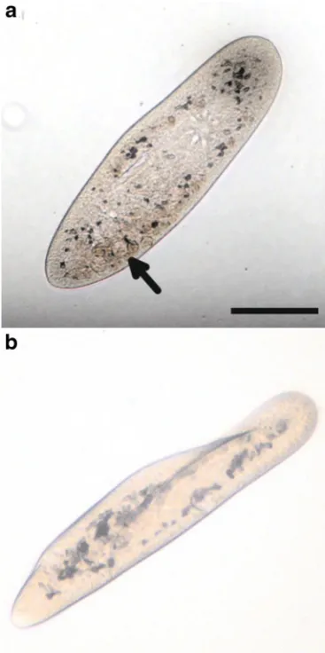
Fig. 1 Photomicrographs to show appearance ofP. caudatumcells (unfixed): (a) Following exposure to MNPs. Multiple rust-coloured deposits (arrowed) are visible in the cytoplasm. (b) Control, demon-strating a lack of rust-coloured deposits.Scale bar100μm
of P. caudatum cells treated with MNPs was not notice-ably altered aside from the inclusion of rust-coloured deposits and more dark objects within intracellular vesi-cles and the cells’ cytoplasm (Fig. 1). Organisms treated with MNPs contained approximately 5% more dark intra-cellular inclusions (including rust-coloured inclusions) than controls. No statistical difference was observed in total cell volume or swimming speed between controls and test organisms.
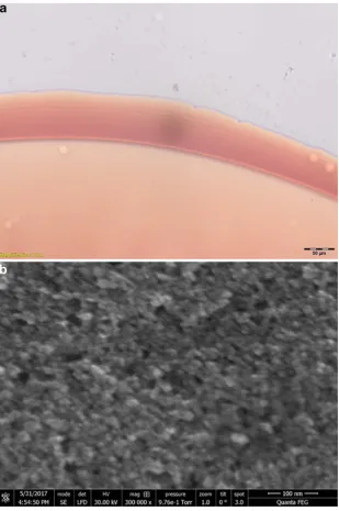
The microscopical appearance of the rust-coloured deposits within the organisms treated with MNPs was sim-ilar to that of suspensions of MNPs in distilled water (Fig.2a). The electron microscopic appearance of the MNPs was consistent with their description as aggregative multi-core objects (Fig.2b).
Furthermore, no statistical difference in growth rates or peak colony density were observed between controls and organisms treated with MNPs. All cultures (test and control) persisted over the duration of the experiment (14 days).
Our results indicate that theP. caudatumcell is highly tolerant to being cultured in the presence of large quantities of MNPs, as indicated by our not observing any delete-rious effects on the health of the organisms with regards to their size, morphology, motility, growth rate or colony density. This indicates that, despite recent evidence sug-gesting that this variety of nanoparticle may be harmful to aquatic microorganisms, they do not appear to induce any readily-observable toxicological effects in our ciliated model organism.
Intriguingly, the observations of MNPs within the organ-isms suggests that they were internalised in the manner of a nutrient source, noting that the size of the individual par-ticle cores, 10 nm, were too large to enter the cell via any other route such as through membrane pores. This apparent uptake of MNPs was likely a result of their starch coat-ing. Interactions betweenP. caudatumand MNPs with other coatings/no coating remain a topic for further study.
[image:3.595.77.261.285.654.2]from that of controls as both varieties were kept in culture for periods exceeding two months (data not shown).
We propose that P. caudatum may detoxify certain environmentally dispersed nanomaterials: that dark/rust-coloured deposits could be easily identified inP. caudatum
cells indicates that they are aggregated in vivo into non-nanoscale objects. This reduction in surface area to volume ratio likely renders the deposits less reactive and therefore less harmful.
This apparent lack of toxicological effects incident of internalising quantities of MNPs allows us to speculate
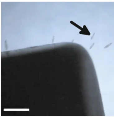
on the potential applications of this process of biological– artificial hybridisation. In further experiments, we exposed 10 μl droplets on glass microscope well slides contain-ing approximately 5 P. caudatumcells treated with MNPs to a 1.28 T 25x40x4.0 mm neodymium magnet. Holding the magnet in close proximity to the margins of the well caused the organisms to be drawn to the edge of the droplet where they were held immobile (Fig.3). By increasing the distance between the magnet and margins of the well, the organisms were able to move but at a significantly reduced speed.
[image:4.595.235.545.252.718.2]Fig. 3 Stereomicrograph showing MNP-treated P. caudatum cells (arrowed) being attracted towards a permanent magnet (black object). Scale bar 500μm
4 Conclusions
Magnetic restraint of ciliates via their hybridisation with biocompatible magnetic nanomaterials would appear to be an attractive alternative the established methods of microorganism immobilisation/quieting, such as: replace-ment of media with inert viscous fluids, induction of hypoxia through hermetically sealing the observation envi-ronment [11, 12], addition of low-concentration toxins (e.g. aliphatic alcohols [13], anaesthetic compounds [14]), ultraviolet light irradiation [15], establishment of fluid pressure gradients/microfluidic compression [16] and adhe-sion to solid surfaces via positively-charged proteins (3-aminopropyltriethoxysilane, protamine sulphate) [17].
The advantage of magnetic restraint over these other quieting methods is that it does not necessitate inducing deleterious health effects on the organism, maintains the chemical composition and hence physical characteristics of the fluid medium (thus minimising interference with natural ciliary beating processes) and allows for momentary adjust-ment of the strength of attraction (i.e. by moving the magnet or using magnets of different strengths). Although magnetic restraint ofParamecium spp.has been previously described via internalised magnetite (particles of ca. 3μm diameter), the authors did not describe the use of microparticles in this context as being a method for fully immobilising the organisms [18].
Finally, this ciliated model organism’s capacity for gath-ering, internalising and concentrating nanomaterials holds exciting possibilities for the prospect of orchestrated bio-logical manipulation and delivery (guided by gradients of attractants, repellents or magnetic fields) of nano and micro-scale compounds of interest, although further research is
required in this area before practical applications can be realised.
Acknowledgments The authors extend their thanks to Dr. David Patton for his electron microscopy expertise.
Compliance with Ethical Standards
Conflict of Interest The authors declare no competing financial interest.
Funding This work was funded by the Leverhulme Trust (grant no. RPG-2013-345).
Open Access This article is distributed under the terms of the Creative Commons Attribution 4.0 International License (http:// creativecommons.org/licenses/by/4.0/), which permits unrestricted use, distribution, and reproduction in any medium, provided you give appropriate credit to the original author(s) and the source, provide a link to the Creative Commons license, and indicate if changes were made.
References
1. Walters, C., Pool, E., & Somerset, V. Toxicology – New Aspects to This Scientific Conundrum (2016-10-26), chap. 3. doi:10.5772/64754.
2. Estelrich, J., Sanch´ez-Mart´ın, M., & Busquets, M. (2015). Inter-national Journal of Nanomedicine,10, 1727. doi:10.2147/IJN.S7 6501.
3. Revia, R.A., & Zhang, M. (2016).Materials Today,19(3), 157. doi:10.1016/j.mattod.2015.08.022.
4. Mayne, R., Patton, D., Costello, B.D.L., Adamatzky, A., & Pat-ton, R.C. (2011). International Journal of Nanotechnology and Molecular Computation,3(3), 1. doi:10.4018/ijnmc.2011070101. 5. R. Mayne, & A. Adamatzky (2015).Nano LIFE,5(1), 140007.
doi:10.1142/S179398441450007X.
6. N. Gizzie, R. Mayne, & A. Adamatzky (2016).Organic Electron-ics, 32, 267. doi:10.1016/j.orgel.2016.02.033. http://linkinghub. elsevier.com/retrieve/pii/S1566119916300775.
7. Berzina, T., Dimonte, A., Cifarelli, A., & Erokhi, V. (2015).
International Journal of General Systems,44, 341.
8. Singh, N., Jenkins, G., Asadi, R., & Doak, S. (2010). Nano Reviews 1.
9. Maher, B.A., Ahmed, I.A.M., Karloukovski, V., Maclaren, D.A., & Foulds, P.G. (2016).PNAS,113(39), 10797. doi:10.1073/pnas.1 605941113.
10. Blinova, I., Kanarbik, L., Irha, N., & Kahru, A. (2015). Hydrobi-ologia pp 1–9,doi:10.1007/s10750-015-2540-6.
11. Mast, S. (1947).Biological Bulletin,92(1), 31.
12. Kirby, H. (1950).Materials and methods in the study of protozoa, (p. 54). Cambridge, UK: Cambridge University Press.
13. Bills, C. (1992).Biological Bulletin,42(1), 7.
14. Zhou, M., Xia, H., Xu, Y., Xin, N., Liu, J., & Zhang, S. (2012).
Journal of Huazhong University of Science and Technology [Med-ical Sciences],32(3), 410. doi:10.1007/s11596-012-0071-1. 15. Boland, J. (1928).Science,67(1748), 654.
16. Yan, Y., Jiang, L., Aufderheide, K.J., Wright, G.A., Terekhov, A., Costa, L., Qin, K., McCleery, W.T., Fellenstein, J.J., Ustione, A., Robertson, J.B., Johnson, C.H., Piston, D.W., Hutson, M.S., Wikswo, J.P., Hofmeister, W., & Janetopoulos, C. (2014).
Microscopy and Microanalysis,20, 141. doi:10.1017/S14319276 13014037.
17. Marsot, P., & Couillard, P. (1973).Journal of Protozoology,20(1), 105.
[image:5.595.76.260.58.246.2]