addenda and errata
Acta Cryst.(2016). E72, 879–880 http://dx.doi.org/10.1107/S2056989016008252
879
Received 20 May 2016 Accepted 20 May 2016
Edited by W. T. A. Harrison, University of Aberdeen, Scotland
Keywords:
Capecitabine from X-ray powder synchrotron
data. Corrigendum
Jan Rohlicek,a* Michal Husak,aAles Gavenda,bAlexandr Jegorov,cBohumil Kratochvilaand Andy Fitchd
aDepartment of Solid State Chemistry, ICT Prague, Technicka 5, Prague, Czech Republic,bIVAX Pharmaceuticals s.r.o., R&D, Opava, Czech Republic,cPharmaceuticals Research and Development, Branisovska 31, Ceske Budejovice, Czech Republic, anddID31 Beamline, ESRF, 6 rue Jules Horowitz, BP 220, F-38043 Grenoble Cedex, France. *Correspondence e-mail: rohlicej@vscht.cz
In the paper by Rohlicek et al.[Acta Cryst.(2009), E65, o1325–o1326], one H atom was placed incorrectly.
Following our powder-diffraction study of capecitabine (Rohlicek et al., 2009), Malin´ska et al. (2014) published the crystal structure of the same molecule based on single-crystal data. Although they modelled the wrong enantiomer [as was pointed out by Kratochvilet al.(2016)], the structures are very similar after inverting the single-crystal structure, including
[image:1.610.207.457.374.459.2]ISSN 2056-9890
Figure 1
Overlay of the capecitabine molecular structures arising from powder diffraction (blue) and from single-crystal diffraction data (red). Only non-H atoms are shown for clarity.
Figure 2
[image:1.610.201.459.514.708.2]the disordered part of the molecule (Fig. 1). Since single-crystal diffraction is more sensitive to H atoms than powder diffraction, Malinskaet al. (2014) were able to locate the H atoms directly. This indicated a different tautomeric form of capecitabine to that assumed in our study, and as they pointed out, we had therefore placed one H atom wrongly.
In our defence, in the powder study, we placed the H atoms geometrically according to a reasonable chemical structure for capecitabine, which shows the tautomeric H atom attached to the N atom of the carbamate group and the plausible forma-tion of an intermolecular N—H O hydrogen bond. As shown by Malin´skaet al.(2014), the H atom is actually located on the N atom of the pyrimidine ring (Fig. 2), thereby forming an intramolecular N—H O link.
With respect to the fact that structure solution from powder diffraction data is based on the proposed molecular structure, readers should beware of the incorrectly placed H atom in Rohliceket al.(2009) and they should be also beware of the wrong enantiomer in a single-crystal study of Malin´skaet al.(2014).
References
Kratochvil, B., Husak, M., Korotkova, E. I. & Jegorov, A. (2016). Chem. Listy,110, 40–47.
Malin´ska, M., Krzecyn´ski, P., Czerniec-Michalik, E., Trzcin´ska, K., Cmoch, P., Kutner, A. & Woz´niak, K. (2014).J. Pharm. Sci.103, 587–593.
Rohlicek, J., Husak, M., Gavenda, A., Jegorov, A., Kratochvil, B. & Fitch, A. (2009).Acta Cryst.E65, o1325–o1326.
880
Rohliceket al. C15H22FN3O6 Acta Cryst.(2016). E72, 879–880
Capecitabine from X-ray powder
synchrotron data
Jan Rohlicek,a* Michal Husak,aAles Gavenda,bAlexandr Jegorov,cBohumil Kratochvilaand Andy Fitchd
aDepartment of Solid State Chemistry, ICT Prague, Technicka 5, Prague, Czech
Republic,bIVAX Pharmaceuticals s.r.o., R&D, Opava, Czech Republic, cPharmaceuticals Research and Development, Branisovska 31, Ceske Budejovice,
Czech Republic, anddID31 Beamline, ESRF, 6 rue Jules Horowitz, BP 220, F-38043 Grenoble Cedex, France
Correspondence e-mail: rohlicej@vscht.cz
Received 3 April 2009; accepted 12 May 2009
Key indicators: powder synchrotron study;T= 293 K; mean(C–C) = 0.004 A˚; disorder in main residue;Rfactor = 0.055;wRfactor = 0.074; data-to-parameter ratio = 5.5.
In the title compound [systematic name 5-deoxy-5-fluoro-N -(pentyloxycarbonyl)cytidine], C15H22FN3O6, the pentyl chain
is disordered over two positions with refined occupancies of 0.53 (5) and 0.47 (5). The furan ring assumes an envelope
conformation. In the crystal, intermolecular N—H O
hydrogen bonds link the molecules into chains propagating along the b axis. The crystal packing exhibits electrostatic interactions between the 5-fluoropyrimidin-2(1H)-one frag-ments of neighbouring molecules as indicated by short O C [2.875 (3) and 2.961 (3) A˚ ] and F C [2.886 (3) A˚ ] contacts.
Related literature
Capecitabine is the first FDA-approved oral chemotherapy for the treatment for some types of cancer, including advanced bowel cancer or breast cancer, see: Wagstaff et al. (2003); Joneset al.(2004).
Experimental
Crystal data
C15H22FN3O6 Mr= 359.35
Orthorhombic,P212121 a= 5.20527 (2) A˚
b= 9.52235 (4) A˚
c= 34.77985 (13) A˚
V= 1723.91 (1) A˚3
Z= 4
Synchrotron radiation = 0.79483 (4) A˚ = 0.15 mm1 T= 293 K
Specimen shape: cylinder 4011 mm
Specimen prepared at 101 kPa Specimen prepared at 293 K
Particle morphology: no specific habit, white
Data collection
ID31 ESRF Grenoble diffractometer
Specimen mounting: 1.0 mm boro-silicate glass capillary
Specimen mounted in transmission mode
Scan method: step Absorption correction: none 2min= 1.0, 2max= 35.0
Increment in 2= 0.003
Refinement
Rp= 0.055 Rwp= 0.074 Rexp= 0.036 RB= 0.102 S= 2.11
Wavelength of incident radiation: 0.79483(4) A˚
Excluded region(s): no Profile function: Pseudo-Voigt
profile coefficients as para-meterized in Thompsonet al.
(1987), asymmetry correction according to Fingeret al.(1994) 499 reflections
91 parameters 77 restraints
H-atom parameters not refined Preferred orientation correction:
[image:3.610.45.259.573.740.2]March–Dollase (Dollase, 1986); direction of preferred orientation 001, texture parameterr= 1.03 (1)
Table 1
Hydrogen-bond geometry (A˚ ,).
D—H A D—H H A D A D—H A
N17—H171 O8i
0.860 1.956 2.797 (5) 170
Symmetry code: (i)xþ1;yþ1 2;zþ
3 2.
Data collection: ESRF SPEC package; cell refinement: GSAS (Larson & Von Dreele, 1994); data reduction: CRYSFIRE2004 (Shirley, 2000) andMOPAC(Dewaret al., 1985); program(s) used to solve structure:FOX(Favre-Nicolin & Cˇ erny´, 2002); program(s) used to refine structure:GSAS; molecular graphics:Mercury(Macraeet al., 2006) andPLATON(Spek, 2009); software used to prepare material for publication:enCIFer(Allenet al., 2004).
This study was supported by the Czech Grant Agency (grant No. GACˇR 203/07/0040), the Institute of Chemical Technology in Prague (grant No. 108–08–0017) and the research program MSM 2B08021 of the Ministry of Education, Youth and Sports of the Czech Republic.
Supplementary data and figures for this paper are available from the IUCr electronic archives (Reference: CV2544).
References
Allen, F. H., Johnson, O., Shields, G. P., Smith, B. R. & Towler, M. (2004).J. Appl. Cryst.37, 335–338.
Dewar, M. J. S., Zoebisch, E. G., Healy, E. F. & Stewart, J. J. P. (1985).J. Am. Chem. Soc.107, 3902–3909.
Dollase, W. A. (1986).J. Appl. Cryst.19, 267–272.
Favre-Nicolin, V. & Cˇ erny´, R. (2002).J. Appl. Cryst.35, 734–743.
Finger, L. W., Cox, D. E. & Jephcoat, A. P. (1994).J. Appl. Cryst.27, 892–900. Jones, L., Hawkins, N., Westwood, M., Wright, K., Richardson, G. & Riemsma,
R. (2004).Health Technol. Assess.8, 1–156.
Larson, A. C. & Von Dreele, R. B. (1994).GSAS. Report LAUR 86-748. Los Alamos National Laboratory, New Mexico, USA.
Macrae, C. F., Edgington, P. R., McCabe, P., Pidcock, E., Shields, G. P., Taylor, R., Towler, M. & van de Streek, J. (2006).J. Appl. Cryst.39, 453–457.
organic compounds
Acta Cryst.(2009). E65, o1325–o1326 doi:10.1107/S1600536809017905 Rohliceket al.
o1325
Acta Crystallographica Section E
Structure Reports
Online
Shirley, R. (2000). CRYSFIRE User’s Manual. Guildford, England: The Lattice Press.
Spek, A. L. (2009).Acta Cryst.D65, 148–155.
Thompson, P., Cox, D. E. & Hastings, J. B. (1987). J. Appl. Cryst. 20, 79–83.
Wagstaff, A. J., Ibbotson, T. & Goa, K. L. (2003).Drugs,63, 217–236.
organic compounds
o1326
Rohliceket al. Csupporting information
sup-1
Acta Cryst. (2009). E65, o1325–o1326
supporting information
Acta Cryst. (2009). E65, o1325–o1326 [doi:10.1107/S1600536809017905]
Capecitabine from X-ray powder synchrotron data
Jan Rohlicek, Michal Husak, Ales Gavenda, Alexandr Jegorov, Bohumil Kratochvil and Andy
Fitch
S1. Comment
Capecitabine is the first FDA-approved oral chemotherapy for the treatment for some types of cancer, including advanced
bowel cancer or breast cancer (Wagstaff et al., 2003; Jones et al., 2004). Capecitabine is 5-deoxy-5-fluoro-N
-[(pentyl-oxy)carbonyl]-cytidine and in vivo is enzymatically converted to the active drug 5-fluorouracil. Crystal structure
determination of capecitabine was not reported yet. In this paper we report crystal structure determination of the title
compound from the powder diffraction data by using synchrotron radiation.
The asymmetric unit consists of one molecule of capecitabine (Fig 1). The crystal packing is stabilized by
intermolecular interactions - electrostatic interactions proved by short O···C and F···C contacts (Table 1) and N—H···O
hydrogen bonds (Table 2).
S2. Experimental
Samples of crystalline capecitabine were prepared by two methods, a and b, respectively. Method a: capecitabine (10 g)
was dissolved in EtOH (80 g). The solution was concentrated under reduced pressure to a residual volume of 25 ml and
kept under stirring overnight. The solid was filtered off and dried at room temperature furnishing capecitabine (6 g).
Method b: capecitabine (18 g) was dissolved in DCM (200 g) and the solution was evaporated to dryness under reduced
pressure. The residue was taken up with toluene (400 g) and about 150 g of solvent were distilled off. The solution was
heated up to 50°C and then allowed to 3 spontaneously cool to 25°C. After cooling to 0°C, the solid was filtered off,
washed with toluene and dried at 60°C under vacuum to constant weight furnishing capecitabine (16.5 g).
S3. Refinement
Both crystallization procedures lead to one polycrystalline form of capecitabine. It was confirmed by measuring on
X-Ray powder diffractometer PANalytical Xpert Pro, Cu Kα radiation (λ = 1.541874 Å). Attempts to determine the structure
from these data were unsuccessful probably due to flexible molecule of capecitabine and low resolution of these data.
The powder obtained by the first "a" procedure was used for structure determination. X-Ray diffraction data were
collected on the high resolution diffractometer ID31 of the European Synchrotron Radiation Facility. The monochromatic
wavelength was fixed at 0.79483 (4) Å. Si (111) crystal multi-analyser combined with Si (111) monochromator was used
(beam offset angle α = 2°). A rotating 1-mm-diameter borosilicate glass capillary with capecitabine powder was used for
the experiment. Data were measured from 1.002°2θ to 34.998°2θ at the room temperature, steps scans was set to
0.003°2θ.
First 20 peaks were used by CRYSFIRE 2004 package (Shirley, 2000) to get a list of possible lattice parameters. The
most probable result was selected (a = 5.21 Å, b = 9.52 Å, c = 34.79 Å, V = 1724 Å3, FOM (20) = 330). If 15 Å3 are used
supporting information
sup-2
Acta Cryst. (2009). E65, o1325–o1326
485 Å3. The found volume of 1724 Å3 suggests that there are four molecules in the unit cell (Z = 4). P2
12121 space group
was selected on the basic of peaks extinction and on the basic of agreement of the Le-bail fit. The structure was solved in
program FOX (Favre-Nicolin & Černý, 2002) using parallel tempering algorithm. The initial model was generated by
AM1 computing method implemented in program MOPAC (Dewar et al., 1985). For the solution process hydrogen
atoms were removed. This model was restrained with bonds and angles restraints, automatically generated by program
FOX. The refinement was carried out in GSAS (Larson & Von Dreele, 1994). Hydrogen atoms were added in positions
based on geometry and structure was restrained by bonds and angles restraints. Five planar restraints for sp2 hybridization
were used (O20/C18/O19/N17, N17/C13/N14/C12, C13/C12/F16/C11, N14/C10/O15/N9 and C4/N9/C10/C11). Due to
relatively high Uiso thermal parameters of alkyl chain (C21—C25) the structure was refined with two disordered chains
(C21—C25 and C21a—C25a) with occupancy factors 0.53 (5) and 0.47 (5). Uiso thermal parameters were constrained
just for atoms in disordered chains by this way (C21/C21a, C22/C22a, C23/C23a, C24/C24a, C25/C25a). At the final
stage atomic coordinates of non-hydrogen atoms were refined to the final agreement factors: Rp=0.055 and Rwp=0.0743.
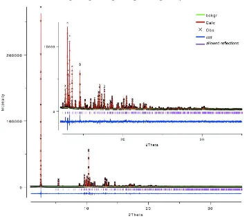
[image:6.610.126.484.269.529.2]The diffraction profiles and the differences between the measured and calculated profiles are shown in Fig. 2.
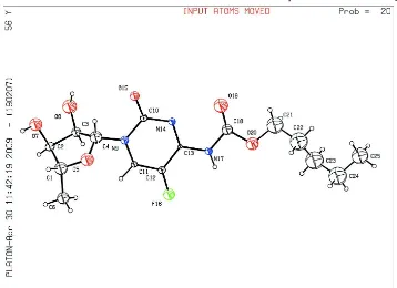
Figure 1
The molecular structure of capecitabine showing the atomic numbering. Displacement spheres are drawn at the 20%
supporting information
sup-3
[image:7.610.127.484.76.395.2]Acta Cryst. (2009). E65, o1325–o1326
Figure 2
The final Rietveld plot showing the measured data (black thin-plus), calculated data (red line) and difference curve (blue
line). Calculated positions of the reflections are shown by verical bars.
5-deoxy-5-fluoro-N-(pentyloxycarbonyl)cytidine
Crystal data
C15H22FN3O6
Mr = 359.35
Orthorhombic, P212121
a = 5.20527 (2) Å
b = 9.52235 (4) Å
c = 34.77985 (13) Å
V = 1723.91 (1) Å3
Z = 4
F(000) = 760
Dx = 1.385 Mg m−3
Synchrotron radiation, λ = 0.79483(4) Å
µ = 0.15 mm−1
T = 293 K
Particle morphology: no specific habit white
cylinder, 40 × 1 mm
Specimen preparation: Prepared at 293 K and 101 kPa
Data collection
ID31 ESRF Grenoble diffractometer Radiation source: X-Ray Si(111) monochromator
Specimen mounting: 1.0 mm borosilicate glass capillary
Data collection mode: transmission Scan method: step
supporting information
sup-4
Acta Cryst. (2009). E65, o1325–o1326
Refinement
Least-squares matrix: full
Rp = 0.055
Rwp = 0.074
Rexp = 0.036
RBragg = 0.102
χ2 = 4.452 11333 data points Excluded region(s): no
Profile function: Pseudo-Voigt profile
coefficients as parameterized in Thompson et al.
(1987), asymmetry correction according to Finger et al. (1994)
91 parameters 77 restraints 6 constraints
H-atom parameters not refined
Weighting scheme based on measured s.u.'s w = 1/σ(Yobs)2
(Δ/σ)max = 0.05
Background function: Shifted Chebyschev Preferred orientation correction: March–Dollase
(Dollase, 1986); direction of preferred orientation 001, texture parameter r = 1.03(1)
Fractional atomic coordinates and isotropic or equivalent isotropic displacement parameters (Å2)
x y z Uiso*/Ueq Occ. (<1)
C1 −0.0205 (8) 0.8964 (3) 0.86415 (10) 0.087 (5)*
C2 0.0063 (7) 0.7423 (4) 0.87424 (8) 0.048 (5)*
C3 0.0924 (6) 0.6753 (3) 0.83655 (8) 0.049 (4)*
C4 −0.0166 (5) 0.7766 (2) 0.80775 (7) 0.081 (5)*
O5 −0.0717 (9) 0.9090 (3) 0.82416 (10) 0.093 (3)*
C6 0.2118 (13) 0.9888 (6) 0.87530 (18) 0.079 (4)*
O7 −0.2355 (9) 0.6775 (5) 0.88107 (14) 0.088 (3)*
O8 0.0594 (11) 0.5279 (3) 0.83793 (13) 0.109 (3)*
N9 0.1175 (4) 0.79531 (18) 0.77283 (7) 0.036 (4)*
C10 0.0276 (4) 0.73076 (17) 0.73805 (7) 0.030 (4)*
C11 0.3307 (5) 0.87392 (18) 0.77201 (7) 0.023 (4)*
C12 0.4772 (3) 0.90315 (14) 0.73950 (6) 0.031 (4)*
C13 0.3691 (3) 0.83732 (13) 0.70512 (6) 0.010 (4)*
N14 0.1675 (4) 0.75150 (16) 0.70410 (6) 0.028 (4)*
O15 −0.1690 (5) 0.6596 (2) 0.73930 (11) 0.046 (3)*
F16 0.6861 (5) 0.98180 (17) 0.74183 (10) 0.072 (2)*
N17 0.4922 (3) 0.86898 (14) 0.67035 (6) 0.030 (3)*
C18 0.4009 (4) 0.8094 (2) 0.63692 (7) 0.063 (5)*
O19 0.2448 (4) 0.7158 (3) 0.63482 (12) 0.108 (3)*
O20 0.5359 (5) 0.8859 (3) 0.60977 (10) 0.087 (4)*
C21 0.491 (4) 0.8346 (15) 0.57240 (14) 0.146 (6)* 0.53 (5)
C22 0.524 (3) 0.957 (2) 0.5449 (2) 0.169 (8)* 0.53 (5)
C23 0.801 (3) 0.9940 (19) 0.5361 (5) 0.174 (9)* 0.53 (5)
C24 0.817 (4) 1.1183 (13) 0.5087 (4) 0.174 (10)* 0.53 (5)
C25 0.700 (5) 1.082 (2) 0.4695 (5) 0.143 (9)* 0.53 (5)
C21a 0.518 (5) 0.8251 (19) 0.57299 (18) 0.146 (6)* 0.47 (5)
C22a 0.680 (3) 0.9142 (19) 0.54603 (17) 0.169 (8)* 0.47 (5)
C23a 0.560 (3) 0.939 (2) 0.5068 (4) 0.174 (9)* 0.47 (5)
C24a 0.764 (5) 0.9452 (15) 0.4756 (2) 0.174 (10)* 0.47 (5)
C25a 0.925 (4) 1.079 (2) 0.4786 (7) 0.143 (9)* 0.47 (5)
H251 0.7123 1.1617 0.453 0.25* 0.53 (5)
H252 0.5245 1.0576 0.4727 0.25* 0.53 (5)
supporting information
sup-5
Acta Cryst. (2009). E65, o1325–o1326
H241 0.7261 1.1953 0.5195 0.25* 0.53 (5)
H242 0.9921 1.1435 0.5053 0.25* 0.53 (5)
H231 0.8866 1.0173 0.5594 0.25* 0.53 (5)
H232 0.8831 0.9152 0.5246 0.25* 0.53 (5)
H221 0.4433 1.0371 0.5559 0.25* 0.53 (5)
H222 0.4406 0.9338 0.5214 0.25* 0.53 (5)
H211 0.3216 0.7981 0.5706 0.25* 0.53 (5)
H212 0.6111 0.7627 0.5664 0.25* 0.53 (5)
H61 0.1794 1.0833 0.868 0.1*
H62 0.2378 0.9842 0.9023 0.1*
H63 0.361 0.9557 0.8624 0.1*
H21 0.1249 0.7267 0.8946 0.075*
H31 0.273 0.6894 0.8356 0.075*
H11 −0.166 0.9315 0.8775 0.12*
H41 −0.1786 0.7386 0.8007 0.12*
H111 0.3869 0.9132 0.7957 0.03*
H171 0.6224 0.9246 0.6699 0.04*
H82 −0.0753 0.5066 0.8272 0.1*
H72 −0.216 0.592 0.883 0.12*
H2511 1.0505 1.0802 0.4588 0.25* 0.47 (5)
H2512 1.008 1.082 0.5029 0.25* 0.47 (5)
H2513 0.8164 1.1589 0.476 0.25* 0.47 (5)
H2411 0.874 0.8661 0.478 0.25* 0.47 (5)
H2412 0.6824 0.943 0.4511 0.25* 0.47 (5)
H2311 0.4682 1.0252 0.5072 0.25* 0.47 (5)
H2312 0.4442 0.8643 0.5013 0.25* 0.47 (5)
H2211 0.7075 1.0029 0.5578 0.25* 0.47 (5)
H2212 0.8402 0.8684 0.5424 0.25* 0.47 (5)
H2111 0.5817 0.7316 0.5736 0.25* 0.47 (5)
H2112 0.3442 0.8245 0.5647 0.25* 0.47 (5)
Geometric parameters (Å, º)
C1—C2 1.515 (5) O20—C21 1.408 (2)
C1—O5 1.421 (5) O20—C21a 1.407 (2)
C1—C6 1.545 (7) C21—C22 1.518 (2)
C1—H11 0.950 C21—H211 0.949 (16)
C2—C3 1.525 (4) C21—H212 0.95 (2)
C2—O7 1.422 (6) C22—C23 1.520 (2)
C2—H21 0.950 C22—H221 0.95 (2)
C3—C4 1.502 (4) C22—H222 0.950 (9)
C3—O8 1.413 (4) C23—C24 1.522 (2)
C3—H31 0.950 C23—H231 0.950 (19)
C4—O5 1.413 (4) C23—H232 0.95 (2)
C4—N9 1.4123 (19) C24—H241 0.949 (19)
C4—H41 0.950 C24—H242 0.95 (2)
C6—H61 0.950 C25—C24 1.530 (2)
supporting information
sup-6
Acta Cryst. (2009). E65, o1325–o1326
C6—H63 0.950 C25—H252 0.95 (3)
O7—H72 0.820 C25—H253 0.95 (2)
O8—H82 0.820 C21a—C22a 1.519 (2)
N9—C10 1.4352 (18) C21a—H2111 0.95 (3)
N9—C11 1.3389 (19) C21a—H2112 0.95 (3)
C10—N14 1.4015 (19) C22a—C23a 1.520 (2)
C10—O15 1.2282 (19) C22a—H2211 0.950 (15)
C11—C12 1.3919 (19) C22a—H2212 0.95 (2)
C11—H111 0.950 C23a—C24a 1.523 (2)
C12—C13 1.4625 (19) C23a—H2311 0.95 (2)
C12—F16 1.3228 (19) C23a—H2312 0.950 (18)
C13—N14 1.3305 (18) C24a—C25a 1.530 (2)
C13—N17 1.4013 (19) C24a—H2411 0.95 (2)
N17—C18 1.3783 (19) C24a—H2412 0.952 (18)
N17—H171 0.860 C25a—H2511 0.950 (19)
C18—O19 1.208 (2) C25a—H2512 0.95 (3)
C18—O20 1.384 (2) C25a—H2513 0.95 (3)
O15···C12i 2.961 (3) O15···C11iii 2.875 (3)
F16···C10ii 2.886 (3)
C2—C1—O5 109.0 (3) O20—C21—H212 110.1 (17)
C2—C1—C6 114.9 (2) C22—C21—H211 110.1 (16)
C2—C1—H11 107.52 C22—C21—H212 109.9 (6)
O5—C1—C6 110.2 (2) H211—C21—H212 109.4 (9)
O5—C1—H11 107.4 C21—C22—C23 114.3 (2)
C6—C1—H11 107.5 C21—C22—H221 108.2 (6)
C1—C2—C3 103.46 (14) C21—C22—H222 108.2 (14)
C1—C2—O7 112.16 (19) C23—C22—H221 108.3 (14)
C1—C2—H21 112.53 C23—C22—H222 108.3 (12)
C3—C2—O7 102.85 (18) H221—C22—H222 109.5 (16)
C3—C2—H21 112.59 C22—C23—C24 110.9 (2)
O7—C2—H21 112.5 C22—C23—H231 109.1 (13)
C2—C3—C4 101.19 (13) C22—C23—H232 109.1 (15)
C2—C3—O8 110.48 (18) C24—C23—H231 109.1 (15)
C2—C3—H31 105.17 C24—C23—H232 109.1 (12)
C4—C3—O8 127.86 (19) H231—C23—H232 109.5 (16)
C4—C3—H31 105.07 C23—C24—C25 111.3 (2)
O8—C3—H31 105.13 C23—C24—H241 109.1 (12)
C3—C4—O5 112.34 (14) C23—C24—H242 109.0 (16)
C3—C4—N9 117.90 (12) C25—C24—H241 109 (2)
C3—C4—H41 105.26 C25—C24—H242 109.0 (17)
O5—C4—N9 109.57 (17) H241—C24—H242 109.4 (11)
O5—C4—H41 105.29 C24—C25—H251 110 (2)
N9—C4—H41 105.37 C24—C25—H252 110 (2)
C1—O5—C4 106.4 (3) C24—C25—H253 109.6 (17)
C1—C6—H61 109.5 H251—C25—H252 109.3 (19)
supporting information
sup-7
Acta Cryst. (2009). E65, o1325–o1326
C1—C6—H63 109.4 H252—C25—H253 109 (2)
H61—C6—H62 109.4 O20—C21a—C22a 107.2 (2)
H61—C6—H63 109.4 O20—C21a—H2111 110.1 (16)
H62—C6—H63 109.6 O20—C21a—H2112 109.9 (18)
C2—O7—H72 109.5 C22a—C21a—H2111 110.2 (17)
C3—O8—H82 109.47 C22a—C21a—H2112 110.1 (13)
C4—N9—C10 120.62 (14) H2111—C21a—H2112 109.4 (6)
C4—N9—C11 119.91 (14) C21a—C22a—C23a 114.4 (2)
C10—N9—C11 119.47 (12) C21a—C22a—H2211 108.3 (6)
N9—C10—N14 118.71 (13) C21a—C22a—H2212 108.2 (15)
N9—C10—O15 118.59 (15) C23a—C22a—H2211 108.2 (19)
N14—C10—O15 122.71 (15) C23a—C22a—H2212 108.3 (10)
N9—C11—C12 125.65 (14) H2211—C22a—H2212 109.4 (10)
N9—C11—H111 117.16 C22a—C23a—C24a 111.0 (2)
C12—C11—H111 117.19 C22a—C23a—H2311 109 (2)
C11—C12—C13 111.59 (12) C22a—C23a—H2312 109.0 (11)
C11—C12—F16 120.89 (15) C24a—C23a—H2311 109.1 (12)
C13—C12—F16 127.52 (14) C24a—C23a—H2312 109.2 (19)
C12—C13—N14 126.04 (12) H2311—C23a—H2312 109.5 (16)
C12—C13—N17 115.94 (14) C23a—C24a—C25a 111.4 (2)
N14—C13—N17 118.02 (18) C23a—C24a—H2411 109.0 (10)
C10—N14—C13 118.29 (13) C23a—C24a—H2412 109 (2)
C13—N17—C18 118.81 (13) C25a—C24a—H2411 109 (3)
C13—N17—H171 120.56 C25a—C24a—H2412 109.0 (16)
C18—N17—H171 120.63 H2411—C24a—H2412 109.4 (11)
N17—C18—O19 125.88 (16) C24a—C25a—H2511 110 (2)
N17—C18—O20 100.60 (15) C24a—C25a—H2512 110 (2)
O19—C18—O20 133.52 (16) C24a—C25a—H2513 109.6 (17)
C18—O20—C21 111.3 (2) H2511—C25a—H2512 109 (2)
C18—O20—C21a 111.7 (2) H2511—C25a—H2513 109 (2)
O20—C21—C22 107.3 (2) H2512—C25a—H2513 110 (2)
O20—C21—H211 110.0 (9)
Symmetry codes: (i) x−1, y, z; (ii) −x+1, y+1/2, −z+3/2; (iii) −x, y−1/2, −z+3/2.
Hydrogen-bond geometry (Å, º)
D—H···A D—H H···A D···A D—H···A
N17—H171···O8ii 0.860 1.956 2.797 (5) 170