Original citation:
Suberu, John O., Romero-Canelón, Isolda, Sullivan, Neil, Lapkin, Alexei A. and Barker,
Guy C.. (2014) Comparative cytotoxicity of artemisinin and cisplatin and their
interactions with chlorogenic acids in MCF7 breast cancer cells. ChemMedChem,
Volume 9 (Number 12). pp. 2791-2797
Permanent WRAP url:
http://wrap.warwick.ac.uk/65535
Copyright and reuse:
The Warwick Research Archive Portal (WRAP) makes this work of researchers of the
University of Warwick available open access under the following conditions.
This article is made available under the Creative Commons Attribution 4.0 International
license (CC BY 4.0) and may be reused according to the conditions of the license. For
more details see:
http://creativecommons.org/licenses/by/4.0/
A note on versions:
The version presented in WRAP is the published version, or, version of record, and may
be cited as it appears here.
DOI: 10.1002/cmdc.201402285
Comparative Cytotoxicity of Artemisinin and Cisplatin and
Their Interactions with Chlorogenic Acids in MCF7 Breast
Cancer Cells
John O. Suberu,
[b]Isolda Romero-Caneln,
[c]Neil Sullivan,
[d]Alexei A. Lapkin,
[b]and
Guy C. Barker*
[a]Introduction
Cancer is a major public health problem with about 7.6 million deaths in 2008; this number is projected to increase to over 13 million in 2030.[1] Although a range of treatment options are
available, in many cases these therapies are fraught with signif-icant levels of toxicity to healthy cells, and drug resistance quickly develops in some treatment regimes. To decrease the current cancer burden, drug discovery is directed at the devel-opment of highly effective and potent medications with re-duced side effects.
Natural products are a rich source of active principles against cancer cells. A major success of pharmacognosy is the isolation of paclitaxel (Taxol) from the bark of the Pacific yew tree (Taxus brevifola). Paclitaxel exerts its anticancer effect by inhibiting mitosis and is now a drug approved by the US Food and Drug Administration (FDA) for ovarian and breast can-cers.[2]Another success is the antimalarial compound
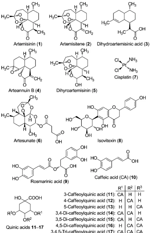
artemisi-nin (1), a sesquiterpene lactone derived from Artemisia annuaL. that possesses a unique trioxane bridge (Figure 1).
The in vitro cytotoxic activity of artemisinin and its deriva-tives has been reported in various cancer cell lines, including drug-resistant lines.[3]Sigh and Lai showed that a combination
In parts of Africa and Asia, self-medication with a hot water in-fusion ofArtemisia annua(Artemisiatea) is a common practice for a number of ailments including malaria and cancer. In our earlier work, such an extract showed better potency than arte-misinin alone against both chloroquine-sensitive and -resistant parasites. In this study, in vitro tests of the infusion in MCF7 cells showed high IC50 values (>200mm). The combination of
[image:2.595.304.548.336.715.2]artemisinin and 3-caffeoylquinic acid (3CA), two major compo-nents in the extract, was strongly antagonistic and gave a near total loss of cytotoxicity for artemisinin. We observed that the interaction of 3CAs with another cytotoxic compound, cispla-tin, showed potentiation of activity by 2.5-fold. The chelation of cellular iron by 3CA is hypothesized as a possible explana-tion for the loss of artemisinin activity.
Figure 1.Structures of some compounds found inArtemisiaaqueous extract (tea) and cisplatin.
[a]Dr. G. C. Barker
School of Life Sciences, University of Warwick, CV4 7AL (UK) E-mail: Guy.Barker@warwick.ac.uk
[b]Dr. J. O. Suberu, Prof. A. A. Lapkin Chemical Engineering and Biotechnology University of Cambridge, CB2 3RA (UK)
[c] Dr. I. Romero-Caneln
Department of Chemistry, University of Warwick, CV4 7AL (UK)
[d]Dr. N. Sullivan
SensaPharm Ltd., 123i Bioscience Centre, Sunderland, SR5 2TA (UK)
of dihydroartemisinin (5) and holotransfferin effectively kills ra-diation-resistant breast cancer cells,[3b] while artemisinin
pre-treated with holotransfferin was also found to be effective toward both drug-sensitive and multi-drug-resistant human lung carcinoma (SCLC) cells.[3c] Artesunate (6) inhibited the
growth of highly angiogenic Kaposi sarcoma cells, showing the anti-angiogenesis effect of artemisinins.[4] In their study, Chen
et al. implanted nude mice with human ovarian cancer cells and found that artesunate decreased tumor growth and signif-icantly lowered vascular endothelial growth factor (VEGF) ex-pression in the cells.[5]The potential of artemisinin to prevent
the development of breast cancer in rats treated with a known carcinogen (7,12-dimethylbenz[a]anthracene, DMBA) has been reported.[6]Artesunate has also been successfully used in
com-bination with standard chemotherapy to treat metastatic mela-noma in human subjects after standard chemotherapy alone was ineffective in stopping tumor growth.[7]
Artemisinin and its derivatives have also been used as che-mosensitizers for conventional treatments in drug-resistant cancer cell lines.[8]Synergistic interaction of dihydroartemisinin
with gemcitabine, a cancer drug, showed a 45 % enhancement in tumor growth inhibition compared with the drug alone.[9]
The improved efficacy of multicomponent combinations in-volving artemisinin in cancer treatment has encouraged inves-tigation of other natural compounds besides artemisinin that may exhibit individual cytotoxic activity or that can be poten-tial artemisinin synergists in the crude extract. Two artemisinin-related compounds, artemisitene (2) and arteannuin B (4), and two unrelated ones, scopoletin and 1,8-cineole, have shown antiproliferative activity.[10] No cross-resistance to artemisinin
was observed with any of these actives, thus showing a poten-tial for use in combination to treat drug-resistant tumors.
In the artemisinin research community, a significant degree of interest has been focused on the activity of aqueous ex-tracts (Artemisia tea).[11] This interest stems largely from the
widely reported use of Artemisia aqueous infusions in folk medicine. Through in vitro tests, we recently showed that some constituents of the extract interact synergistically with artemisinin, resulting in increased antiplasmodial activity.[12]
Consequently, we were interested in the interactions of artemi-sinin with co-metabolites in the extract, in view of improved cytotoxicity.
Carbonara et al. observed that the major constituents of Ar-temisiatea are chlorogenic acids (11–17).[13]They also detected
a number of feruloylquinic acids together with some flavo-noids in the extract. Chlorogenic or caffeoylquinic acids (CQAs) are esters of caffeic (10) and quinic acids. The pharmacological properties of these catechols include antioxidant, hepatopro-tectant, antibacterial, antihistiminic, chemopreventive, and other biological effects.[14] Lee and Zhu showed that
chloro-genic acids and other catechol-containing dietary polyphenols can inhibit the methylation of synthetic DNA substrates in vitro and can inhibit the methylation of the promoter region of the RAbgene in human breast cancer cells; both are nor-mally hypermethylated in neoplastic cells.[15]In their study,
Nor-atto et al. showed the chemopreventive potential of dietary chlorogenic and neochlorogenic acids.[16]These compounds
ex-erted relatively high growth inhibition on the estrogen-inde-pendent breast cancer cell line and low toxicity in normal cells. Chlorogenic acid derivatives were also found to inhibit hepato-cellular carcinoma cell line proliferation and induced apoptosis in leukemia cell lines.[17]
This study was therefore undertaken to evaluate the in vitro cytotoxicity toward breast cancer cells ofArtemisiatea and ar-temisinin in combination with co-metabolites present in the tea extract. It specifically looks at the interaction of artemisinin with chlorogenic acid (3-caffeoylquinic acid, 3CA) to assess possible implications for the use of Artemisia tea in cancer therapy and compares this with cisplatin’s interaction with 3CA in MCF7 cells.
Results and Discussion
The metabolite profile and cytotoxic activity of Artemisia hot water infusion (tea) in MCF7 breast cancer cells was evaluated and compared with the activity of artemisinin alone and in combination with chlorogenic acid, a co-metabolite in the ex-tract. A combination study of chlorogenic acid with the drug cisplatin [cis-diamminedichloroplatinum(II)] was carried out for comparative analysis and possible elucidation of the interac-tions inArtemisiatea extract.
Composition ofArtemisiatea
The profile of metabolites in aqueous extract is listed in Table 1. These were analyzed by both MS–MS and HPLC
meth-ods. The profiling is based on the extensive analysis by Carbo-nara et al.[13]and on our earlier work[18]with artemisinin-related
compounds in the extracts.
The levels of artemisinin reported in the tea extracts are varied and the values obtained in this study (47.50.8 mgL1)
are within that range. This corresponds to about 16 % of clini-cal exposure to the drug in reported treatment regimes, which also showed that the bioavailability of artemisinin in tea is very similar to pure artemisinin administered as a capsule.[13, 19]
[image:3.595.305.549.461.579.2]Van der Kooy and Verpoorte[19a] have shown that the method
Table 1.Metabolites in the aqueousArtemisiaextract analyzed by both MS–MS and HPLC methods.
Compound Amount [mg (Ltea)1][a]
artemisinin 47.500.80
arteannuin B 1.300.01
caffeic acid 0.800.03
3,5-dicaffeoylquinic acid 57.001.70
3-caffeoylquinic acid 72.001.60
4-caffeoylquinic acid 20.401.60
4,5-dicaffeoylquinic acid 31.604.00
5-caffeoylquinic acid 9.000.70
isovitexin 65.007.20
rosmarinic acid 1.100.01
[a] Values are the meanSD ofn=2 determinations of triplicate meas-urements.
employed in preparing the hot water infusion does affect the amount of artemisinin and other co-metabolites extracted. This study, as well as others,[19a, b] used the therapeutically
recom-mended ratio of 1:200 w/v or 5 gL1.[20] Arteannuin B (5)
(1.3 mgL1), a biosynthetic precursor of artemisinin, was also
detected in the tea extract using our method.[18]
The most abundant of the caffeic derivatives11–17was 3-caffeoylquinic acid (11) (72 mgL1) in the analyzed extract,
fol-lowed by 3,5-dicaffeoylquinic acid (15) (57 mgL1). A
compara-tively lower amount (0.8 mgL1) was observed for caffeic acid
(10). The only flavonoid analyzed was isovitexin (8) (65 mgL1)
and was relatively abundant in our extract. Rosmarinic acid (9) was lower (1.1 mgL1) in our samples than the levels found by
de Magalh¼es and co-workers.[19c]
Cytotoxicity of cisplatin, artemisinin, and 3CA
Table 2 shows the 50 % inhibitory concentration for artemisinin, cisplatin and 3CA in MCF7 breast cancer cells. This cell line is derived from breast adenocarcinoma tissues and is a common model employed in carcinogenesis and chemopreventive stud-ies.[21]
Cytotoxicity of artemisinin
The cytotoxicity of artemisinin (Table 2 and Figure 2) in the MCF7 cells shows it to be potent against invasive breast ductal carcinoma that is estrogen sensitive. The IC50values obtained
for the compound (9.130.07mm) are within a range of values
(IC500.17–87.10mm) reported by Efferth and Oesch for
artemisi-nin and its derivatives determined for the tumor panel of 60 cell lines of the National Cancer Institute (NCI) screening pro-gram.[22]Artemisinin had the highest IC
50value (least potent) of
all the related derivatives reported. Artemisinin is metabolized into dihydroartemisinin (DHA), which has a lower IC50 value
(2.3mm) in MCF7 cells[22, 23]
Several researchers have investigated the mechanism of se-lective cytotoxicity of artemisinin and its derivatives toward neoplastic cells. Mercer et al. showed that selective activation of the trioxane bridge via carbon-centered radicals occurs in rapidly dividing or susceptible cells.[24]This then results in
mito-chondrial membrane depolarization, leading to induction of apoptosis by the chemical stress pathway and activation of caspases-3 and -7 in HL-60 cells, resulting in degraded DNA or hypodiploidy. Li et al. also showed that artemisinin derivatives induce apoptosis mainly through G1arrest.[25] The G1 phase is
associated with increased iron intake and transfferin receptor expression. Down-regulation of anti-apoptotic Bcl-2 proteins and up-regulation of pro-apoptotic Bax proteins have been as-sociated with artesunate-treated human vein endothelial cells. Artemisinins have also been associated with lowered vascular endothelial growth factor (VEGF) expression. VEGFs are potent angiogenic factors.[26] These studies suggest that the
mecha-nism(s) for the cytotoxicity of artemisinins involves many differ-ent pathways.
Activity of cisplatin
Cisplatin showed superior cytotoxicity in MCF7 cells compared with artemisinin (Table 2 and Figure 3). The mean IC50 value
obtained (5.750.07mm) is similar to values reported by Isik-Table 2.IC50values for artemisinin, cisplatin and 3CA in MCF7 cells.
Compound IC50[mm][a]
artemisinin 9.130.07
cisplatin 5.750.02
3-caffeoylquinic acid 126.980.13
[image:4.595.309.544.64.243.2][a] Values are the meanSD ofn=2 determinations of triplicate meas-urements.
[image:4.595.49.289.372.419.2]Figure 2.A dose–response curve for artemisinin in MCF7 cells. Percentage cell survival is plotted against the logarithm of treatment concentrations. Data points are the meansSD of duplicate determinations of triplicate measurements.
[image:4.595.309.542.521.700.2]dag et al. (IC508.6mm) using MCF7 cells and the same duration
of drug exposure.[27] Although cisplatin is very effective with
solid-type carcinoma, drug resistance and toxic side effects have also been reported.[28]
As a platinum-based drug, cisplatin (7) exerts its cytotoxic effect through multiple mechanisms of which the most impor-tant and the best understood involves interaction with DNA to form GG intrastrand DNA cross-links, leading to the activation of several signal transduction pathways and culminating in the induction of mitochondrial apoptosis.[29] Consistent initial
re-sponses have been obtained by cisplatin treatment. However, these often result in the development of chemoresistance and therapeutic failure.[30]The combination of cisplatin with a
che-mosensitizer or a synergist can potentially improve efficacy and restore sensitivity to cisplatin.[31]
Cytotoxicity of 3CA
The IC50 for 3CA in MCF-7 cells (127.00.8mm) was highest
among the three single agents tested (Figure 4). This is similar to the observation by Lee et al., who reported that the growth inhibition of MCF-7 cells by 3CA was insignificant up to 20mm
and only inhibited by about 15 % at 50mm concentration.[15a]
Therefore, 50 % growth inhibition at a concentration of 126.9
0.1mm, which we obtained, is in the range of the reported
values. The chemopreventive and antiproliferation effects of 3-caffeoylquinic acid along with other dietary derivatives have also been reported.[16, 17]
The cytotoxicity of 3CA is dose dependent and is observable only above a certain concentration.[15a] Controversial and
con-flicting experimental results have been observed[32]in trials
in-volving endogenous antioxidants such as 3CA because of their “double-edged sword” effect at cellular redox sites. Depending on the dosage level and the in situ matrix, these compounds can either be pro-oxidative or antioxidative.
Cytotoxicity ofArtemisiatea
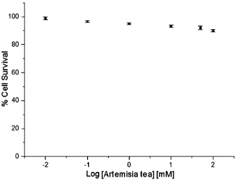
The dose–response forArtemisiatea is shown in Figure 5. Sev-eral repeat analyses gave values above 200mm. The high IC
50
values observed with tea and the complex composition of the same necessitated examination of the interactions among fewer metabolites. The combination of artemisinin with 3CA was subsequently investigated. In our analysis of the tea, 3CA was one of the most abundant components, second only to isovitexin (Table 1). Our choice of 3CA was also informed by its prominent dietary profile.
Cytotoxic combination studies
The cytotoxicity ofArtemisiatea was investigated to assess its possible role in cancer therapy. The unexpectedly high IC50
value for the phyto complex led to the investigation of simpler combinations in the extract. The combination of artemisinin and 3CA resulted in a drastic modification of artemisinin’s ac-tivity (Table 3). Consequently, we investigated the interaction of 3CA with cisplatin to see if a similar effect is reproduced in another anticancer drug.
[image:5.595.310.542.216.395.2]The combination of caffeic and chlorogenic acids with che-motherapeutic agents as chemosensitisers has been reported. An increased sensitivity of multidrug-resistant breast cancer cells (MCF-7/Dox) to doxorubicin was observed with caffeic
Figure 5.A dose–response curve forArtemisiatea extract in MCF7 cells. Per-centage cell survival is plotted against the logarithm of treatment concentra-tions. Data points are the meansSD of duplicate determinations of tripli-cate measurements.
Table 3.IC50values for 3CA in combination with artemisinin and cisplatin in MCF7 cells.
Compounds IC50[mm][a]
artemisinin+3CA >200
cisplatin+3CA 2.270.06
[image:5.595.50.292.530.711.2][a] Values are the meanSD ofn=2 determinations of triplicate meas-urements.
Figure 4.An IC50curve for 3CA in MCF-7 cells. Percentage cell survival is plotted against the logarithm of treatment concentrations. Data points are the meansSD of duplicate determinations of triplicate measurements.
acid.[33]A US patent for the use of chlorogenic acid as a
sensitiz-er for chemothsensitiz-erapeutic agents reported a 30 % decrease in the viability of cancer cells sensitized by chlorogenic acids to doxorubicin compared with cells administered with doxorubi-cin alone.[34]
Artemisinin combination with 3CA
The dose–response curve for the combination of artemisinin and 3CA at a 1:1 molar ratio is shown in Figure 6. The IC50in
MCF-7 cells for the combination is >200mm. This represents
a near complete loss of cytotoxicity for the compound in the presence of 3CA (Table 2). A similar loss of activity was ob-served in combinations involving lower 3CA concentrations with artemisinin (artemisinin/3CA, 1:0.5 and 1:0.01). This sug-gests an antagonistic interaction between artemisinin and 3CA when combined and may partly explain the high IC50 values
observed.
Cisplatin combination with 3CA
To investigate if the strong antagonistic interaction observed for artemisinin and 3CA is reproduced in other anticancer agents, an equimolar combination of cisplatin and 3CA was tested. An IC50 value of 3.6mm was obtained from the dose–
response curve (Figure 7). This represents a 2.5-fold improve-ment in potency relative to cisplatin alone (Table 2). Kim re-ported the chemosensitizing effect of chlorogenic acids, and an improvement in activity of doxorubicin was observed when combined with chlorogenic acid in a range of combinations.[34]
The mechanism for the interaction of 3CA in MCF-7 with ar-temisinin (antagonism) and cisplatin (potentiation) seem to be pharmacokinetic in nature, where the observed change in ac-tivity for a combination relative to the single agent is due to
modification in the absorption, distribution, metabolism, or ex-cretion of a compound (cisplatin and artemisinin) by another (3CA).[35] The improvement in cytotoxicity of the combination
of cisplatin and 3CA over cisplatin alone is less likely due to 3CA cytotoxicity, which is shown to be relatively inactive (Table 2).
The activation of artemisinin and the cleavage of the endo-peroxide bridge to form a carbon-centered radical and/or reac-tive oxygen species (ROS) is a key to the compound’s cytotox-icity and antiplasmodial activities. This activation has been sug-gested to be initiated by endogenous iron, which is relatively abundant in actively dividing cells relative to normal cells.[3d]
Kono et al. and others reported that 3CA has iron-chelating properties and forms a complex with the metal.[36]In a
combi-nation with artemisinin, 3CA may chelate and complex with endogenous iron, and as a result depletes the iron pool avail-able for the activation of artemisinin. This effect will be more pronounced in the cytotoxic activity of artemisinin relative to its antiplasmodial activity, because the erythrocytic iron pool is severalfold more abundant than the neoplastic cell iron pool.[36a]
This is consistent with our observations for the combination in both antiplasmodial and cytotoxic assays. In the previous work, a mild antagonism was observed for the antiplasmodial activity of 3CA and artemisinin combination.[12] In the above
cytotoxicity assay, strong antagonism (or a near total loss of ac-tivity) was observed (Figure 6). Williamson has reviewed similar adverse reactions (ADRs) in herbals and the methods by which antagonism arises in these mixtures.[37]
In contrast, cisplatin is activated in the cell by aquation of the molecule, resulting in the loss of one or both of its chloride ions. The activation is enhanced by a lower intracellular chlo-ride ion concentration than its extracellular concentration.[28b]
[image:6.595.310.542.66.247.2]Metal ions do not seem to play any role in cisplatin activation and are thus unaffected by the metal-chelating properties of 3CA. It would be interesting to investigate whether cisplatin
[image:6.595.53.286.310.493.2]Figure 6.A dose–response curve for MCF-7 cells treated with a combination of artemisinin and 3CA. Percentage cell survival is plotted against the loga-rithm of treatment concentrations. Data points are the meansSD of dupli-cate determinations of triplidupli-cate measurements.
can chelate with CO2 and deprotonated OH (six-membered
ring) of 3CA.
Conclusions
This study investigated in vitro the use of Artemisia tea as a chemotherapeutic agent using MCF7 cells. The high IC50 value observed for the tea extract led to the investigation of the combinations of 3-caffeoylquinic acid (3CA), a major com-ponent of the tea, with artemisinin, the main active ingredient in the extract. The combination showed a near total loss (strong antagonism) of cytotoxicity. This was in contrast to a 2.5-fold improvement observed when 3CA was combined with cisplatin, another anticancer agent. An explanation was suggested for these observations and also a possible reason was advanced for the difference in antiplasmodial and cytotox-icity of 3CA combination with artemisinin via endogenous iron-mediated activation of the artemisinin molecule.
Based on these results, the use of Artemisia tea in cancer therapeutics seems at best unpredictable and at worst ineffec-tive. Further in vivo and in vitro investigations of the interac-tions between artemisinin with 3CA and other dietary antioxi-dants is imperative before any recommendation for the use of artemisinin and its derivatives as antiproliferative drugs with the possible avoidance of antioxidant food and drink immedi-ately before and after intake of the drugs in single or combina-tion therapies can be established.
Experimental Section
Chemicals: Artemisinin (98 %), dimethyl sulfoxide (DMSO), chloro-genic acids, trichloroacetic acid (99 %), sulforhodamine B (SRB; 75 %), sodium phosphate monobasic monohydrate (99 %), sodium phosphate dibasic heptahydrate (99 %), acetic acid ( 99 %), and cisplatin were obtained from Sigma–Aldrich (Dorset, UK). Arteannuin B was gifted by Walter Reed Army Institute of Re-search (WRAIR) USA. LC–MS-grade formic acid in water, acetonitrile, and HPLC-grade acetonitrile were obtained from Fisher Scientific, UK. Purified water (~18 MWcm1
) was dispensed from a Milli Q system (Millipore, UK). For the in vitro assays, RPMI 1640 medium, as well as fetal bovine serum,l-glutamine, a penicillin/streptomy-cin mixture, trypsin, and phosphate-buffered saline (PBS) were pur-chased from PAA Laboratories GmbH (Germany).
Plant materials: High-yielding dried A. annua biomass was ob-tained from BIONEX Madagascar and stored under dark, cool con-ditions until use.
Plant extracts:Artemisiatea was prepared according to published methods with a slight modification.[11a, 38]
Briefly, 1 L of boiling water was added to 5 g of dried plant material, stirred and stored in the dark for 1 h. The extract was filtered in vacuo and lyophi-lized after freezing to obtain the dried tea extract. The ethanolic extract was obtained by sonication for 30 min in ethanol at 1:10 (w/v) biomass-to-solvent ratio. The sonication bath was kept cool with ice, and the extract was filtered and concentrated in vacuo at 308C, and further dried under a gentle stream of nitrogen gas. These extracts were used in the antiproliferation assays.
MS–MS method for artemisinins: The MS–MS method is described in detail elsewhere.[18] Briefly, the MS–MS system was operated
with an ESI interface in positive ionization mode (ESI+). The cone and desolvation gas flow rates were set at 45 and 800 L h1, re-spectively. MS parameters were automatically defined using Waters IntelliStart software for the tuning and calibration of the TQD and subsequently manually optimized for all analytes. Capillary voltage was set at 2.8 kV, collision voltage at 7 V, source temperature was 1508C, and cone voltage was set at 24 V. A multiple reaction-moni-toring (MRM) transition of 283!219+229+247+265 was used for artemisinin. Quantification was determined by using MRM modes for the above transitions. The dwell time was automatically set at 0.161 s. Data were acquired by MassLynx ver. 4.1 software and processed for quantification with QuanLynx ver. 4.1 (Waters Corp., Milford, MA, USA).
The HPLC system consisted of a binary pump, a cooling autosam-pler with an injection loop of 10mL set at 108C. The column heater was set at 308C and a GenesisLightn C18 column (100 2.1 mm, 4mm) (Grace, IL, USA) protected by an Acquity-LC column in-line filter unit (0.2mm in-line frit) was used for separation of metabo-lites. The mobile phase consisted of A: 0.1 % formic acid in water and B: 0.1 % formic acid in acetonitrile used in the following gradi-ent: 0–7.00 min, 25!98 % B; 7–9.5 min, 98 % B; 9.5–10 min, 98! 25 % B; 10–15 min, 25 % B at a flow rate of 0.4 mL min1. Weak wash solvent was 10 % acetonitrile, strong and needle wash sol-vent was a mixture of acetonitrile, propan-2-ol, methanol, and water (30:30:30:10v/v/v/v).
Cell culture: MCF7 human breast carcinoma cells were obtained from the European Collection of Cell Cultures (ECACC) and used between passages 5 and 18. The cells were grown in RPMI 1640 supplemented with 10 % fetal calf serum, 1 % 2 mm l-glutamine, and 1 % penicillin/streptomycin, as adherent monolayers at 310 K in a 5 % CO2humidified atmosphere and passaged at approximate-ly 70–80 % confluence.
In vitro growth inhibition assays: Briefly, 5000 cells were seeded per well in 96-well plates. The cells were pre-incubated in drug-free media at 310 K for 48 h before adding various concentrations of the compounds to be tested. Stock solutions of the compounds were first prepared in 5 % DMSO and a mixture 0.9 % saline and medium (1:1) following serial dilutions in RPMI 1640. The drug ex-posure period was 24 h. After this, supernatants were removed by suction, and each well was washed with PBS. A further 72 h was al-lowed for the cells to recover in drug-free medium at 310 K. The SRB assay was used to determine cell viability.[39]Absorbance meas-urements of the solubilized dye (on a BioRad iMark microplate reader using a 470 nm filter) allowed the determination of viable treated cells relative to untreated controls using the inflection point of a dose–response graph. IC50 values (concentrations that caused 50 % cell growth inhibition) were determined as duplicates of triplicate readings in two independent sets of experiments and their standard deviations were calculated.
IC50 modulation experiments: Experiments to investigate the
effect of co-administration of artemisinin and 3CA were carried out as described above, with the following modifications: cells were pre-incubated in drug-free medium for 48 h at 310 K, before adding artemisinin together with 3CA. To prepare stock solutions of the drug, the solid artemisinin was dissolved first in 5 % DMSO and then diluted in a 1:1 mixture of 0.9 % saline and the cell cul-ture medium. This stock was further diluted using RPMI 1640 until working concentrations were achieved. Separately, a stock solution of 3CA was prepared in a similar manner. Both solutions were added to each well independently, but within 5 min of each other. Once again the drug exposure time was 24 h and the drug-free
covery time was 72 h. The SRB assay was used to determine cell vi-ability. IC50values were determined as duplicates of triplicates in two independent sets of experiments and their standard deviations were calculated.
Acknowledgements
This study was funded by the Engineering and Physical Sciences Research Council (EPSRC, UK) and SensaPharm Ltd. via an Indus-trial CASE PhD studentship. The award was allocated competi-tively by the Chemistry Innovation Knowledge Transfer Network (CIKTN, UK). The authors acknowledge Prof. Peter Sadler for help-ful comments, Dr. Carol Jenner for editing the manuscript, and BIONEXX (Madagascar) and Charles Giblane for the supply of A. annua biomass.
Keywords: antagonism · Artemisia tea · artemisinin ·
chlorogenic acid·cisplatin·synergy
[1] J. Ferlay, H. R. Shin, F. Bray, D. Forman, C. Mathers, D. M. Parkin,Int. J. Cancer2010,127, 2893 – 2917.
[2] a) S. Nobili, D. Lippi, E. Witort, M. Donnini, L. Bausi, E. Mini, S. Capaccioli,
Pharmacol. Res.2009,59, 365 – 378; b) W. W. Hsiao, L. Liu,Planta Med. 2010,76, 1118 – 1131.
[3] a) A. M. Gravett, W. M. Liu, S. Krishna, W.-C. Chan, R. K. Haynes, N. L. Wilson, A. G. Dalgleish,Cancer Chemother. Pharmacol. 2011, 67, 569 – 577; b) N. P. Singh, H. C. Lai,Anticancer Res.2004,24, 2277 – 2280; c) D. Sadava, T. Phillips, C. Lin, S. E. Kane,Cancer Lett. 2002,179, 151 – 156; d) T. Efferth, A. Benakis, M. R. Romero, M. Tomicic, R. Rauh, D. Steinbach, R. Hfer, T. Stamminger, F. Oesch, B. Kaina,Free Radical Biol. Med.2004,
37, 998 – 1009.
[4] R. Dell’Eva, U. Pfeffer, R. Ven, L. Anfosso, A. Forlani, A. Albini, T. Efferth,
Biochem. Pharmacol.2004,68, 2359 – 2366.
[5] H.-H. Chen, H.-J. Zhou, G.-D. Wu, X.-E. Lou,Pharmacology2004,71, 1 – 9. [6] H. Lai, N. P. Singh,Cancer Lett.2006,231, 43 – 48.
[7] T. G. Berger, D. Dieckmann, T. Efferth, E. S. Schultz, J.-O. Funk, A. Baur, G. Schuler,Oncol. Rep.2005,14, 1599 – 1604.
[8] a) P. Reungpatthanaphong, S. Mankhetkorn,Biol. Pharm. Bull.2002,25, 1555 – 1561; b) W. M. Liu, A. M. Gravett, A. G. Dalgleish,Int. J. Cancer 2011,128, 1471 – 1480.
[9] S.-J. Wang, Y. Gao, H. Chen, R. Kong, H.-C. Jiang, S.-H. Pan, D.-B. Xue, X.-W. Bai, B. Sun,Cancer Lett.2010,293, 99 – 108.
[10] T. Efferth, F. Herrmann, A. Tahrani, M. Wink, Phytomedicine 2011, 18, 959 – 969.
[11] a) A. De Donno, T. Grassi, A. Idolo, M. Guido, P. Papadia, A. Caccioppola, L. Villanova, A. Merendino, F. Bagordo, F. P. Fanizzi,Trans. R. Soc. Trop. Med. Hyg.2012,106, 696 – 700; b) J. Mouton, O. Jansen, M. Frdrich, F. van der Kooy,Planta Med.2013,79, 468 – 470; c) C. H. Blanke, G. B. Nai-sabha, M. B. Balema, G. M. Mbaruku, L. Heide, M. S. Muller,Trop. Doct. 2008,38, 113 – 116; d) L. Heide,Trans. R. Soc. Trop. Med. Hyg.2006,100, 802.
[12] J. O. Suberu, A. P. Gorka, L. Jacobs, P. D. Roepe, N. Sullivan, G. C. Barker, A. A. Lapkin,PLoS ONE2013,8, e80790.
[13] T. Carbonara, R. Pascale, M. P. Argentieri, P. Papadia, F. P. Fanizzi, L. Villa-nova, P. Avato,J. Pharm. Biomed. Anal.2012,62, 79 – 86.
[14] a) A. Belkaid, J.-C. Currie, J. Desgagnes, B. Annabi,Cancer Cell Int.2006,
6, 7; b) B. Zhang, R. Yang, C. Z. Liu,Sep. Purif. Technol.2008,62, 480 – 483; c) R. Feng, Y. Lu, L. L. Bowman, Y. Qian, V. Castranova, M. Ding,J. Biol. Chem. 2005, 280, 27888 – 27895 ; d) P. Miketova, K. H. Schram, J.
Whitney, E. H. Kearns, B. N. Timmermann, J. Mass Spectrom. 1999,34, 1240 – 1252.
[15] a) W. J. Lee, B. T. Zhu,Carcinogenesis2006,27, 269 – 277; b) S. M. Sirchia, A. T. Ferguson, E. Sironi, S. Subramanyan, R. Orlandi, S. Sukumar, N. Sacchi,Oncogene2000,19, 1556.
[16] G. Noratto, W. Porter, D. Byrne, L. Cisneros-Zevallos,J. Agric. Food Chem. 2009,57, 5219 – 5226.
[17] a) U. H. Jin, J. K. Lee, S. K. Kang, J. K. Kim, W. H. Park, J. G. Kim, S. K. Moon, C. H. Kim,Life Sci.2005,77, 2760 – 2769 ; b) G. Bandyopadhyay, T. Biswas, K. C. Roy, S. Mandal, C. Mandal, B. C. Pal, S. Bhattacharya, S. Rak-shit, D. K. Bhattacharya, U. Chaudhuri,Blood2004,104, 2514 – 2522. [18] J. Suberu, L. Song, S. Slade, N. Sullivan, G. Barker, A. A. Lapkin,J. Pharm.
Biomed. Anal.2013,84, 269 – 277.
[19] a) F. van der Kooy, R. Verpoorte,Planta Med.2011,77, 1754 ; b) K. Rath, K. Taxis, G. Walz, C. H. Gleiter, S. M. Li, L. Heide,Am. J. Trop. Med. Hyg. 2004,70, 128 – 132; c) P. Melillo de Magalh¼es, I. Dupont, A. Hendrickx, A. Joly, T. Raas, S. Dessy, T. Sergent, Y.-J. Schneider,Food Chem.2012,
134, 864 – 871; d) C. W. Wright, P. A. Linley, R. Brun, S. Wittlin, E. Hsu,
Molecules2010,15, 804 – 812.
[20] M. Willcox,J. Alternative Compl. Med.2009,15, 101 – 109.
[21] J. Paluszczak, V. Krajka-Kuz´niak, W. Baer-Dubowska,Toxicol. Lett.2010,
192, 119 – 125.
[22] T. Efferth, F. Oesch,Biochem. Pharmacol.2004,68, 3 – 10.
[23] R. Hooft van Huijsduijnen, R. K. Guy, K. Chibale, R. K. Haynes, I. Peitz, G. Kelter, M. A. Phillips, J. L. Vennerstrom, Y. Yuthavong, T. N. C. Wells,PLoS ONE2013,8, e82962.
[24] A. E. Mercer, J. L. Maggs, X.-M. Sun, G. M. Cohen, J. Chadwick, P. M. O’Neill, B. K. Park,J. Biol. Chem.2007,282, 9372 – 9382.
[25] Y. Li, F. Shan, J.-M. Wu, G.-S. Wu, J. Ding, D. Xiao, W.-Y. Yang, G. Atassi, S. Lonce, D.-H. Caignard,Bioorg. Med. Chem. Lett.2001,11, 5 – 8. [26] G.-D. Wu, H.-J. Zhou, X.-H. Wu,Vasc. Pharmacol.2004,41, 205 – 212. [27] I. Isikdag, Y. Ozkay, Z. Incesu,Turkish J. Pharm. Sci.2011,8, 178 – 188. [28] a) B. Tegze, Z. Szllsi, I. Haltrich, Z. Pnzvlt, Z. Tth, I. Lik, B. Gyo˝rffy,
PLoS ONE2012,7, e30804; b) A. Florea, D. Bsselberg,Cancers2011,3, 1351 – 1371.
[29] Z. H. Siddik,Oncogene2003,22, 7265 – 7279.
[30] L. Galluzzi, L. Senovilla, I. Vitale, J. Michels, I. Martins, O. Kepp, M. Caste-do, G. Kroemer,Oncogene2012,31, 1869 – 1883.
[31] D. Chirnomas, T. Taniguchi, M. de La Vega, A. P. Vaidya, M. Vasserman, A.-R. Hartman, R. Kennedy, R. Foster, J. Mahoney, M. V. Seiden, Mol. Cancer Ther.2006,5, 952 – 961.
[32] a) E. G. Yordi, E. M. Prez, M. J. Matos, E. U. Villares inNutrition, Well-Being and Health(Ed.: J. Bouayed), InTech, DOI: 10.5772/29471. Avail-able from: http://www.intechopen.com/books/nutrition-well-being-and- health/antioxidant-and-prooxidant-effect-of-polyphenol-compounds-and-structure-activity-relationship-eviden 2012; b) A. Nemeikaite˙-Cˇe˙n-iene˙, A. Imbrasaite˙, E. SergedNemeikaite˙-Cˇe˙n-iene˙, N. Cˇe˙nas, Arch. Biochem. Biophys. 2005,441, 182 – 190.
[33] C. Ahn, W. Choi, J. Kong,Anticancer Res.1997,17, 1913.
[34] S. Y. Kim (National Cancer Center), EP Patent No. 2211853 A2,2010. [35] “Phytocomplexes versus Single-Entity Drugs : The Never-Ending
Dilem-ma in Herbal Medicine” E. M. Williamson in Herbal Medicines: Develop-ment and Validation of Plant-Derived Medicines for Human Health, Vol. 1
(Eds.: G. Bagetta et al.), CRC Press, Boca Raton,2011, pp. 401 – 409. [36] a) Y. Kono, S. Kashine, T. Yoneyama, Y. Sakamoto, Y. Matsui, H. Shibata,
Biosci. Biotechnol. Biochem.1998,62, 22 – 27; b) J. Psotov, J. Lasovsky, J. Vicar, Biomed. Pap. Med. Fac. Univ. Palacky. Olomouc. Czech. Repub. 2003,147, 147 – 153.
[37] E. M. Williamson,Phytomedicine2001,8, 401 – 409. [38] E. Hsu,Trans. R. Soc. Trop. Med. Hyg.2006,100, 505 – 508. [39] V. Vichai, K. Kirtikara,Nat. Protoc.2006,1, 1112 – 1116.
Received: July 10, 2014