University of Warwick institutional repository: http://go.warwick.ac.uk/wrap
A Thesis Submitted for the Degree of PhD at the University of Warwick
http://go.warwick.ac.uk/wrap/66426
This thesis is made available online and is protected by original copyright. Please scroll down to view the document itself.
Molecular Dynamics Simulation
Joanne Frances Thalassinou BSc (Hons), MRes
A thesis submitted for the degree of Doctor of Philosophy
University of Warwick
Department of Chemistry
List of Figures viii
List of Tables x
Declaration xi
Acknowledgements xii
Summary xiii
Abbreviations xv
1 Introduction 2
1.1 Nonribosomal Peptides . . . 3
1.2 Nonribosomal Peptide Synthetases . . . 5
1.2.1 Polyketide Synthetases . . . 10
1.3 Molecular Engineering Approaches . . . 10
1.4 The Adenylation Domain . . . 14
1.4.1 A Domain Reaction mechanism . . . 15
1.4.2 A domain substrate specificity . . . 16
1.4.3 Domain Alternation . . . 20
1.5 The Peptidyl Carrier Protein Domain . . . 35
1.6 The Condensation Domain . . . 40
1.7 The Epimerisation Domain . . . 45
1.8 The Thioesterase Domain . . . 47
1.9 Additional Tailoring Domains . . . 50
1.10 Linear NRPSs . . . 51
1.11 Aims of Thesis . . . 51
2 Computational Methods 54 2.1 Secondary Structure Prediction . . . 55
2.2 Homology Modelling . . . 57
2.2.1 Template Identification . . . 58
2.2.2 Target-Template Alignment and Refinement . . . 59
2.2.3 Model Building . . . 60
2.2.4 Rigid-Body Assembly . . . 61
2.2.5 Satisfaction of Spatial Restraints . . . 61
2.2.6 Loop Modelling . . . 62
2.2.7 Side-chain Modelling . . . 62
2.2.8 Model Optimisation, Validation and Assessment . . . 62
2.3 Docking Methods . . . 65
2.3.1 Matching Methods . . . 65
2.3.2 Docking Simulation Methods - Flexible Ligand Search Algorithms 66 2.3.3 AutoDock . . . 68
2.4 Statistical Mechanics . . . 72
2.5 Molecular Dynamics . . . 75
2.5.1 Finite Difference Methods . . . 75
2.5.2 The Verlet Algorithm . . . 77
2.5.3 The Leap-frog Algorithm . . . 78
2.5.4 Time Step . . . 79
2.6 Setting up a Molecular Dynamics Simulation . . . 80
2.6.1 The Initial Configuration . . . 80
2.6.2 Energy Minimisation . . . 80
2.6.3 Generating the Initial Velocities . . . 82
2.6.4 Equilibration . . . 83
2.7 Periodic Boundary Conditions . . . 83
2.8 Force Fields . . . 85
2.8.1 Bonded Interactions . . . 86
2.8.2 Bond Stretching Potential . . . 86
2.8.3 Angle Potential . . . 86
2.8.4 Proper Dihedrals . . . 88
2.8.5 Proper Dihedrals: Periodic Function . . . 88
2.8.6 Proper Dihedrals: Ryckaert-Bellemans Function . . . 88
2.8.7 Improper Dihedrals . . . 89
2.8.8 Non-Bonded Interactions . . . 89
2.8.9 Lennard-Jones nteraction . . . 90
2.8.10 Coulomb Interaction . . . 91
2.8.11 Coulomb Interaction: Particle-Mesh Ewald . . . 91
2.8.12 Special Interactions: Position Restraints . . . 93
2.8.13 LINCS . . . 94
2.8.14 Simple Point Charge Water . . . 95
2.9 Temperature Coupling . . . 95
2.10 Pressure Coupling . . . 97
2.11 Molecular Dynamics of Biomolecules . . . 99
2.11.1 MD Simulations of the Complete Satellite Tobacco Mosaic Virus . 100 2.11.2 MD Simulations and Markovian State Models: Folding of the Villin Headpiece . . . 101
2.11.3 Coarse-Grained Models . . . 102
2.12 Computational Methods Used in this Thesis . . . 103
3 Molecular dynamics simulations of the phenylalanine activating adenylation domain, PheA, fromBacillus brevis 104 3.1 Introduction . . . 105
3.2 Methods . . . 107
3.2.1 Simulation System . . . 107
3.2.2 Simulation Setup . . . 108
3.2.3 Energy Minimisation . . . 110
3.2.4 PheA1-apo and -holo Energy Minimisation Protocol . . . 110
3.2.5 PheA2-apo and -holo Energy Minimisation Protocol . . . 111
3.2.7 Development of AMP Force Field Parameters . . . 112
3.2.8 MD Simulation Analysis Methods . . . 113
3.3 Results and Discussion . . . 115
3.3.1 Global Structural Stability . . . 115
3.3.2 Radius of Gyration . . . 126
3.3.3 Secondary Structure . . . 127
3.3.4 Structural Flexibility . . . 130
3.3.5 Principal Modes of Motion . . . 134
3.3.6 Principal Modes of Motion - Holo Simulations . . . 137
3.3.7 Principal Modes of Motion - Apo Simulations . . . 142
3.3.8 Principal Modes of Motion - Comparison . . . 147
3.3.9 Intramolecular Hydrogen Bonding . . . 149
3.3.10 Interdomain Hydrogen Bonding . . . 150
3.3.11 Interdomain Hydrogen Bonding - Holo Simulations . . . 150
3.3.12 Interdomain Hydrogen Bonding - Apo Simulations . . . 153
3.3.13 Ligand Binding . . . 154
3.3.14 AMP Binding . . . 163
3.3.15 Mg Coordination . . . 170
3.4 Conclusions . . . 170
3.4.1 Summary of Domain Motion . . . 175
4 Molecular dynamics simulations of the phenylalanine activating adenylation domain, PheA, with Noncognate Substrates 179 4.1 Overview . . . 180
4.2 Introduction . . . 180
4.3 Methods . . . 182
4.3.1 System Preparation . . . 182
4.3.2 Docking . . . 182
4.3.3 Energy Minimisation Protocol prior to Simulations . . . 185
4.3.4 Simulation Preparation . . . 186
4.3.5 MD Simulation Analysis Methods . . . 187
4.4 Results and Discussion . . . 187
4.4.1 Docking Results . . . 188
4.4.2 Global Structural Stability . . . 190
4.4.3 Radius of Gyration . . . 195
4.4.4 Secondary Structure . . . 195
4.4.5 Structural Flexibility . . . 197
4.4.6 Principal Modes of Motion . . . 199
4.4.7 Intramolecular Hydrogen Bonding . . . 204
4.4.8 Interdomain Hydrogen Bonding . . . 204
4.4.9 Substrate Hydrogen Bonding . . . 205
4.4.10 AMP Substrate Hydrogen Bonding . . . 212
4.4.11 Mg Coordination . . . 221
4.5 Conclusions . . . 222
4.5.1 Summary of Domain Motion . . . 223
5 Molecular Modelling of the Module 2 Adenylation Domain, CchH2, from Strep-tomyces Coelicolor 226 5.1 Overview . . . 227
5.2.1 Coelichelin . . . 228
5.2.2 Molecular Modelling of the CchH A domains . . . 233
5.3 Methods . . . 234
5.3.1 Homology Modelling . . . 234
5.3.2 Docking . . . 237
5.3.3 MD Simulations of the CchH2 Homology model . . . 239
5.4 Results . . . 243
5.5 CchH2 Homology Model . . . 243
5.6 Docking Results . . . 258
5.6.1 Global Structural Stability . . . 265
5.6.2 Secondary Structure . . . 274
5.6.3 Structural Flexibility . . . 275
5.6.4 Principal Modes of Motion . . . 282
5.6.5 Intramolecular Hydrogen Bonding . . . 285
5.6.6 Ligand Binding . . . 285
5.6.7 AMP Hydrogen Bonding . . . 290
5.6.8 Magnesium ion coordination . . . 292
5.6.9 Conclusions . . . 293
6 Other Studies 296 6.1 Introduction . . . 297
6.2 Point Mutation Simulations . . . 297
6.2.1 Simulation Set Up . . . 298
6.2.2 Structural Drift . . . 299
6.2.3 Residue by Residue Fluctuations . . . 301
6.2.4 Principal Modes of Motion and DynDom Analysis . . . 303
6.2.5 Hydrogen Bonding of Substrate with PheA . . . 307
6.2.6 Summary . . . 317
6.3 Metdynamics Calculations - Set Up . . . 317
7 Conclusion 319 7.1 Conclusions . . . 320
7.1.1 Summary of Project . . . 320
7.1.2 Future Directions . . . 323
Appendix I 325 Appendix II 334 Appendix III 344 Appendix IIII 350 7.2 Modeller Parameter and Run Files . . . 351
7.2.1 Script to check alignment . . . 351
7.2.2 Script to build models . . . 351
7.2.3 Script to assess model using ga341 . . . 351
7.2.4 Add in and refine loop residues . . . 352
7.3 AMP Hydrogen Bonding . . . 353
1.1 Nonribosomally synthesised peptides . . . 4
1.2 The surfactin synthetase SrfA-C termination module . . . 7
1.3 The ten NRPS domains . . . 8
1.4 The reaction sequence of peptide chain elongation . . . 10
1.5 The reactions catalysed by the NRPS domains . . . 11
1.6 The two half-reactions of the adenylate forming superfamily of enzymes . . 14
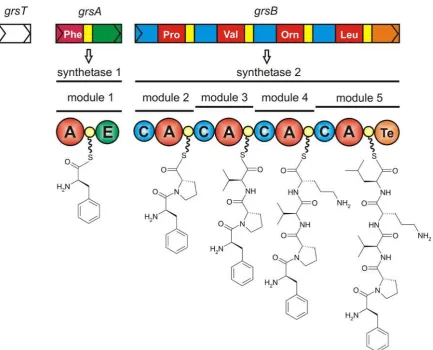
1.7 The Gramicidin S biosynthetic gene cluster (grs) . . . 17
1.8 The structure of the A domain PheA . . . 19
1.9 First and second half-reaction conformers of the adenylate forming super-family . . . 22
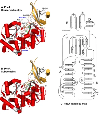
1.10 PheA conserved motifs, subdomains and topology map . . . 25
1.11 The structures of A domains PheA and SrfA-C . . . 34
1.12 The structures of PCP domain TycC3 . . . 38
1.13 Partner domain interaction residues NRPS CP domains TycC3 and EntB . . 42
1.14 Structure of the condensation domain VibH . . . 44
1.15 Structures of the SrfA-C and FenB thioesterase domains. . . 49
1.16 Structure of PA1221 A domain and PCP domain. . . 52
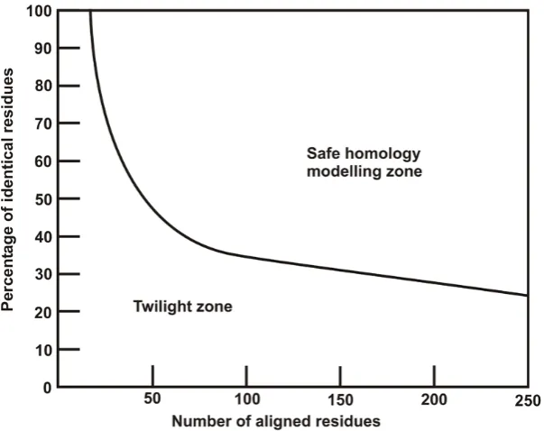
2.1 The two zones of sequence alignments. . . 58
2.2 A two dimensional schematic of periodic conditions. . . 84
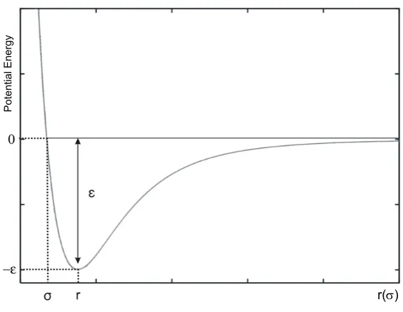
2.3 Schematic representation of the Lennard-Jones potential function. . . 90
3.1 All atom CαRMSDs: PheA-apo and -holo simulation . . . 116
3.2 RMSD PheA1-apo simulation . . . 118
3.3 RMSD PheA2-apo simulation . . . 119
3.4 RMSD PheA1-holo simulation . . . 123
3.5 RMSD PheA2-holo simulation . . . 124
3.6 RMSF PheA1-apo, PheA2-apo, PheA1-holo and PheA2-holo simulations . 131 3.7 Comparison of PheA1-apo and PheA2-apo RMSF with PheA B-factors . . 133
3.8 Comparison of PheA1-holo and PheA2-holo RMSFs with PheA B-factors . 134 3.9 PCA analysis of the PheA apo and holo simulations . . . 135
3.10 Domain motion in PheA1-holo . . . 138
3.11 Domain motion in PheA2-holo . . . 140
3.12 Domain motion in PheA1-apo . . . 143
3.13 Domain motion in PheA2-apo . . . 145
3.14 PheA interdomain hydrogen bond inreaction groupings . . . 151
3.15 Interdomain hydrogen bonding in the PheA apo and PheA holo simulations 152 3.16 The structure of the A domain PheA . . . 156
3.17 Hydrogen bonding between L-Phe and PheA, PheA1-holo simulation . . . 157
3.19 Hydrogen bonding between L-Phe and PheA, PheA1- and PheA2-holo
sim-ulations over time . . . 162
3.20 Hydrogen bonding between AMP adenine and PheA, PheA1-holo simulation 164 3.21 Hydrogen bonding between AMP adenine and PheA, PheA2-holo simulation 165 3.22 Hydrogen bonding between AMP ribose and phosphate, and PheA, PheA1-holo simulation . . . 167
3.23 Hydrogen bonding between AMP ribose and phosphate and PheA, PheA2-holo simulation . . . 168
3.24 Extreme motion from PheA1-holo . . . 173
3.25 Extreme motion from PheA2-holo . . . 174
3.26 Extreme motion from eigenvector 2 in PheA2-holo . . . 176
3.27 Schematic of the domain motion observed in PheA-holo simulations . . . . 178
4.1 Docked structures: PheA with L-Tyrosine, Aspartic acid and Arginine . . . 189
4.2 RMSD PheA-Tyr simulation . . . 191
4.3 RMSD PheA-Asp simulation . . . 193
4.4 RMSD PheA-Arg simulation . . . 194
4.5 RMSFs of the PheA-Tyr, PheA-Asp and PheA-Arg simulations . . . 198
4.6 PCA analysis of the PheA-Tyr, -Asp and -Arg simulations . . . 200
4.7 Domain motion in the PheA-Tyr simulation . . . 201
4.8 Domain motion in the PheA-Asp simulation . . . 203
4.9 Interdomain hydrogen bonding: PheA-Tyr, PheA-Arg and PheA-Asp sim-ulations . . . 206
4.10 Hydrogen bonding between L-Tyr and PheA, PheA-Tyr simulation . . . 208
4.11 Hydrogen bonding between L-Asp and PheA, PheA-Asp simulation . . . . 209
4.12 Hydrogen bonding between L-Asp sidechain and PheA, PheA-Asp simulation210 4.13 Hydrogen bonding between L-Asp and PheA, PheA-Asp simulation, at 0, 2, 5, 8 and 11 ns. . . 211
4.14 Hydrogen bonding between L-Arg and PheA, PheA-Arg simulation . . . . 213
4.15 Hydrogen bonding between L-Arg sidechain and PheA, PheA-Arg simulation214 4.16 Hydrogen bonding between AMP adenine and PheA, PheA-Tyr simulation . 215 4.17 Hydrogen bonding between AMP adenine and PheA, PheA-Asp simulation 216 4.18 Hydrogen bonding between AMP adenine and PheA, PheA-Arg simulation 217 4.19 Hydrogen bonding between AMP ribose and phosphate, and PheA; PheA-Tyr simulation . . . 218
4.20 Hydrogen bonding between AMP ribose and phosphate, and PheA; PheA-Asp simulation . . . 219
4.21 Hydrogen bonding between AMP ribose and phosphate, and PheA; PheA-Arg simulation . . . 220
4.22 Schematic of the domain motion in PheA-Tyr and PheA-Asp simulations . 225 5.1 Organisation of the coelichelin,cch, biosynthetic gene cluster and NRPS. . 229
5.2 Predicted substrate specificity determining residues and substrates of the CchH A domains. . . 230
5.3 MODELLER alignment of PheA and CchH2 . . . 245
5.4 Options for insertion locations 1-4, PheA and CchH2 alignment . . . 246
5.5 Options for insertion locations 5-8, PheA and CchH2 alignment . . . 247
5.6 CchH2 homology model evaluation . . . 250
5.7 Summary of PheA-CchH2 sequence alignments . . . 251
5.9 Prosa2003 PheA and CchH2 energy profiles . . . 254
5.10 Optimised PheA and DhbE-CchH2 alignment . . . 256
5.11 Prosa2003 PheA, DhbE, PheA-CchH2, and PheA,DhbE-CchH2 profiles . . 257
5.12 Structure of final CchH2 homology model and PheA . . . 259
5.13 CchH2 Magnesium ion positioning . . . 260
5.14 Docking results: L-Thr and CchH2 . . . 262
5.15 Docking results: L-Ser and CchH2 . . . 264
5.16 Docking results: L-Val and CchH2 . . . 265
5.17 RMSD CchH2-apo simulation . . . 266
5.18 RMSD CchH2-Thr simulation . . . 267
5.19 RMSD CchH2-Ser simulation . . . 268
5.20 RMSD CchH2-Val simulation . . . 269
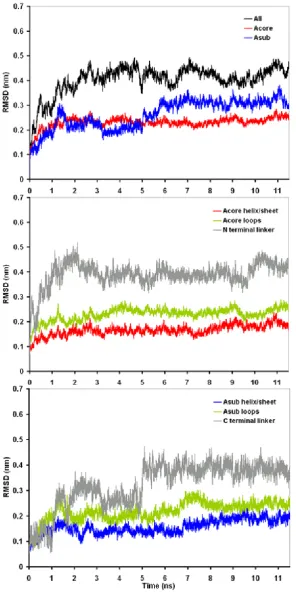
5.21 RMSD N-terminal and C-terminal domain; CchH2-apo simulation . . . 270
5.22 RMSD secondary structure of C-terminal domain; CchH2-apo simulation . 271 5.23 RMSD N-terminal and C-terminal domain; CchH2-Thr simulation . . . 272
5.24 RMSD N-terminal and C-terminal domain; CchH2-Ser simulation . . . 273
5.25 RMSD N-terminal and C-terminal domain; CchH2-Val simulation . . . 274
5.26 RMSF CchH2-apo simulation . . . 277
5.27 RMSF CchH2-Thr simulation . . . 278
5.28 RMSF CchH2-Ser simulation . . . 280
5.29 RMSF CchH2-Val simulation . . . 281
5.30 Domain motion in CchH2-Thr . . . 283
5.31 Domain motion in CchH2-Ser eigenvector 1 . . . 284
5.32 Domain motion in CchH2-Ser eigenvector 2 . . . 284
5.33 Hydrogen bonding between L-Thr substrate and CchH2 . . . 286
5.34 Hydrogen bonding between L-Thr sidechain and CchH2 . . . 287
5.35 Hydrogen bonding between L-Ser and CchH2 . . . 288
5.36 Hydrogen bonding between L-Ser sidechain and CchH2 . . . 289
5.37 Hydrogen bonding between L-Val and CchH2 . . . 291
6.1 RMSD PheA-Phe-Lys simulation . . . 298
6.2 RMSD PheA-Asp-Lys simulation . . . 299
6.3 RMSD PheA-Phe-His simulation . . . 300
6.4 RMSD PheA-Asp-His simulation . . . 300
6.5 RMSF of the PheA-Phe-Lys simulation . . . 301
6.6 RMSF of the PheA-Asp-Lys simulation . . . 302
6.7 RMSF of the PheA-Phe-His simulation . . . 302
6.8 RMSF of the PheA-Asp-His simulation . . . 303
6.9 PCA PheA-Phe-Lys, PheA-Asp-Lys, PheA-Phe-His and PheA-Asp-His . . 304
6.10 Domain motion PheA-Phe-Lys simulation . . . 307
6.11 Domain motion PheA-Asp-Lys simulation . . . 308
6.12 Domain motion PheA-Asp-His simulation . . . 309
6.13 Hydrogen bonding L-Phe amino and PheA; PheA-Phe-Lys simulation . . . 310
6.14 Hydrogen bonding L-Phe carboxyl and PheA; PheA-Phe-Lys simulation . . 311
6.15 Hydrogen bonding L-Asp amino and PheA; PheA-Asp-Lys simulation . . . 312
6.16 Hydrogen bonding L-Asp carboxyl and PheA; PheA-Asp-Lys simulation . . 312
6.17 Hydrogen bonding L-Asp sidechain and PheA; PheA-Asp-Lys simulation . 313 6.18 Hydrogen bonding L-Phe amino and PheA; PheA-Phe-His simulation . . . 314
6.19 Hydrogen bonding L-Phe carboxyl and PheA; PheA-Phe-His simulation . . 315
6.21 Hydrogen bonding L-Asp carboxyl and PheA; PheA-Asp-His simulation . . 316
6.22 Hydrogen bonding L-Asp sidechain and PheA; PheA-Asp-His simulation . 316 7.1 The mechanism of PPTase and A domain action . . . 327
7.2 The structure of Sfp and of TubCdd . . . 328
7.3 The average NMR solution structures of the TycC3-PCP conformers . . . . 329
7.4 Mechanism of condensation and cyclization . . . 330
7.5 Mechanism of epimerization and methylation . . . 331
7.6 Three strategies for chain termination in NRPSs . . . 332
7.7 Mechanism of type II thioesterase action . . . 333
7.8 RMSD AMP 1 ns simulation . . . 336
7.9 Mg ion coordination in the PheA1-holo system simulation . . . 337
7.10 Mg ion coordination in the PheA2-holo system simulation . . . 338
7.11 Docking flowchart - part 1 . . . 345
7.12 Docking flowchart - part 2 . . . 346
7.13 Mg ion coordination in the PheA-Tyr system simulation . . . 347
7.14 Mg ion coordination in the PheA-Asp system simulation . . . 348
7.15 Mg ion coordination in the PheA-Arg system simulation . . . 349
7.16 Hydrogen bonding between AMP and CchH2; CchH2-Thr simulation . . . 354
7.17 Hydrogen bonding between AMP Ribose and Phosphate, and CchH2; CchH2-Thr simulation . . . 355
7.18 Hydrogen bonding between AMP and CchH2; CchH2-Ser simulation . . . 356
7.19 Hydrogen bonding between AMP Ribose and Phosphate, and CchH2; CchH2-Ser simulation . . . 357
7.20 Hydrogen bonding between AMP and CchH2; CchH2-Val simulation . . . 358
7.21 Hydrogen bonding between AMP Ribose and Phosphate, and CchH2; CchH2-Val simulation . . . 359
7.22 Mg ion coordination; CchH2-Thr simulation . . . 360
7.23 Mg ion coordination; CchH2-Ser simulation . . . 361
1.1 Conserved motifs of the NRPS Adenylation domains . . . 16
1.2 Structures of the adenylate forming superfamily of enzymes. . . 23
1.3 PCP domain residues that interact with partner domains . . . 41
3.1 Summary of PheA-apo and -holo simulation systems. . . 109
3.2 AMP PO−42 partial charges, calculated using HF/6-31G* and scaled ac-cording to GROMOS force field conventions. . . 113
3.3 Average values of radius of gyration for the apo and holo PheA simulations. 127 3.4 Average secondary structure contents in PheA apo and holo simulations . . 128
3.5 RMSIP between the first ten eigenvectors for the PheA-apo and holo simu-lations. . . 136
3.6 Average number of intramolecular hydrogen bonds (P-P H bonds) for the apo and holo PheA simulations. . . 149
4.1 Disocciation constants for binding of various amino acids to the adenylation domain PheA. Table adapted from Luoet al.1. . . 182
4.2 Kinetic Constants for Amino Acid-Dependent ATP Hydrolysis by ApoPheATE and HoloPheATE Measured by Continuous Spectrophotometric Pyrophos-phate Assay (PheATE) and ATP-PPi Exchange Assay (apoPheATE). . . 183
4.3 Summary of the noncognate simulation systems. . . 186
4.4 Average values of the radius of gyration (Rg) for the PheA noncognate substrate simulations. . . 195
4.5 Intramolecular (protein-protein) hydrogen bonds for the PheA noncognate substrate simulations. Standard deviation in parentheses . . . 204
5.1 Average secondary structure contents in CchH2 apo and holo simulations . 276 5.2 PCA analysis of the CchH2 simulations . . . 282
5.3 Average number of intramolecular hydrogen bonds (P-P H bonds) for the apo and holo CchH2 simulations. . . 285
6.1 Comparison of the L-Phe and L-Asp Adenylation domain binding pocket specificity conferring code. . . 297
6.2 Summary of the domain motion identified by DynDom from the first two eigenvectors of the PheA-Phe-Lys simulation. . . 305
6.3 Summary of the magnitude of the domain motion identified by DynDom from the first two eigenvectors of the PheA-Phe-Lys simulation. . . 306
7.1 Conserved motifs of the NRPS domains. . . 326
7.2 AMP ff43a2 topology file - atoms . . . 335
7.3 AMP ff43a2 topology file - bonds . . . 339
I hereby declare that this thesis, submitted in partial fulfilment of the requirements for the degree of Doctor of Philosophy and entitled ”Structural Study of the Adenylation Domain by Molecular Dynamics Simulation”, represents my own work and has not been previously submitted to any other institution for any degree, diploma or other qualification.
I would like to thank my supervisor Professor Mark Rodger for his support and guidance. This work would not have been possible without the love and support of my family and friends. In particular I would like to thank my parents, Andrea, Lee, Ethan and Vib.
Most importantly I would like to acknowledge my husband, Kostas, for his love, sup-port and patience.
As antibiotic resistance is increasing more rapidly than new antibiotics are produced and/or discovered, there is an increasing need to identify new ways to design novel antibiotics. A potential avenue for this, is the exploitation of Nonribosomal Peptide Synthetases (NRPSs) from bacteria and fungi which biosynthesise structurally complex biologically active pep-tide products, including numerous potential antibiotics and other molecules with pharma-cologically attractive properties. In order to do so, however, a detailed molecular under-standing of NRPSs is required.
NRPSs are modular proteins, with each module comprising domains that each perform specific functions to select, activate, alter (optional) and combine amino/hydroxyl acid sub-strates to form a specific peptide product. The Adenylation domain (A domain) specifically selects and activates the substrate through a two step reaction. In the first half reaction, a highly reactive aminoacyl adenylate is formed by reaction with Mg-adenosine triphosphate (ATP) resulting in the release of pyrophosphate. In the second half reaction the A domain binds the phosphopantetheinyl (PPant) arm of the downstream domain, the Peptidyl Carrier Protein (PCP) domain. The terminal thiol of the PPant arm attacks the activated aminoa-cyl group displacing adenosine monophosphate (AMP), leaving the amino acid substrate tethered to the PCP domain as a thioester.
The A domain is of particular interest as a target for engineering approaches as it is consid-ered to be the primary determinant of substrate specificity. Little is understood, however, about the molecular basis of substrate selectivity or how the dynamics of the domain enable the two part reactions to take place.
In 1997, the first A domain structure was determined; the L-phenylalanine (L-Phe) activat-ing A domain (PheA) of the Gramicidin S synthetase from Bacillus brevis. All of the A domain structures determined to date are either unligated (apo form) or co-crystallised with reactants or products from the first half reaction. The NRPS A domains are members of the adenylate-forming superfamily which have been structurally characterised in three states, apo, with the first half reaction and second half reaction ligands. Comparison between these structures, suggested these enzymes use a domain alternation strategy to reconfigure a single active site to perform two different reactions. While the A domains have only been determined in the adenylate-forming conformation, similarities between members of the adenylate-forming superfamily suggest NRPS A domains may exploit of a similar strategy of domain alternation to reconfigure the enzyme’s single active site.
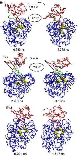
Inter-domain rotation was observed in the apo and cognate holo simulations and with one of the noncognate substrates, L-Thr. This motion occurred between the Acore domain and Asub domain or part of the Asub domain. The rotation observed in the simulations with the cognate substrate creates a widening between the two domains of PheA on the side of the enzyme where the PPant arm is thought to bind. Results from one of the cognate holo simulations suggests the A3 motif loop may be important in stabilising the A domain to increase the domain rotation or maintaining the opening through with PPant is proposed to access the active site.
Results from one of the noncognate substrate simulations, L-Asp substrate, suggests a role for the A3 motif loop in the removal of noncognate ligands from the binding site. Results from the simulation with noncognate substrate L-Tyr also suggest that interaction of the substrate with the key Asp and Lys binding pocket residues may be required for rotation of the Asub domain can occur.
A homology model of the second A domain of the NRPS that forms Coelichelin has built and it is shown that the core regions of the model are stable in the MD simulations carried out in the apo form, with the cognate ligand (L-Thr) and noncognate ligands (L-Ser and L-Val). Some domain rotation was observed in the simulations with L-Thr and L-Ser. The findings from this study support the suggestion that interaction between the key Asp and Lys binding pocket residues and the substrate may be required for domain rotation.
A
A Adenylation.
AMP Adenosine Monophosphate.
atm Atmospheres.
ATP Adenosine Triphosphate.
B
BLOSUM BLOcks SUubstitution Matrix.
BPTI Bovine Pancreatic Trypsin Inhibitor.
C
C Condensation.
C-Mt C-Methylation.
CG Conjugate Gradient.
Cy Heterocyclisation.
D
DOPE Discrete Optimized Protein Energy.
E
E Epimerisation.
F
F Formylation.
G
GA genetic algorithm.
GOR Garnier, Osgusthorpe and Robson.
GROMACS Groningen Machine for Chemical Simualtions.
H
HMM Hidden Markov Model.
K
K Kelvin.
L
LGA Lamarckian genetic algorithm.
LS local search.
M
MC Monte Carlo.
MD Molecular Dynamics.
MSM Markovian state models.
N
N-Mt N-Methylation.
nm nanometre.
NOE nuclear Overhauser effect.
NRP Nonribosomal peptide.
NRPS Nonribosomal peptide synthetase.
O
P
PAM Percentage Accepted Mutation.
PBC Periodic Boundary Conditions.
PCA principal components analysis.
PCP Peptidyl Carrier Protein.
PDB Protein Data Bank.
PDF Probability Density Function.
PME particle-mesh Ewald.
Ppant 4’-phosphopantetheinyl.
ps Picoseconds.
PSSM Position Specific Score Matrix.
R
Red Reduction.
RMSD Root Mean Square Deviation.
RMSF root mean square fluctuations.
S
SAM S-adenosyl methionine.
SD Steepest Descent.
SPC Simple Point Charge.
STMV satellite tobacco mosaic virus.
T
T Thiolation domain.
Te Thioesterase.
V
vdW van der Waals.
Small peptide natural products have a range of powerful biological activities and are
criti-cal elements of modern therapy. Mainly synthesised by microorganisms they are produced
either by the ribosomal machinery or by gigantic multi-domain enzymes called
nonriboso-mal peptide synthetases (NRPS) using a thiotemplated mechanism. Nonribosononriboso-mal peptide
(NRP) natural products are structurally diverse secondary metabolites thought to be
pro-duced primarily to offer the host organism a survival advantage and which have been
opti-mised to perform a certain function(s) over years of evolution. The diverse sphere of action
they possess includes antibiotic, antifungal, immunosuppressive and cytostatic activity2. NRPs, especially those with antibiotic activity, have been and continue to be of
tremen-dous pharmacological importance either as therapeutic agents or as promising scaffolds for
the development of substances with novel activities. NRPSs are composed of catalytic
do-mains arranged into modules. Each module is responsible for the specific incorporation of
a proteinogenic or non-proteinogenic amino acid monomer into the peptide product. This
relatively simple biosynthetic logic generates peptides of high structural complexity3. The modular multi-domain architecture of these synthetases makes them amenable to genetic
manipulation and is one strategy for the production of “novel natural products”. Many
of the key principles of nonribosomal peptide synthesis have been determined using
bio-chemical and genetic studies. A comprehensive understanding of the molecular basis of
the numerous protein-protein recognition events underpinning the mechanism of
nonribo-somal peptide synthesis is necessary to realise the potential of producing novel products
with predefined characteristics by genetic modification of the synthetases4.
1.1
Nonribosomal Peptides
Nonribosomal peptides (NRP) are structurally diverse complex peptide secondary
metabo-lites of low molecular weight (see figure 1.1 for examples). The sheer bulk of the
multi-domain NRPSs and the rate at which the products are synthesised places a limitation on the
gramicidin S8–10, vancomycin11, daptomycin12–14, and the penicillin and cephalosporin tripeptide precursor ACV15. Cyclosporin A produced by Tolypocladium niveumis a NRP with immunosuppressive activity routinely used in transplant aftercare16. Thiocoraline17 has potent antitumor activity and is currently undergoing clinical trials for use in cancer
therapy18. Yersiniabactin19, vibriobactin20 and enterobactin21 are all siderophores, iron scavengers that are produced under iron limiting conditions. This chelation of iron by
bac-teria is vital for their survival and is often a virulence determinant in pathogens21.
The great structural diversity exhibited by NRPs distinguishes them from peptides
synthe-sised ribosomally and can in part, yet not exclusively, be attributed to the array of
pre-cursors NRPSs can utilise. Unlike ribosomal protein synthesis which is limited to the 22
proteinogenicα-amino acid building blocks, NRPSs have been shown to incorporate
sev-eral hundred substrates22, including many non-proteinogenic amino, aryl carboxylic and
α-hydroxy acids. These include L-hf ornithine found in coelichelin23, dihydroxyphenyl-glycine (DHPG) in vancomycin, (4R)-4-[(E)-2-butenyl]-4-methyl-L-threonine (Bmt) in
cy-closporin A and 2,3-dihydroxybenzoate (DHB) in vibriobactin. The structures can be linear
(myxothiazol), macrocyclic (tyrocidin A24), branched macrocyclic (fengycin), or dimers (gramicidin S) or trimers (enterobactin) of identical structural elements. NRPs often
con-tain small heterocyclic rings such as thiazole (epothilone) and oxazoline (vibriobactin),
and may containN-formylations (anabaenopeptilide 90-A25),N-methylations (cyclosporin A), acylations and glycosylations (vancomycin). The majority of known NRPs contain
either unusual or modified amino acids either at their N- or C-termini, which suggests
pre-selectivity of these compounds for stability and biological activity5. The vast structural diversity of these natural products is strictly associated with their biological function.
1.2
Nonribosomal Peptide Synthetases
In ribosomal protein synthesis, protein structure is determined by the genetic code. In
contrast, the multi-domain proteins called nonribosomal peptide synthetases (NRPS) act
Found in bacteria and fungi, NRPSs assemble peptides by the repetitive condensation of
simple monomers using a strategy termed the multiple thiotemplated mechanism26–28.
The complete synthesis of a NRP can be performed either by a single synthetase, as is
usu-ally the case in fungi, or by a series of structurusu-ally distinct synthetases, as is often seen in
bacteria, the encoding genes of which are almost always organised in an operon. NRPSs
are organised into modules, each one responsible for the specific recognition, activation
and incorporation of one monomer into the nascent peptide chain. The number and order
of modules in the synthetase usually dictates the primary sequence of the peptide product;
these NRPSs are referred to as type A. NRPSs that do not follow this linear logic however,
cannot be considered as rare exceptions but rather as variations of the common NRPS
reper-toire designed to increase the biosynthetic potential of the synthetases5. The modules of an iterative NRPS (type B) are used in a sequential repeated manner to produce peptides
con-taining multiple copies of identical structural elements. Iterative NRPSs include those that
synthesise gramicidin S, a pentapeptide dimer, and enterobactin, a dipeptide trimer.
Non-linear NRPSs can use one or more of their modules more than once to generate peptides that
contain repeats of specific precursors. Iterative NRPSs commonly contain unusual
arrange-ments of the NRPS domains. The first modules of nonlinear synthetases syringomycin and
coelichelin ofStigmatella aurantiacaandStreptomyces coelicoloris used twice to produce
the tripeptide and tetrapeptide products, respectively5.
NRPS modules are composed of individual domains with defined functions that, when
present on the same synthetase, are separated by short spacer regions of about 15 amino
acids. These regions are homologous to classical protein linkers, see figure 1.2 for the
lo-cation and structure of these linker regions in the surfactin synthetase SrfA-C termination
module. These physically linked NRPS domains retain their functionality when excised and
expressed heterologously as separate units. NRPSs that synthesise siderophores commonly
have fewer physical linkages between the domains. Such domains are referred to as
stand-alone or free-standing. The interactions between physically linked domains are termed
intramolecular or in cis interactions, and those between distinct stand-alone domains, or
Figure 1.3: The ten NRPS domains. Where; F - formylation, A - adenylation, C - condensation, Cy cyclisation, E epimerisation, Ox oxidation, Te thioesterase, and Red -Reductase. PCP domains are coloured yellow. Mt - represents both N- and C-Methylation domains. Image adapted from figure 1 in reference30.
A typical round of peptide initiation and elongation is illustrated in figure 1.4 and
pro-ceeds as follows. The Adenylation (A) domain of module 1, A1, specifically selects and activates the amino acid substrate, forming a highly reactive aminoacyl adenylate by
reac-tion with Mg-Adenosine triphosphate (ATP). The A domain is therefore considered to be
the primary determinant of substrate specificity. After pyrophosphate (PPi) is released the A domain binds the phosphopantetheinyl (Ppant) arm of the downstream Peptidyl Carrier
Protein (PCP) domain1, PCP1. The terminal thiol of this arm attacks the activated aminoa-cyl group displacing adenosine monophosphate (AMP), leaving the amino acid substrate
tethered to the PCP domain as a thioester. The downstream Condensation (C) domain of
the adjacent module, module 2, catalyses peptide bond formation between substrates bound
to the PCPs of modules 1 and 2. The C domain catalyses the nucleophilic attack of the
substrate bound to the PCP2 domain on the activated thioester of the substrate bound on the upstream PCP1 domain31. This condensation reaction results in the covalent attachment of the peptidyl product to the PCP2 domain and the release of the sulfhydryl group of the PPant moiety of the PCP1 domain. The C3 domain then forms a peptide bond between the peptidyl product from the last reaction, now tethered to PCP2, and the substrate attached to the PCP3 module. The tripeptide produced from this reaction is now tethered to the PCP3 domain. The peptidyl chain continues to grow in this fashion until all of the substrates
are incorporated, at which point the chain is released from the last domain in the final (or
termination) module either by hydrolysis or by cyclisation. This is usually achieved by a
thioesterase (Te) domain in a two stage reaction. An acyl-O-TE-enzyme intermediate is
formed, then subsequently attacked either by water32 or a peptide-internal nucleophile33. This produces either a linear or a macrocyclic peptide. The favoured mechanism appears to
be macrocyclic release.
A schematic of the modular organisation of the ten known NRPS domains is shown in figure
1.3. Of these ten domains, only three - the A, PCP and C domains - are needed to perform
all the functions required for one complete cycle of elongation. These core domains are
arranged in the order C-A-PCP in a minimal elongation module. A minimal initiation, or
starter module, consists of A-PCP, and a standard termination module consists of an
elon-gation module followed by a Te domain, C-A-PCP-Te. Optional NRPS domains include
the Epimerisation (E), Heterocyclisation (Cy), N- and C-Methylation (N-Mt and C-Mt),
Oxidation (Ox), N-formyltetrahydrofolate-dependent formyltransferase (F) and Reduction
(Red) domains. While the majority are optional editing domains that can be present in
any elongation module, the Red domain can only be located in a termination module as
a replacement of a Te domain. All of the NRPS domains can be identified from primary
sequence data by the location of a series of highly conserved motifs (shown in table 7.1
in appendix 7.1.2). A complete summary of the reactions catalysed by each of the NRPS
domains, including the optional domains, is shown in figure 1.5.
The speed, order and uni-directionality of the peptide elongation reaction are controlled
by the C domain34. This domain possesses a donor site for the electrophile (the substrate from the upstream PCP domain) and an acceptor site for the nucleophile (the substrate on
the downstream PCP domain). Strong stereoselectivity31,34,35 and a degree of selectivity towards the side chain of the aminoacyl thioester35 are observed at the C domain acceptor site. The donor site exhibits broader substrate specificity31,36.
Structural models have been determined for each of the main NRPS domains (A, PCP, C
A
B
C
aa2 aa1
aa2
aa1
Figure 1.4: The reaction sequence of peptide chain elongation. The Ppant prosthetic group is represented by the zigzagged line.A1 The A domain selects a specific substrate and catalyses formation of the amino acyl adenylate by ATP hydrolysis. A2 The acyl moiety is transferred to the thiol group of the PCP PPant prosthetic group.BTransfer of the substrate to the acceptor site of the upstream C domain is facilitated by movement of the acyl-S-Ppant. Peptide bond formation with the amino acyl or peptidyl chain of the preceding PCP domain is then catalysed. C The donor site of the downstream C domain (from the subsequent module) is where the elongation cycle is completed. Image adapted from5.
1.2.1
Polyketide Synthetases
The biosynthetic strategy of the modular architecture of NRPSs is comparable to that of the
polyketide synthetases (PKS) of secondary metabolism and fatty acid synthetases (FAS) of
primary metabolism. These similarities facilitate the transition between NRPS and PKS
modules in NRPS-PKS hybrid megaenzymes39. The PKS modules integrate acetate and propionate into the peptide chain. Numerous hybrid NRPS-PKS systems have been
dis-covered and the ratio of NRPS to PKS modules can vary greatly. These hybrid systems
produce structures with biological activities similar to NRPs. NRPS-PKS products include
the anticancer molecules bleomycin A240and epothilone41,42.
1.3
Molecular Engineering Approaches
Almost all peptide-based antibiotics are made by NRPSs43. The growing number of pathogenic bacterial strains resistant to antibiotics, especially in hospitals, is of great medical, societal
and governmental concern. Historically the introduction and use of newly developed
ef-fective and safe antibiotics is followed by the rapid development of resistance mechanisms
de-Figure
1.5:
Reactions
catalysed
by
the
NRPS
domains.
Image
adapted
from
figure
2
of
veloped resistance mechanisms and the way in which bacteria transfer genetic information
between each other has lead to the formation of multi-drug resistant pathogenic bacterial
strains. Thus the average pre-resistance life span of any antibiotic is short and the
develop-ment and discovery of new antibiotics a constant requiredevelop-ment.
The multidomain, modular, assembly line architecture of NRPSs makes them amenable
to reprogramming in order to synthesise novel antibiotic products. Strategies to generate
designer products include the formation of hybrid NRPSs by specifically recombining the
domains and modules, or altering domain specificity. Hybrid NRPSs have been formed
by: fusing domains together, e.g. A-PCP unit exchange in surfactin synthetase45; deleting or rearranging the order of entire modules, e.g. module deletions and fusion in tyrocidine
synthetase46,47; and altering the specificity of the A domain in situ, e.g. the L-Glu activating domain of surfactin synthetase was modified to preferentially select L-Gln48.
As almost every event in nonribosomal peptide synthesis is governed by protein-protein
recognition or substrate specific events, alteration of the protein domains or peptide
sub-strates within a biosynthetic complex can have repercussions elsewhere in the assembly
line. NRPS reprogramming experiments have already revealed that the positioning of
do-main or module fusion sites and alteration of the substrate specificity of one dodo-main within
a module (either by point mutation or by complete domain replacement) has implications
on the productivity of the synthetase and the activity downstream domains.
In the first experiment of its kind, exchange of A-PCP units in surfactin synthetase yielded
the expected products but the productivity of the synthetases was drastically reduced45. This decrease in productivity was subsequently attributed to the substrate specificity
re-quirements of the C domains31. A better understanding of domain borders and the iden-tification and characterisation of the NRPS domain linkers has already been exploited to
generate hybrid NRPSs, using module deletion and fusion, with improved yields47.
The selective association and communication between the individual synthetases of a
biosyn-thetic complex is critical for the synthesis of the predefined peptide. The identification,
(COM) domains, at the termini of the interacting tyrocidine synthetases (TycA, TycB and
TycC) determined their decisive role in the correct protein-protein recognition of
associ-ated peptide synthetases49. Further experiments have identified key residues important for maintaining the correct, or preventing the incorrect, interaction between the tyrocidine
syn-thetases. Mutation of these residues has proven successful in switching the specificity of
one synthetase for another, has aided the formation of an artificial hybrid NRPS complex
and the combinatorial biosynthesis of various designed peptide products49.
Compared to domain and module swapping the alteration of the substrate specificity of the
A domain by targeted mutation of the active site residues is a relatively small modification.
To date the majority of changes in A domain substrate specificity have been fairly trivial, i.e.
the native and achieved substrates have had side chains of very similar size, overall polarity
and shape. Altering an A domain from one that recognizes a large substrate to one that
recognizes a small substrate, or vice versa; from one that is specific for a hydrophobic
sub-strate to a hydrophilic subsub-strate, or vice versa; and engineering an A domain with relaxed
substrate selectivity capable of utilising a wide range of substrates are the major challenges
in this area. An A domain with broad substrate selectivity would help to achieve one of
the major goals of the engineered biosynthesis field - the truly combinatorial synthesis of
peptides30. The possible degree of A domain substrate specificity switching may however be dictated by the substrate specificity imposed by downstream domains, particularly the C
domain.
Although the A domains have been studied extensively, knowledge of the selectivity
mech-anism is still relatively rudimentary. Understanding the molecular basis of this selectivity is
critical for informed reprogramming of these domains. Determining the substrate
selectiv-ity mechanism for the other NRPS domains is similarly important, as if they are controlled
by a relatively minor number of residues the potential to alter them in concert with the A
domains may arise. The following aspects of NRP synthesis are as yet unknown and the
answers are likely to further the progress of synthetase reprogramming. How do the PCP
domains maintain the correct order of interactions when there is a choice of domain
Superfamily enzymatic half-reaction 1: RCOOH + Mg-ATP RC-OAMP + PPi
O
H3N
O
R O
H3N
O
R O
AMP Mg-ATP
+ PPi (1)
Superfamily enzymatic half-reaction 2: RC-OAMP + CoASH RC-SCoA + AMP O
O
+
H3N
O
R O
AMP
- AMP
PCP
SH
PCP
S
O
NH3
R
[image:32.595.104.537.64.388.2](2)
Figure 1.6: The two half-reactions of the adenylate forming superfamily of enzymes. Adapted from figure 1 of54.
aiding domain interactions?4
1.4
The Adenylation Domain
The adenylation domains (A domains) of NRPSs; acyl-, acetyl- and aryl-Coenzyme A
(A) synthetases/ligases; and insect luciferases are the three subfamilies that constitute the
adenylate-forming superfamily of enzymes (PFAM00501) which catalyse two sequential
half-reactions via a ping-pong mechanism9,50–53. The first half-reaction is the conversion of a carboxylic acid substrate to an acyl-adenylate by consumption of Mg-ATP. In the second
half-reaction the acyl-adenylate intermediate is esterified with either CoA (acetyl-, acyl- and
aryl-CoA synthetases/ligases) or the enzyme bound CoA derivative PPant (A domains), or
oxidised with molecular oxygen (insect luciferases).
The adenylate-forming superfamily of enzymes share between 20 and 40 % sequence
mo-tifs, denoted A1-A10, are shown in table 1.17,9,22,50,55. The superfamily is characterised by a glycine/serine/threonine rich motif (motif A3 in the A domains)56 that is homologous to the Walker type A motif57. The Walker motif forms a traditional phosphate binding loop (P loop) found in all guanosine and some adenosine nucleotide-binding proteins58–60. Adenylate-forming enzymes are, on average, 500–700 residues in length and adopt a
com-mon fold consisting of a large 400–550 residue N-terminal subdomain and a smaller 100–
130 residue C-terminal subdomain52,61–63.
Enzymes from this superfamily are thought to exploit a “domain alternation” strategy to
catalyse the two half-reactions. Comparison of structures co-crystallised with the first
and second half-reaction structures revealed a large difference in the orientation of the
C-terminal domain relative to the N-terminal domain. The change in C-terminal domain
orientation between the two states presents different sets of residues from the smaller
do-main to the active site. The alternation between the two conformations reconfigures the
enzymes single active site enabling catalysis of the two half-reactions52,53.
1.4.1
A Domain Reaction mechanism
A domains select the amino acid substrate and form a highly reactive aminoacyl adenylate
intermediate by reaction with Mg-ATP (half-reaction 1). Following PPi release, the thiol at the end of the PPant arm attacks the activated aminoacyl group displacing AMP and
resulting in the covalent tethering of the substrate to the PPant arm as a thioester
(half-reaction 2). The first half of this (half-reaction can be studied by the carboxyl substrate-dependent
reversal of adenylation with labelled PPi64. The two stage reaction and mechanism of the A domains can be seen in figure 1.6 and figure 7.1 from appendix 7.1.2 respectively.
In ribosomal synthesis, aminoacyl-tRNA synthetases perform a role equivalent to that of
the NRPS A domain, however these enzymes share neither sequence nor structural
Core motifa Consensus sequence
A1 L(TS)YxEL
A2 (core 1) LKAGxAYL(VL)P(LI)D A3 (core 2) LAYxxYSTG(ST)TGxPKG
A4? FDxS
A5aa NxYGPTETTxx
A5aryl† QVxFMAEGLVN A6 (core 3) GELxJGx(VL)ARGYL A7 (core 4) Y(RK)TGDL
A8 (core 5) GRxDxQVKIRGxRIELGEIE
A9 LPxYM(IV)P
A10 NGK(VL)DR
Table 1.1: Conserved motifs of the NRPS Adenylation domains6. a Former nomencla-ture is given in brackets. ? This motif differs in aryl activating domains. † A5 motif from
aryl activating domains73
specificity. Some A domains demonstrate higher substrate specificity than others24and for some A domains the substrate incorporated has been shown to depend on those available in
the growth media67–72.
As NRPSs are large multimodular proteins comprised of structurally and functionally
in-dependent domains acting in an assembly line manner, A domains usually represent one of
the constituent parts of these multimodular proteins. Precise dissection at the boundary of
the structural A domain74,75 has shown excised domains are soluble and catalytically ac-tive when expressed heterologously as separate units24,74,76. A domains that naturally occur and function as distinct enzymes primarily incorporate aromatic carboxy acids. NRPSs that
produce bacterial siderophores, often incorporate aryl acid derivatives at the N-terminal end
of the peptide chain. In these synthetases the A domain from the first module that selects
and activates the aryl-acid substrate exists as a distinct stand-alone A domain, e.g. DhbE
and EntE in bacillibactin and enterobactin synthesis respectively77.
1.4.2
A domain substrate specificity
The gramicidin S biosynthesis operon from Bacillus brevis, shown in figure 1.7, contains
the structural genes grsT, grsA and grsB. GrsAand grsBcode for synthetases GrsA and
ing A domain of GrsA, was determined co-crystallised with L-Phe and AMP; the hydrolysis
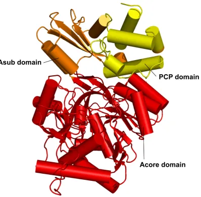
products of the adenylate intermediate62. The 514 residue polypeptide chain folds into two compact domains; a large 412 residue N-terminal domain (Acore domain) and smaller 102 residue C-terminal domain (Asub domain). Few direct protein-protein contacts are formed between the domains. The Acore domain contains three subdomains: subdomains A and B are bothβ-sheets, and subdomain C is a distortedβ-barrel. These subdomains pack together
to form a five-layeredαβαβαtertiary structure. No interpretable electron density was
ob-tained for the highly conserved A3 motif residues. The Asub domain comprises two sub-domains, D and E; a small two strandβ-sheet (subdomain D) and two helices which pack
against one side of a three-stranded anti-parallel β-sheet (subdomain E). The ligands are
bound in a cleft at the domain interface that is lined mainly by polar and charged residues.
Determination of the structure of PheA greatly facilitated the study of A domain specificity.
Co-crystallisation with L-Phe identified the location of the substrate binding pocket and
allowed determination of the residues that line the pocket and make contact with L-Phe.
The structure of PheA, and the L-Phe and AMP binding pockets can be seen in figure 3.16.
Of the ten PheA L-Phe substrate binding pocket residues, nine (Asp 235, Ala 236, Trp 239,
Thr 278, Ile 299, Ala 301, Ala 322, Ile 330, Cys 331) are contributed by the Acore domain and are located between, and inclusive of, motifs A4 and A5. D235 and I330 line the top;
W239, T278 and I299 the bottom; and A236, A301, A322 and C331 the sides of the PheA
binding pocket.
The first residue, D235, is well positioned to form hydrogen bonds with the substrate α
-amino group. The tenth residue is the strictly invariant lysine residue (Lys 517) from the
A10 motif (KA10) that is contributed by the C-terminal domain and resides on a long loop that projects into the active site. In the PheA structure the KA10 residue is well placed to form key polar interactions with both ligands; theαcarboxy group of the Phe substrate and
fAB) valine-activating domain which resulted in a 94 % reduction in activity as compared
to the wild-type enzyme80.
Using these structural data and exploiting the relatively high sequence identity of the A
domains, two independent bioinformatics studies identified an empirical correlation
be-tween the ten residues corresponding to those lining each A domain binding pocket and
the substrate activated81,82. The profile of ten residues, determined using sequence anal-ysis, for each substrate activating A domain is commonly referred to as the “specificity
conferring code”. Subsequent analyses of additional A domain sequences have shown that
identical substrates can be activated by domains with different predicted selectivity pocket
residues83,84. This apparent degeneracy in the ten residue specificity conferring code is thought to arise from including residues lining the bottom of the binding pocket in the
specificity profiles of the domains that activate the smaller substrates (e.g. proline and
thre-onine) which are thought to utilise only residues lining the top of the binding pocket30,85. Determination of the structure of theBacillus subtilis stand-alone A domain DhbE which
activates the aryl acid 2,3-dihydroxybenzoate (DHB) enabled refinement of the specificity
conferring code for these non amino acid activating domains. The authors’ comprehensive
study of the three determined DhbE structures: apo (pdb 1MDF), complexed with AMP
and DHB (pdb 1MD9), and complexed with the adenylate (pdb 1MDB), in tandem with
se-quence alignment and modelling studies identified a structural basis for discerning between
A domains that activate DHB and those that activate salicylic acid (SAL)73.
1.4.3
Domain Alternation
Members of the adenylate-forming family have been structurally characterised in three
states: without ligands (apo); with substrates, products or analogues of the first half-reaction;
or with substrates, products or analogues of the second half-reaction. A representative list of
these structures is shown in table 1.2. Comparison of the structures of family members
de-termined in the presence of first and second half-reaction ligands has identified two distinct
conformations of these enzymes, which differ in the orientation of the C-terminal domain
into two groups depending on whether they are in the conformation thought productive for
catalysing the first half-reaction (conformation 1) or second half-reaction (conformation 2).
Figure 1.9 shows structures in both conformations. The N-terminal domains have been
superimposed, highlighting the difference in C-terminal domain positioning. Apo state
enzymes have been determined in both conformational states. While the position of the
C-terminal domain relative to the N-C-terminal domain is equivalent within the structures of the
two groups, the degree of rotation of the C-terminal domain varies in the first half-reaction
structures and this group can be sub divided into three further groups (called 1.1, 1.2 and
1.3 in table 1.2). The varying C-terminal domain rotation exhibited by the first half-reaction
structures does not however affect the residues presented to the active site. Only one residue
from the C-terminal domain - the invariant Lys residue from motif A10 located on a long
loop that projects into the active site - is critical for binding the substrates of the first
half-reaction. This is in direct comparison to the numerous C-terminal domain residues which
interact in the second half-reaction with either CoA or the PPant portion of CoA, which are
located on the opposite face of the C-terminal domain, in theβ-hairpin region of motif A8.
The variation observed in the conformation 1 structures may therefore be a direct result of
the fewer C-terminal domain residues participating in the reaction and therefore stabilising
the conformation.
Conformation 1
The PheA62 and DhbE73 A domain structures,Saccharomyces cerevisiaeacetyl-CoA syn-thetase (yAcS) structure co-crystallised with AMP (pdb 1RY2)87, and the Alcaligenes sp. AL3007 chlorobenzoate:CoA ligase (CBL) apo (pdb 3CW8) and co-crystallised with
4-chlorobenzoate (pdb 3CW9) structures63, are representative of the conformation thought productive for formation of the adenylate (conformation 1). In this conformation the
Examination of the PheA structure reveals the Lys residue is capable of forming hydrogen
bonds with both theαcarboxy group of the Phe substrate and the ribose O40and O50 atoms of AMP62.
In the structure ofSalmonella entericaacetyl-CoA synthetase (bAcS) determined co-crystallised
with adenosine-50-propylphosphate and Coenzyme A (pdb 1PG4), which is representative of the second half-reaction conformation, the equivalent KA10residue is located∼27 ˚Afrom the active site52. The exclusive importance of this residue in the first half-reaction has been biochemically determined in numerous enzymes of this superfamily. Acetylation of
the KA10 residue (K609) in S. enterica bAcS has been shown to inhibit catalysis of the first reaction without affecting the ability of the enzyme to catalyse the second
half-reaction92,93. Determination of the acetylated form of bAcS showed no conformational changes were induced by acteylation of this residue (pdb 1PG3)52. Mutation of this Lys residue inPhotinus pyralis firefly luciferase (Luc)94, in propionyl-CoA synthetase (PrpE) fromS. enterica95, andSalmonella typhimuriumbAcS91dramatically reduced the ability of the enzyme to catalyse the first half-reaction while having little effect on the ability of the
enzyme to catalyse the second half-reaction.
Mapping the A domain motifs onto the PheA structure revealed the majority are located
adjacent to the enzyme active site, see figure 1.10. Of the ten conserved motifs biochemical
characterisation has indicated a role for residues from eight of the motifs (A3-A10) in the
first half-reaction. Motifs A1 and A2 are both far from the enzyme active site and it has
been proposed that they are conserved for structural reasons. The A1 motif residues form
part of a large helix that links strand β-B2toβ-A1. This helix strongly contributes to the
fold of the N-terminal domain22. Motif A2 immediately follows strand β-A1 forming a short helix and strandβ-A2. The strandβ-A2residues interact with the A3 motif residues
that form strandβ-A5.
Motifs A4, A5 and A10 all contribute residues to the substrate binding pocket. The
sec-ond and third residues of motif A4 (FDxS); second, tenth and eleventh residues of A5
(NxYGPTETTxx); and third residue of A10 (NGK(VL)DR) together constitute six of the
A PheA
Conserved motifs
B PheA Subdomains
[image:43.595.115.520.150.619.2]C PheA Topology map
Figure 1.10:PheA conserved motifs, subdomains and topology map. A) The conserved A domain motifs mapped onto the structure PheA62. The A
the α amino group of the Phe substrate62. This aspartic acid residue is only invariant in amino acid activating A domains, in DhbE the neutral amino acid Asn replaces this Asp
residue73.
In the first half-reaction conformation ofA. sp. AL3007 CBL, His 207 forms a hydrogen
bond to the oxygen atom bridging the 4-CB and AMP portion of the adenylate. His 207 is
the first residue of the A4 motif, the equivalent residue in PheA is Phe 234. The position of
H207 in conformation 1 structure occludes the PPant arm thiol binding site thus preventing
binding of CoA to the wrong conformation. In the conformation 2 structure Glu 410 rotates
into the active site interacting with His 207 and pulling it from the active site. The thiol of
the PPant arm binds in the space vacated by His 20753.
In PheA, as well as lining the Phe substrate binding pocket, additional A5 motif residues are
well placed to interact with the AMP ligand. The main chain carbonyl of A322 may accept
a hydrogen bond from the amino group of the adenine base and the main chain carbonyl
oxygen of Gly 324 may accept a hydrogen bond from theα-amino of the Phe substrate62. The product of aB. brevisNagano E-4 strain mutant gene was found to contain a mutation
of the A5 motif glycine residue (G1793D) in the valine activating A domain of gramicidin
synthetase 2 (GrsB)96which was responsible for abolishing enzyme activity97.
Residues from motifs A3, A6, A7, A8 and A9 have been biochemically determined to be
important for binding the Mg-ATP substrate or forming the amino acyl-adenylate. As the
core 2 (A3) motif defines the superfamily, the function of numerous motif residues have
been investigated. The A3 motif is located in a disordered loop region connecting the
anti-parallel strandsβ-A5andβ-A6. These strands are flanked by strandsβ-A2andβ-A7formed
by the residues of motifs A2 and A6 respectively. In the P loop57,98,99 the conserved Lys residue aids binding of the ATP γ-phosphate atoms, by analogy a similar role was
sug-gested for the invariant A3 lysine residue (LAYxxYSTG(ST)TGxPKG), KA3. Mutation of this conserved Lys to Arg and Thr in TycA reduced the enzyme activity to 90% and 99.5%
that of the wild type enzyme respectively100. In an independent study the mutation of KA3 to Arg in TycA resulted in a 75% reduction in activity when compared to the wild-type
valine activating domain of surfactin synthetase 2 (SrfAB) had no significant effect on
en-zyme activity (activity was reduced to 91% of the wild type); mutation of the conserved Lys
to Gln however, reduced activity of the enzyme to 39% that of the wild-type enzyme80. In 4CL fromArabidopsis thialanamutation of KA3to Ser reduced enzyme activity to 3% that of the wild type enzyme101. Separate mutation of each of the three A3 motif Gly residues to Ala (YSTG(ST)TGxPKG), and Pro to Val (LAYxxYSTG(ST)TGxPKG) in theB. brevis
TycA102 Phe activating A domain103 had no significant effect on the adenylation activ-ity of the enzyme100. Direct participation of the first Gly residue (YSTG(ST)TGxPKG) in the adenylation reaction was demonstrated in the Val activating A domain of GrsB,
however, when a mutant with a point mutation of this residue to Asp was found to be
completely inactive97. Mutation of the core 2 loop residues G163, G166, P168 and K169 (LAYxxYSTG(ST)TGxPKG) inPseudomonas sp. CBS3CBL resulted in impaired
cataly-sis of the CBA adenylation partial reaction104. In the PheA complex the side chain of the first Thr residue (LAYxxYSTG(ST)TGxPKG) is well placed to form a hydrogen bond to
the α-phosphate oxygen atom62. Although residues 192GTTGN196 of this motif - which would form the loop - were not determined in the PheA structure, the orientation and
prox-imity of these residues to the AMP binding site suggest an interaction with the PPileaving group22.
The structure of human medium chain acyl-CoA (MC-ACS) was determined in complex
with Mg-ATP (pdb 3C5E) by the Structural Genomics Consortium in Toronto90. This MC-ACS structure represents the first superfamily enzyme co-crystallised with ATP and
pro-vides insight into the role of the A3 motif and additional residues in Mg-ATP binding. The
A3 motif residues that form a hydrogen bonding network with the α β and γ-phosphate
oxygen atoms are shown in italic: 215LAYxxYTSG(T)SGxPKG230 - where the TSG(ST)S sequence replaces STG(ST)T of the standard core 2 motif. The hydroxyl side chain of S222
interacts with theα-phosphate atoms. The amino backbone and hydroxyl side chain groups
of T224 interact with theβ-phosphate oxygen atoms. The hydroxyl side chain groups of
T221 and S225, amino backbone groups of G223 and S225 and amino side chain group of
K229 interact with the γ phosphate oxygen atoms. Additionally an oxygen from each of
four water molecules, two of which interact with the A5 Glu residue (NxYGPTETTxx).
Motif A6 forms strands β-A7 and β-C3 of the N-terminal subdomain. Photoaffinity
la-belling of theB. brevistyrocidine synthetase 1 (TycA)102 Phe activating A domain103with 2-azidoadenosine triphosphate (2-azido-ATP) identified residues (373GYWWRPDLTAEK384) which include a region of this motif, thus indicating its involvement in catalysing aminoacyl
adenylate formation105.
In PheA the A7 motif residues link strands β-C4 and β-C5 and are adjacent to both the
adenine binding site and the conserved Arg residue of motif A8. This motif bears homology
to the ATPase motif106–110 which plays a role in nucleotide binding108. Mutation of the invariant Asp residue (Y(RK)TGDL) from A7 motif to Asn and Ser in TycA decreased the
phenylalanine-dependent ATP-PPiexchange activity to 78% and 12% that of the wild-type level respectively100. In PheA the position of the Asp side chain enables acceptance of hydrogen bonds from the 20 and 30 ribose hydroxyl groups62.
In the PheA structure residues from the the A8 motif link the Acore and Asub domains and form the β-hairpin111 that is subdomain D of the A
sub domain. Sequencing of a mutant B. brevis Nagano BII-3 strain gene coding for the Pro activating A domain of
GrsB defective in Pro activation identified a point mutation of the second A8 motif Gly
(GRxDxQVKIRGxRIELGEIE) to Glu. Further mutation of this residue to Ala, Val, Arg
and Trp resulted in scarcely active enzymes. These results suggest this residue is essential
for aminoacyl-adenylation112. Additionally, photoaffinity labelling of TycA with 2-azido-ATP and fluorescein 50-isothiocyanate indicated this region was involved in catalysing aminoacyl adenylate formation105and that the Lys residue (GRxDxQVKIRGxRIELGEIE) is involved in Mg-ATP binding79.
Mutation of the first Arg in motif A9 (LPxYM(IV)P) to Thr in TycA resulted in profound
loss of activity113. The A9 motif residues connect strands β-E2 and β-E3 and adopt a helix conformation. These residues are located on the opposite face of the Asub domain to the active site and motif A10. Photoaffinity labelling of TycA with 2-azido-ATP identified
involvement of this motif in aminoacyl adenylate formation catalysis105.
Conformation 2 Structures
TheSalmonella entericabAcS structures cocrystallised with adenosine-50-propylphosphate and Coenzyme A52,Salmonella typhimuriumbAcS structures determined with various lig-ands91, andA. sp. AL3007CBL structure co-crystallised with 4-chlorophenacyl-Coenzyme A (4-CP-CoA)53, are representative of the conformation of the enzyme used to catalyse the thioester forming second half-reaction (conformation 2). Re-orientation of the C-terminal
domain in conformation 2 removes the KA10 residue from the active site positioning it
∼27 ˚A away from the domain interface binding pocket. The alternate C-terminal domain
orientation relative to the N-terminal domain presents the A8 motif residues to the active
site52. Analysis of the second half-reaction structures coupled with the results of biochem-ical experiments has identified A5 and A8 motif residues as important for the
thioester-forming half-reaction.
In the S. enterica bAcS second half-reaction structure (pdb 1PG4) the binding site for
adenosine-50-propylphosphate, a mimic for the acyl-adenylate intermediate, is almost com-pletely buried. The position of the AMP moiety and propyl group of adenosine-50-propylphosphate is comparable to that of AMP and Phe, respectively, in the PheA structure. The nucleotide
portion of CoA binds on the surface of the protein and the pantetheine moiety, which is
less well-ordered than the nucleotide moiety, passes through a channel between the two
domains and points into the AMP binding site. In this structure the A5 motif Glu (E417)
residue (NxYGPTETTxx) forms a salt bridge with the third A8 motif Arg (R526) residue
(GRxDxQVKIRGxRIELGEIE) stabilising the conformation 2 structure52.
This A5 motif Glu residue also has a role in the first half-reaction as illustrated in the
recently determined MC-ACS structure complexed with Mg-ATP and an unknown acyl
ligand (pdb 3C5E). This structure provides insight into the positioning of Mg2+ and the role of the A5 motif Glu residue in the coordination of this ion. An oxygen from each of