MS detection, the increasing use of a combined CE-MS technique can be expected.
To be more widely accepted in the area of biomedi-cal research, CE-based protein separations must dem-onstrate a number of features that match the success of conventional (gel) electrophoretic systems. Besides proRling complex protein samples, these systems allow for immunological and enzymatic assaying of separated proteins as well as for simultaneous trans-fer of sample components into another separation dimension without altering the separation in theRrst one. All of these are achieved with minimal distur-bance of zone integrity. Thus, the major efforts will probably be made in developing both multidimen-sional separation systems involving CE and CE-based separation systems permitting post- or on-column enzymatic and immunological analysis of the separ-ated components of complex biological samples. In-corporating immobilized enzymes or antibodies into CE-MS systems will revolutionize the analysis of pro-tein structure and, especially, glycopropro-tein analysis.
Further Reading
Camilleri P (ed.) (1998)Capillary Electrophoresis:Theory and Practice, 2nd edn. Boca Raton: CRC Press.
El Rassi Z (ed.) (1997) Electrophoresis 18: No. 12/13. Special Issue on Capillary Electrophoresis and Elec-trochromatography.
Hjerten S (1996) Capillary electrophoretic separation in open and coated tubes with special reference to proteins. Methods in Enzymology270: 296.
Karger BL, Chu YH and Foret F (1995) Capillary electrophoresis of proteins and nucleic acids. Annual Review of Biophysics and Biomolecular Structure 24: 579.
Khaledi MG (ed.) (1998) High-performance Capillary Electrophoresis:Theory,Techniques,and Applications. New York: Wiley.
Landers JP (ed.) (1997) Handbook of Capillary Elec-trophoresis, 2nd edn. Boca Raton: CRC Press. Lunte SM and Radzik DM (eds) (1996)Pharmaceutical and
Biomedical Applications of Capillary Electrophoresis. Oxford: Pergamon.
Righetti PG (ed.) (1996)Capillary Electrophoresis in Ana-lytical Biotechnology. Boca Raton: CRC Press. Righetti PG and Deyl Z (eds) (1997)Journal of
Chroma-tography B 699, Special Volume: Proteins: Advanced Separation Technologies.
Wehr T, Rodriguez-Diaz R and Zhu M (1999) Capillary Electrophoresis of Proteins. New York: Marcel Dekker.
Centrifugation
A. Yamazaki, Kresge Eye Institute, Wayne State
University, Detroit, MI, USA
Copyright^ 2000 Academic Press
Introduction
Modern technological developments have made cen-trifugation one of the most important and widely applied techniques in experimental research. In bio-logical studies centrifugation is used for the extrac-tion and isolaextrac-tion of biological materials and for the measurement of physical properties of macro-molecules. Indeed, biological materials have been ex-tracted and isolated for more than a thousand years using centrifugal forces. In the 1920s, Svedberg and other researchers developed motor-driven centrifuges which had an optical system to observe sedimentation of macromolecules during centrifugation, and used these instruments for the measurement of physical properties of macromolecules, especially proteins. The molecular mass of most proteins was determined using these analytical centrifuges until 1970, but they
have not been recently used for that purpose because much easier methods for the measurement of molecu-lar mass, such as size exclusion chromatography and sodium dodecyl sulfate (SDS)-gel electrophoresis, have been developed. More recently, centrifugation has become an indispensable tool for the isolation of proteins, nucleic acids and subcellular particles. The use of centrifuges has also been revived for the measurement of physical properties of proteins, espe-cially for the characterization of protein associations and protein}protein interactions. In this section, im-portant points of theory and practice for the separ-ation and isolsepar-ation of proteins by centrifugsepar-ation are summarized.
Theoretical Basis of Centrifugation
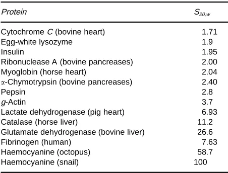
Table 1 Sedimentation coefficients of some proteins
Protein S20,w
CytochromeC (bovine heart) 1.71
Egg-white lysozyme 1.9
Insulin 1.95
Ribonuclease A (bovine pancreases) 2.00
Myoglobin (horse heart) 2.04
-Chymotrypsin (bovine pancreases) 2.40
Pepsin 2.8
g-Actin 3.7
Lactate dehydrogenase (pig heart) 6.93
Catalase (horse liver) 11.2
Glutamate dehydrogenase (bovine liver) 26.6
Fibrinogen (human) 7.63
Haemocyanine (octopus) 58.7
Haemocyanine (snail) 100
macromolecular particles in a solution do not show any perceptible sedimentation in a uniform gravi-tational Reld. However, these macromolecular par-ticles do sediment under a centrifugal force. If the effect of diffusion is neglected, in a solution (density ) the motion of a particle (mass m and volumeVp) that is located a distancerfrom the axis revolving with angular velocitycan be expressed by the following equation:
vf"m2r!2rVp [1]
wherevis the velocity of the sedimenting particle,fits frictional coefRcient;m2rthe centrifugal force and 2rVp the buoyant force. This equation may be rearranged to give:
v"dr/dt"s2r [2]
where:
s"m!Vp f
This is the well-known sedimentation equation in whichsis the sedimenting coefRcient and has dimen-sions of time. For most biological macromolecules, the magnitude of sis about 10\13s. Therefore, the unit of sedimentation, the Svedberg (S), has been deRned as being equal to 10\13s. The standard sedi-mentation coefRcient (S20,w) is deRned as that equiva-lent to sedimentation in water at 203C. The sedimentation coefRcients (S20,w) of some proteins are shown inTable 1.
The sedimentation coefRcient s may be trans-formed to a more practical form. The mass of 1 mole of particles,M, is
M"mNo [3]
where No is Avogadro’s number. Thus, a particle’s volume,Vp, may be expressed in terms of its molar mass:
Vp"m"M/No [4]
whereis the particle’s partial speciRc volume. Eqns [1], [3] and [4] may be combined to give:
vf"M(1!) 2r
No [5]
When eqns [2] and [5] are combined,smay be ex-pressed as:
s" v 2r"
M(1!)
Nof [6]
Since the particle’s partial volume, , may be ex-pressed by the reciprocal of the buoyant density of the particles,p, as"1/p,smay also be expressed as:
s" v 2r"
M(1!/p) Nof
[7]
Sincef"6rp, whereis the viscosity of the liquid medium andrp is the radius of unsolvated spherical particle, these equations indicate that the sedimenta-tion velocity, v, is related to the sedimentation co-efRcients, which is mostly a function of particle size, density of the particlep, density of the mediumand the viscosity of the liquid medium,. In other words, for a given particle, sedimentation is directly related to particle size, particle density and the centrifugal
Reld, and inversely to the viscosity and density of the liquid medium.
Centrifugation for Protein Separation
Figure 1 Fractionation of particles by differential centrifugation. Reproduced with permission from Griffith (1979).
required. Modern ultracentrifuges can generate ap-proaching 1 000 000g, which is sufRcient to pellet even small proteins. Ultracentrifuges can be divided into two types: analytical and preparative. Analytical ultracentrifuges have a device by which the sedimenta-tion rate of molecules can be optically measured dur-ing centrifugation and can be used to obtain data on the sedimentation properties of particles. The masses of most proteins were determined by these ultracen-trifuges before development of simpler molecular mass determination methods. Eqn [4] indicates that the particle’s mass m"M/No can be determined from its sedimentation coefRcient s, if its frictional coefRcientf, is known, as indicated in eqn [6].
Preparative ultracentrifuges are designed for sample preparation. This kind of ultracentrifuge is also commonly used for quantitative estimations of sedimentation coefRcients of particles in a density gradient, although the data obtained are not as accu-rate as those obtained using analytical ultracen-trifuges. Preparative ultracentrifugation can be divided into two methods, namely differential ultra-centrifugation and density gradient ultra-centrifugation. Differential centrifugation is based on the differences in the sedimentation rates of particles in samples. If a suspension of particles is centrifuged in a tube without a density gradient, each particle will move toward the bottom of a tube. In this case, the rate of sedimentation,v, is dependent upons(eqn [2]). Since s is mostly a function of particle size, the rate of sedimentation is proportional to particle size. In the course of the ultracentrifugation, two fractions can be obtained from a solution of particles: a pellet contain-ing sedimented particles and a supernatant solution of the unsedimented fraction. A given particle in the solution may sediment to the pellet or near the bot-tom, as illustrated inFigure 1. As might be expected, this centrifugation willRrst sediment the largest par-ticles in the sample solution to the bottom of the tube. The only particle that is in puriRed form is the most slowly sedimenting one, but the yield is very low. The major problem with differential centrifugation is that the centrifugal force necessary to pellet the larger particles is also often sufRcient to pellet the smaller particles initially near the bottom of the tube (Figure 1). To separate one particle from another effectively, a 10-fold difference in mass is usually required. Thus, this centrifugation is recommended for the separation of proteins from large particles such as cells or organelles. However, it cannot be used for the isolation of one protein from another because the partial speciRc volume, , of most pro-teins (in eqn [6]) is not sufRciently different.
Eqn [6] assumes that centrifugation is performed in a homogeneous medium. However, centrifugation
can be carried out in a solution of an inert substance in which the concentration increases from the top to the bottom of the centrifuge tube, i.e. density in-creases from top to bottom. In such density gradient centrifugation of a mixture of particles with different sizes or buoyant densities, a particle will become stationary when (1!) in eqn [6] is zero. Thus, various components will separate according to size or buoyant densities, and form bands or zones of par-ticles with similar densities. Thus, the use of such density gradients greatly enhances the resolving power.
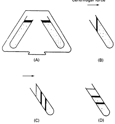
Figure 2 Types of density gradient centrifugation. (A) Rate-zonal centrifugation. (B) Isopycnic centrifugation using a preformed density gradient. (C) Isopycnic centrifugation using a self-forming gradient. Reproduced with permission from Rick-wood (1992) by permission of Oxford University Press.
Figure 3 Operation of fixed-angle rotors. (A) The gradient is prepared, the sample is loaded and the centrifuge tubes are placed into the rotor. (B) Both sample and gradient reorient during acceleration. (C) Bands form as particles sediment. (D) Bands and gradient are both reoriented when the rotor is at rest. Repro-duced with permission from Rickwood (1992) by permission of Oxford University Press.
RbCl, NaBr or KBr can also be used to form shal-lower gradients for better resolution of these proteins. Rate-zonal centrifugation is ideal for particles of deRned size such as protein and RNA. In the rate-zonal ultracentrifugation, a mixture containing particles is layered on top of a density gradient. Loading the concentrated samples to the top of the gradient increases the eventual resolution of re-covered particles. Sucrose is commonly used to form a density gradient. During centrifugation, particles move through the gradient at their characteristic sedimentation rates, forming zones that can be re-covered at the end of the run (Figure 2). Because the
sedimentation rate is more affected by molecular size, the rate-zonal ultracentrifugation separates similarly shaped macromolecules largely on the basis of their molecular masses. It should be noted that particles separated by the rate-zonal centrifugation may not be homogeneous because particles with similar mass, even proteins, are sometimes heterogeneous.
Practical Aspects for Protein
Separation by Centrifugation
Since rate-zonal centrifugation is commonly used for the separation of proteins, the following discussion will focus on a practical approach for this technique. Types of Rotor
[image:4.568.295.513.400.635.2]Figure 4 Operation of swinging-bucket rotors. (A) The gradient is performed and the sample is loaded on the top of the gradient. (B) Centrifuge bucket reorients as rotor accelerates to lie perpen-dicular to the axis of rotation. (C) Bands form as the particle sediment. (D) Rotor decelerates. Centrifuge bucket comes to rest in its original vertical position. Reproduced with permission from Rickwood (1992) by permission of Oxford University Press. EfRciency for the pelleting of particles is high due to the short sedimentation path. However, Rxed-angle rotors are not common for protein separation because the pelleting process also disrupts sample zones as particles sediment through the gradient. Thus,
Rxed-angle rotors are mainly used for the pelleting of materials.
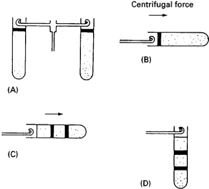
For the separation of proteins from other proteins, especially for small scale separation, the swinging-bucket rotor is widely used for rate-zonal centrifu-gation. This type of rotor is also used for the estima-tion of sedimentaestima-tion coefRcients of proteins. As shown inFigure 4, in the swinging-bucket rotor, the sample tubes are loaded into individual buckets which hang vertically while the rotor is at rest. When the rotor begins to rotate, the buckets swing out perpendicular to the axis of rotation. In these rotors, resolution of particles is high because particles sedi-ment with a relatively long path length. For the same reason, run times are generally longer. Many types of swinging-bucket rotors are commercially available. The centrifuge tube should be as long as possible if high resolution is the objective. For large volume samples, swinging-bucket rotors with wider tubes should be used because the sample can be loaded in a narrow zone while still reducing particle interac-tions during sedimentation.
Vertical rotors are suitable for isopycnic as well as for rate-zonal separations. However, this type of rotor is not practical for the separation of proteins. As a result of diffusion and reorientation during
centrifugation, sample bands will be signiRcantly broader than analogous bands in swinging-bucket rotors. In addition, if the sample contains pellets or
Soats, these materials will distribute along the length of the tube and can subsequently contaminate the supernatant during reorientation at the end of run. Choice of Density Gradient
A density gradient is essential for rate-zonal centrifu-gation to support the zones of particles as they sedi-ment. In addition, the sample can be loaded on to the top of the gradient as a narrow zone and the increas-ing density from the top to the bottom of the density gradient suppresses mechanical disturbances. More-over, the presence of a gradient of increasing viscosity serves to sharpen the sample zones during centrifu-gation. The density gradient material for protein sep-aration requires the following properties.
1. The materials should be sufRciently soluble in water to produce the range of densities needed. 2. Solutions of the gradient materials should be
ad-justable to a pH and ionic strength that are not harmful to proteins in the sample.
3. The materials should not interfere with methods of analysis of the target protein.
Sucrose has most often been used as a gradient material. Sucrose is inexpensive and extremely sol-uble in aqueous media and can be used to produce density gradients ranging up to 1.35 g mL\1. Thus, it is suitable for separation of almost all proteins in cells. Although concentrated solutions of sucrose have high osmotic potential that cause shrinkage of certain cells and organelles, the high osmotic pressure has relatively less effect on the biological properties of proteins. Generally, sucrose is relatively inert to pro-teins, although contaminants in many commercial sources of sucrose may interact with proteins. Such impurities can be removed by treatment with ac-tivated charcoal. However, it is best to purchase specially puriRed sucrose for density gradient work. To sterilize sucrose solutions, autoclaving (1003C or above) of the solution should be avoided and treat-ment with 0.1% diethylpyrocarbonate is recommen-ded. As described above, isopycnic centrifugation can be used for the separation of different types of pro-teins. However, it should be noted that the density of sucrose, even of a saturated solution, is too low for the separation. For this purpose, RbCl, NaBr or KBr can be used to form shallower gradients for better resolution.
of proteins by rate-zonal separations. However, it should be noted that the high viscosity of glycerol reduces the effective density range and glycerol ap-pears to inhibit some enzyme activities.
Preparation of Gradients
Density gradients can be divided into two types: con-tinuous and disconcon-tinuous. For protein separation, continuous gradients are usually used in rate-zonal centrifugation. The most common continuous gradi-ent for protein separation is a linear gradigradi-ent in swinging-bucket tubes. A linear gradient is a gradient in which the density increases linearly in a tube of constant cross-sectional area with increasing distance from the centre of rotation. Thus, in this conR gura-tion the linear gradient can be deRned as one where the density increases linearly with volume.
When designing a linear gradient in swinging-bucket rotors, several points should be emphasized. The density at the top of the gradient must be sufR -cient to support the sample while the density of the bottom of the gradient must not exceed the density of proteins to be separated. In general, the greater the slope of the gradient, the better the resolution ob-tained because the viscous drag rises rapidly as the sucrose concentration increases. Usually, as a Rrst attempt, a 5}30% or 10}40% sucrose gradient should be used. It should be emphasized that the sample volume is related to the slope of the gradient because a given slope of gradient can only tolerate a limited amount of sample before gradient inversion occurs. Poor resolution during rate-zonal centrifu-gation almost always results from overloading.
Linear gradients are prepared using gradient makers. Many conRgurations of gradient maker are available. The simplest gradient makers consist of two vessels of equal cross-sectional area joined by a connecting channel with a stopcock. One chamber is a reservoir and the other chamber has a mixing device and an exit connected to the centrifuge tube. There are two methods for preparing linear gradients: 1. The reservoir contains the less dense solution, the mixing chamber contains the denser solution, and the gradient is routed to the wall of the centrifuge tube at the top. This method is readily applicable to centrifuge tubes made of hydrophilic materials such as cellulose nitrate and cellulose acetate butyrate.
2. The reservoir contains the denser solution, the mixing chamber contains the less dense solu-tion, and the gradient is routed to the bottom of the centrifuge tube. This method can be applied to any type of centrifuge tube and it is much easier to prepare the gradient without disturbance.
The gradient should be prepared and maintained at 43C.
Preparation of Sample
The sample should be ready for loading before the gradient is prepared and should be kept cold for many preparations. The sample is usually prepared in the same buffer as the gradient. In addition, several points are important in sample preparation:
1. The sample solution must have a density less than that of the gradient.
2. Gradients should be centrifuged as soon as poss-ible after the sample has been loaded to prevent so-called droplet sedimentation.
3. For optimal resolution in rate-zonal centrifu-gation, the sample must be loaded on to the top of gradient and the sample volume should not exceed 2}3% of the gradient volume.
Loading of the sample on to the density gradient is one of the most crucial steps in rate-zonal centrifu-gation. The simplest method is to use a pipette to load the sample directly to the meniscus at the tube wall. Conditions During Centrifugation
Smooth acceleration and deceleration are important for all gradient work. In addition, control of the temperature of the sample and gradient are important for reliable and reproducible sedimentation. Fortu-nately, most modern ultracentrifuges are equipped with programmed acceleration and deceleration modes which minimize the disturbance of gradient and temperature control. It should be emphasized that, during the gradient reorientation phase of a run using a swinging-bucket rotor, the rotor should be accelerated as slowly as possible up to 1000 rpm, and the brake switch should be off below 1000 rpm dur-ing deceleration.
Recovery of Fractions from the Gradient
After centrifugation, gradients are fractionated to re-cover protein bands. Great care must be taken at this stage to avoid loss of resolution. Several points should be emphasized.
1. All operations should be designed to minimize disturbance of the gradient.
2. The volume of the tubing from the gradient to the fraction collector should be minimized.
3. Care must be taken to avoid contamination of the recovered fractions by pelleted materials.
In order to collect the entire gradient in a series of fractions, several methods may be applied. The simplest is to pierce the bottom of the tube with a needle, and collect the gradient as it drops out. Another method is to pump the gradient from the bottom of the tube with a narrow capillary tube. However, this method is not recommended because of the potential to disturb the gradient and resulting loss of resolution.
See also: III/Proteins: Capillary Electrophoresis; Crystal-lization; Electrophoresis; Field Flow Fractionation; High-Speed Countercurrent Chromatography; Ion Exchange.
Further Reading
GrifRth OM (1979) Ultracentrifuge Rotors:A Guide to Their Selection. Palo Alto, Beckman Instruments.
Hsu HW (1981) Separation by Centrifugal Phenomena. New York: John Wiley.
Laskin AI and Last JA (eds) (1974)Subcellular Particles, Structures,and Organelles. New York: Marcel Dekker. Neurath N and Hill RL (eds) (1975)The Proteins, 3rd edn.
New York: Academic Press.
Price CA (1982)Centrifugation in Density Gradient. New York: Academic Press.
Rickwood D (ed.) (1983)Iodinated Density Gradient Me-dia:A Practical Approach. Oxford: IRL Press.
Rickwood D (ed.) (1984)Centrifugation, 2nd edn,A Prac-tical Approach. Oxford: IRL Press.
Rickwood D (ed.) (1992) Preparative Centrifugat-ion,A Practical Approach. Oxford: Oxford University Press.
Schachman HK (1959)Ultracentrifugation in Biochemis-try. New York: Academic Press.
Sheeler P (1981) Centrifugation in Biology and Medical Science. New York: John Wiley.
Crystallization
M. Y. Gamarnik, Nanoscale Phases Research,
Bensalem, PA, USA
Copyright^ 2000 Academic Press
Introduction
The Rrst protein crystals described in the literature were obtained by Hunefeld in 1840. Hunefeld ob-served hemoglobin crystals after slow drying of blood pressed between two slides of glass. It is remarkable that thisRrst result demonstrated the basic principle used today, that protein crystals similar to inorganic crystals may be produced by concentration of a pro-tein in solution through slow dehydration. Through-out the history of protein crystal growth, the rationale for protein crystallization has been,Rrstly, separation of proteins from complex extracts, and then, starting in the 1930s, as puriRcation as deter-mination of the three-dimensional structure of pro-tein molecules.
Knowledge about the three-dimensional structure is necessary to better understand the functions of protein molecules in living systems and plants. Three-dimensional structure can be determined by X-ray diffraction. For X-ray diffraction, good quality pro-tein crystals of appropriate sizes are required. Crystal sizes in each direction should be at least 0.1 mm, if using a strong beam of synchrotron radiation, or at least 0.3 mm for conventional sources of X-rays.
Protein molecules in the crystalline state are more stable than in solution. Therefore, crystallized
pro-teins are more stable against denaturation and may be preserved for a signiRcantly longer period of time than in solution. That is the reason that protein crys-tallization is often directed as much on preservation as on separation and puriRcation.
This article comprises a brief description of general principles of protein crystal growth and a description of various techniques of protein crystallization with the emphasis on methods using a small amount of a crystallizing solution, from about 1 to 20L. The consumption of small amounts of protein is of value, since screening and optimization tests of determina-tion of crystallizadetermina-tion condidetermina-tions typically require many portions of protein solution.
General Principles of Protein
Crystallization
Intermolecular Interaction
To crystallize a protein it should be Rrst of all dis-solved to give a solution where the protein molecules become close one to another to create a nucleus that grows into a crystal.