10.4236/pp.2010.11001 Published Online July 2010 (http://www.SciRP.org/journal/pp)
Anti-Amnesic Activity of Vitex Negundo in
Scopolamine Induced Amnesia in Rats
Abhinav Kanwall,2, Jogender Mehla3, Madhusudana Kunchal, Vegi Ganga Modi Naidul, Yogendra Kumar Gupta3, Ramakrishna Sistla1*
1Division of Pharmacology, Indian Institute of Chemical Technology (IICT), Hyderabad, India; 2National Institute of Pharmaceutical
Education and Research (NIPER), Hyderabad, India; 3Department of Pharmacology, All India Institute of Medical Sciences (AIIMS),
New Delhi, India. Email: sistla@iict.res.in
Received June 8th, 2010; accepted July 12th, 2010.
ABSTRACT
In the present study we investigated the anti-amnesic activity of Vitex negundo in scopolamine induced amnesia in rats. Wistar rats (180-200 g) were trained on active avoidance task. Each animal received session of 15 trials with inter trial duration of 15 s for 5 days. Scopolamine (3 mg/kg, i.p) was administered at different time periods on the basis of stages of memory i.e acquisition, consolidation and retention in different groups (n = 6). Effect of Vitex negundo extract was evaluated and compared to a standard drug, Donepezil. Significant (p < 0.05) increase in the avoidance response on the 5th session has been observed as compared to 1st session in control group. Scopolamine treatment significantly (p < 0.05) reduced the avoidance response compared to control. Extract treated groups shown significant (p < 0.05) in-crease in number of avoidance responses as compared to scopolamine treated groups. Inin-creased oxidative stress in brain after scopolamine treatment, as observed by increase in MDA & decrease in GSH & SOD, was lowered in the groups treated with extracts. AChE activity was also improved after V. negundo treatment. Results of the study have shown that V. negundo treated groups decrease the phenomenon of amnesia by increasing learning of memory through antioxidant effect and decreasing AChE activity.
Keywords: Vitex Negundo, Amnesia, Acetylcholinestrase, Scopolamine, Learning and Memory, Oxidative Stress
1. Introduction
The Memory is the most important function of the brain. Memory is the process by which organisms are able to record their experiences and use this information to adapt their responses to the environment. Hence it is vital for survival [1]. Central cholinergic system is considered as the most important neurotransmitter involved in regula-tion of cognitive funcregula-tions [2]. Impaired cognitive func-tions are the major features of Alzheimer disease (AD) [3]. Presence of acetylcholine within the neocortex is sufficient to ameliorate learning deficits and restore memory [4]. The prevalence of AD increases with the age (65 yrs) from 2% to 30-45% in those over 85 yrs [5]. AD and stroke together rank as the third most common causes of death [6]. The incidence of AD for those aged 65yrs and older was 3.24 per 1000 individuals in a year [7]. One study in India showed that, the median survival time determined to be 3.3 yrs for patients with dementia and 2.7 yrs for patients with AD [8]. Scopolamine, a
nonselective muscarinic cholinergic antagonist, is a well- known centrally acting cholinergic probe, which causes impairment in learning [9]. In addition, scopolamine also causes increase in cognitive impairment in healthy eld-erly subjects compared to young adults [10]. The treat-ment with AChE inhibitors and muscarinic receptors agonists which increases cholinergic neurotransmission causes an improvement in cognitive deficits in AD [11]. Besides reducing cholinergic activity, oxidative stress plays an important role and is one of the major causes for memory loss in AD [12,13]
Extensive research is going on different plants all around the world as plant extracts have a relatively higher therapeutic window, lesser side effects and are economical. Plant extracts may also provide a source of new compound as many synthetic drugs have been
originated from herbal sources. Vitex negundo, a
parts of the plant are used, the extract from leaves and the roots is the most important in the field of phytomedi-cine and is sold as drugs. The leaf extract is used in Ay-urvedic and Unani system of medicine [14]. Water ex-tract of mature fresh leaves exhibited anti-inflammatory, analgesic and antihistamine properties [15]. Literature survey of V. negundo revealed the presence of volatile oil, triterpenes, diterpenes, sesquiterpenes, lignan, flavonoids, flavones glycosides, iridoid glycosides, and stilbene de-rivative [14]. Lignans, one class of natural compounds present in V. negundo,showed anti-cholinesterase activ-ity in in-vitro [14]. However no studies were conducted to explore the effect of V. negundo extract against mem-ory impairment in in-vivo.
In the process of learning and memory, three important stages have been suggested viz., acquisition, consolida-tion and recall of the learned task [16]. The Scopolamine hydrobromide is an anticholinergic drug, which produces amnesia by reducing the levels of acetylcholine, which is considered to be an important neurotransmitter for the learning and memory. Therefore, the present study was aimed to investigate the anti-amnesic effect of V. ne-gundo aqueous extract on scopolamine administered at different stages of active avoidance learning in rats.
2. Materials and Methods
2.1 Materials
Aqueous extract of the plant Vitex negundo was obtained from Amruta herbals Pvt Limited, Indore (M.P), India, (Batch no. AHVN/556.) along with the copy of certifi-cate of analysis. Scopolamine hydrobromide, Thiobarbi-turic acid (TBA), Glutathione, DTNB, Acetylthiocholine all were purchased from Sigma-Aldrich (Bangalore, In-dia). SOD kit was purchased from Fluka. Other chemical and reagents are of analytical grade.
2.2 Animals
Male Wistar rats weighing between 180-200 g were ob-tained from National Institute of Nutrition, Hyderabad. The animals were housed in an animal facility of Indian Institute of Chemical Technology (IICT). The animal house maintained at 20 ± 2°C and 50-60% relative hu-midity. A 12-hour dark/light cycle was maintained throughhout the study. Air changes were maintained with 5µ HEPA filter. Rats had free access to food (pellet diet supplied from M/s Petcare India Ltd., Bangalore) and
water ad libitum. This study protocol was approved by
the Institutional Animal Ethics Committee of Indian In-stitute of Chemical Technology, Hyderabad.
2.3 Behavioral Test
2.3.1 Two-Way Active Avoidance with Negative (Punishment) Reinforcement
The animals were trained on Active Avoidance Task in
an automatic reflex conditioner with two-way shuttle box (Ugo Basile, Italy). The rats were treated orally with the standard drug through an intragastric feeding tube. Simi-larly the plant extract were administered for 14 days. For this purpose each rat is placed in a compartment sepa-rated from the other one by a guillotine door in the shut-tle box. Exploration period of 2 min is given initially. There after, the trial start. In each trial the animal is sub-jected to a light for 30 s followed by a sound stimulus for 10s. Immediately after the sound stimulus, the rat re-ceives a single low intensity foot shock (0.5 mA; 3 s) from 10th day to 14th through the floor grid if it does not
transfer to the other shock free compartment. Infrared sensors monitor the transfer time from one compartment to another, which is recorded as avoid (after the stimulus of either light alone or both light and sound) and escape (after the foot shock) response. Each animal received a daily session of 15 trials with an inter-trial duration of 15 s for 5 days i.e., a maximum of 75 trials. The rats were evaluated on the basis of their performance in the last session i.e., in the 5th session for their decrease in amne-sic activity and increased learning and memory. The cri-terion for improved cognitive activity was taken as sig-nificant increase in the avoidance response on 5th session
(retention) compared to 1st session.
2.4 Scopolamine Induced Loss of Memory in Rat Acquisition: scopolamine was administered 5 min prior to 1st Trial on 1st session
Consolidation: scopolamine was administered 5 min after the 15th (
i.e., last) trial on 1st session (Training ses-sion)
Retention: scopolamine was administered 5 min prior to the 1st trial on the last session
i.e., 5th session (Training session)
Dementia effect of scopolamine was evaluated on the basis of significant decrease in number of avoidance re-sponse in the treated groups as compared to that of con-trol group in the last session i.e., 5th session.
2.5 Treatment Schedule
The animals were divided into eight different groups (n = 6). Scopolamine (3 mg/kg, i.p) was administered at dif-ferent time periods in the three groups (GR-2, GR-3, and GR-4) as follows:
Group I (GR-1)–Saline (control),
Group II (GR-2)–scopolamine was administered 5 min prior to 1st Trial on 1st session (Training session)
Group III (GR-3)–scopolamine was administered 5 min after the 15th (
i.e., last) trial on 1st session (Training session).
Group IV (GR-4)–scopolamine was administered 5 min prior to the 1st trial on the last session
i.e., 5th ses-sion.
is given to GR-2 rats prior to 1 hour of 1st trial.
Group VI (GR-6)–Similar to GR-2 but rats were pre-treated with plant extract for 14 days.
Group VII (GR-7)–Similar to GR-3 but rats were pre-treated with plant extract for 14 days.
Group VIII (GR-8)–Similar to GR-4 but rats were pre-treated with plant extract for 14 days.
2.6 Biochemical Estimation of Markers of Oxidative Stress
On day 14th following the behavioral testing, animals
were sacrificed and the brain tissues were quickly re-moved, cleaned with ice-cold saline and stored at –80°C for biochemical estimation.
2.6.1 Preparation of Brain Homogenate
Brain-tissue samples were thawed and homogenized with 10 times (w/v) ice-cold 0.1 M phosphate buffer (pH 7.4). Aliquots of homogenates from the rat brains were sepa-rated and used to measure protein, lipid peroxidation and glutathione. The remaining homogenates were centri-fuged at 10,000 rpm for 15 min and the supernatant was then used for enzyme assay. Superoxide dismutase was determined within 24 h.
2.6.2 Estimation of Malondialdehyde (MDA)
Aliquotes of 0.5 ml distilled water and 1.0 ml 10% TCA were added to a volume of 0.5 ml brain tissue homoge-nate, mixed well and centrifuged at 3000 rpm for 10 min. To 0.2 ml supernatant, 0.1 ml thiobarbituric acid (TBA) (0.375%) was added. The total solution was placed in a water bath at 80ºC for 40 min and then cooled to room temperature. The absorbance of the clear supernatant was measured at 532 nm in spectrophotometer [17].
2.6.3 Estimation of Superoxide Dismutase (SOD) The SOD activity of the brain tissue was analyzed by using the SOD Assay kit (Fluka). For the assay, 200 µl of working solution, 20 µl of dilution buffer and 20 µl of enzyme working solution was added. Incubate the plate at 37°C for 20 min. Absorbance was read at 450 nm us-ing a microplate reader.
2.6.4 Measurement of Glutathione
Pipette out 100 µl of the brain supernatant and add 50 µl of O-ophthaldehyde (100 µl/ml). Incubate at room tem-perature for 15 min. The flouroscent complex formed was read at an excitation wavelength of 350 nm and emission wavelength of 420 nm [18].
2.6.5 Estimation of Cholinergic Status in the Rat Brain The cholinergic marker, acetylcholinesterase was esti-mated in the whole brain according to the method of [19]. Briefly, the brains of the rats were removed over ice and the brain was separated using fine forceps. The tissue was then homogenized in 100 mM phosphate buffer. 0.1
ml of this homogenate was incubated for 5 min with 2.7 ml of phosphate buffer and 0.1 ml of DTNB. Then, 0.1 ml of freshly prepared acetylthiocholine iodide, pH 8 was added and the absorbance was read at 412 nm for 3 min at 30, 60, 90, 120, 150 and 180 sec.
2.7 Statistical Analysis
All data were expressed as mean ± SD. The significance of difference among the values of control, scopolamine treated, standard drug and extract treated groups for each session was determined by ANNOVA (one-way) fol-lowed by Dunnett’s test. The difference between values on 1st session and 5th session of the same group was
ana-lyzed by student’s t-test.
3. Results
3.1 Selection of the Dose
One single dose (300 mg/kg) of the herbal extract has been selected after the initial pilot study. This pilot study was done by taking limited number of Wistar rats. In the pilot study three different doses (100, 300 & 900 mg/kg) were taken. Based on initial data (data not shown) from active avoidance test 300 mg/kg was selected for the main study. It was also seen that animals with higher dose (900 mg/kg) tolerated the shock and remained at one place, which is not acceptable for the avoidance test. However, with lower dose (100 mg/kg) there was no significant difference in the number of avoidances be-tween different groups of animals.
3.2 Automatic Reflex Conditioner
There was a significant (p < 0.05) increase in avoidance response on 5th session (6.4 ± 1.67) as compared to 1st
session (3.0 ± 1.00) in the control group (Table 1). All groups except GR-3 have shown significant (p < 0.05) increase in avoidance response compared to their first session data. Significant (p < 0.05) reduction of avoid-ance response was observed in scopolamine treated group (GR-2) compared to control group (GR-1). How-ever standard drug (donepezil) treatment and extract feeding (GR-5 and GR-6) significantly (p < 0.05) in-creased the avoidance response in their first session compared to their corresponding scopolamine treated group (GR-2). This reflects the effectiveness of donepe-zil as well as aqueous extract during scopolamine in-duced memory loss. However donepezil group (GR-5) showed improved response compared to extract treated group (GR-6) (5.8 ± 0.83 vs 4.8 ± 0.44) at the end of 5th
session. While extract treatment showed significant (p < 0.05) improvement of avoidance response at the end of
5th session in GR-7, no improvement was observed in
Table 1. The number of avoidance responses in control (GR-1), scopolamine (GR-2, 3, 4) and drug treated (GR-6, 7, 8) groups (Mean ± SD, n = 6)
S.NO. GROUPS DAY1 DAY2 DAY3 DAY4 DAY5
1 GR-1 03 ± 1.00 3.6 ± 1.14 4.2 ± 0.83 05 ± 0.70 6.4 ± 1.67**
2 GR-2 2.4 ± 0.55 03 ± 1.00 3.4 ± 1.14 3.8 ± 0.83 4.2 ± 0.83**,†
3 GR-3 3.2 ± 1.09 2.6 ± 0.54 3.4 ± 0.54 3.4 ± 0.89 3.2 ± 0.44
4 GR-4 3.4 ± 0.54 3.6 ± 0.89 4.2 ± 0.83 4.6 ± 0.54 4.8 ± 0.44*
5 GR-5 03 ± 0.70 04 ± 100 4.4 ± 0.54 5.4 ± 0.89 5.8 ± 0.83**, Ψ
6 GR-6 3.4 ± 0.54 3.8 ± 0.83 4.2 ± 0.83 4.8 ± 0.83 4.8 ± 0.44**,†,Ψ
7 GR-7 3.6 ± 0.54 3.6 ± 0.54 3.8 ± 0.44 4.4 ± 1.52 4.6 ± 0.54*,Ψ
8 GR-8 3.6 ± 0.89 3.8 ± 0.44 4.4 ± 0.54 4.8 ± 0.44 5.4 ± 0.54**
*p < 0.05 vs. Day 1; **p < 0.01 vs. Day 1; †p < 0.05 vs. Standard drug (GR-6); Ψp < 0.05 vs. corresponding scopolamine treated group
3.3 Markers of Oxidative Stress in Rat Brain 3.3.1 Malondialdehyde (MDA) levels
Scopolamine treatment (GR-2, GR-3 and GR-4) signifi-cantly (p < 0.05) increased the brain MDA level com-pared to control (GR-1) group (Figure 1). However only GR-3 showed significant (p<0.05) change compared to
GR-1. Standard drug (GR-5) and aqueous extract of V.
Negundo (GR-6, GR-7 and GR-8) treatment significantly (p < 0.05) decreased brain MDA level compared to their corresponding scopolamine treated groups (GR-2, GR-3 and GR-4).
3.3.2 Glutathione (Gsh) Levels
Brain GSH level was decreased significantly (p < 0.05) in scopolamine treated groups (GR-2, GR-3 and GR-4) compared to control (GR-1) (Figure 2). However
stan-dard drug (GR-5) and aqueous extract of V. Negundo
(GR-6, GR-7 and GR-8) treatment significantly (p < 0.05) increased brain GSH level compared to their corre-sponding scopolamine treated groups (GR-2, GR-3 and GR-4).
3.3.3 SOD Activity
SOD Activity has been expressed in % inhibition rate. Scopolamine treatment decreased brain SOD activity significantly (p < 0.05) in GR-2 and GR-3 groups but not in GR-4 (Figure 3). No improvement of SOD activity
was observed in V. Negundo extract treated groups
(GR-6, GR-7 and GR-8) compared to their corresponding scopolamine treated groups (GR-2, GR-3 and GR-4). However standard drug (GR-5) increased the SOD activ-ity significantly (p < 0.05) compared to the correspond-ing scopolamine treated group (GR-2).
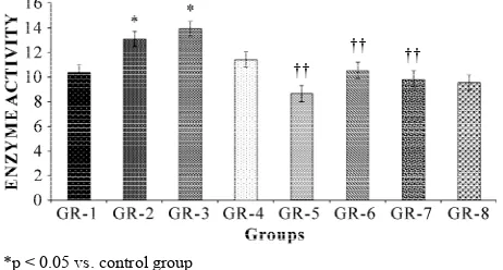
3.3.4 AChE Activity
Acetylcholinestrase activity was estimated by the Vmax
values as shown in Figure 4. The scopolamine treated
groups have more Vmax values as compared to control
group. Significant (p < 0.05) increased of AChE activity
† † †† ††
*p < 0.05 vs. control group
[image:4.595.308.533.364.472.2]†p < 0.05 vs. corresponding scopolamine treated group ††p < 0.01 vs. corresponding scopolamine treated group
Figure 1. Effect of aqueous extracts of Vitex Negundo (300 mg/kg body wt.) on MDA levels in brains on Session 5 on different groups (Mean ± SD, n = 6)
† †† †† ††
*p < 0.05 vs. control group
†p < 0.05 vs. corresponding scopolamine treated group ††p < 0.01 vs. corresponding scopolamine treated group
[image:4.595.310.535.553.654.2]††
*p < 0.05 vs. control group
[image:5.595.59.285.84.222.2]†p < 0.05 vs. corresponding scopolamine treated group ††p < 0.01 vs. corresponding scopolamine treated group
Figure 3. Effect of aqueous extracts of Vitex Negundo (300 mg/kg body wt.) on SOD levels in brains on Session 5 on different groups (Mean ± SD, n = 6)
†† †† ††
*p < 0.05 vs. control group
†p < 0.05 vs. corresponding scopolamine treated group ††p < 0.01 vs. corresponding scopolamine treated group
Figure 4. Effect of aqueous extracts of Vitex Negundo (300 mg/kg body wt.) on AChE levels in brains on Session 5 on different groups (Mean ± SD, n = 6)
was observed in scopolamine treated groups (GR-2 and GR-3, not in GR-4) compared to control (GR-1) (Figure 4). However standard drug (GR-5) and aqueous extract of V. Negundo (GR-6 and GR-7, not in GR-8) treatment significantly (p < 0.01) decreased brain AChE activity compared to their corresponding scopolamine treated groups (GR-2, GR-3 and GR-4).
4. Discussion
V. negundo possesses many medicinal properties. Leaves of V. negundo have been investigated for its anti-in- flammatory activity [15,20]. Telang et al first noticed
non-steroidal anti-inflammatory (NSAID) activity of V.
negundo. Similarly, fresh leaves of V. negundo have been suggested to possess anti-inflammatory and pain sup-pressing activities. Antinociceptive activity study of ethanolic leaf extract of V. negundo showed that it pos-sesses both central and peripheral analgesic activity [21]. V. negundo has been also used in adjuvant therapy to standard anti-inflammatory drugs [22]. Literature survey of V. negundo also revealed the presence of lignans de-rivative, which is responsible for anti-cholinesterase
ac-tivity in in-vitro [14]. LD50 dose of V. negundo leaf
ex-tract is 7.58 g/kg which practically falls in the non- toxic dose range [23].
The administration of the antimuscarinic agent polamine produces transient memory deficit. Also, sco-polamine has been shown to impair memory retention when given to rat shortly before training in an avoidance task. The ability of a range of different cholinergic ago-nist drugs to reverse the amnesic affects of scopolamine is now well documented in animals and human volun-teers [24]. The scopolamine amnesia test is widely used as primary screening test for so called anti-Alzheimer drugs [24]. Here, scopolamine is given at different time of training sessions and trials. Such protocol is adapted to distinguish between the three different stages of memory i.e. acquisition, consolidation and retention.
In the preliminary screening of the present study showed that the improvement in learning and memory tasks in the shuttle-box was only observed at a dose of 300 mg/kg body wt. Therefore, the aqueous extract with 300 mg/kg was evaluated in more details. The avoid-ance responses shown by the animals were due to their ability to learn the task, which reflects the cognitive function. The task was investigated by using the sco-polamine-induced dementia with the aqueous herbal ex-tract of V. negundo. The animals were treated with sco-polamine at different time intervals of trial in the sessions according to the different stages of the memory. Sco-polamine administered 5 min prior to 1st trial on 1st
ses-sion was for the acquisition whereas scopolamine ad-ministered 5 min after the 15th (
i.e., last) trial on 1st ses-sion was for the consolidation stage of the memory. Similarly for the requisition (recall) the scopolamine was administered 5 min prior to the 1st trial on the last session
i.e., 5th session. To evaluate the effect of the herbal
aqueous extract of V. negundo, scopolamine was
admin-istered in the similar pattern in the pretreated herbal ex-tract animals. The efficacy and potency of the exex-tract was compared with the vehicle control and standard drug (Donepezil) group.
From the behavioral test i.e. two way shuttle box ac-tive avoidance test, it is clearly seen that there was a general decrease in the performance in the active avoid-ance in the scopolamine treated groups. The memory loss effect of scopolamine is more prominent compared
to the control group. The aqueous herbal extract of V.
negundo improved the memory loss effect of scopola-mine in all three events like acquisition, consolidation and retention. As scopolamine-induced memory loss was more prominent in acquisition period, we administered standard drug, donepezil with this group. In comparison with Donepezil, the extract treated group had almost equal avoidance responses which indicates therapeutic efficacy of V. negundo against memory loss.
[image:5.595.55.286.274.398.2]mechanism by which V. negundo enhanced the anti-am- nesic activity by increasing the performance of learning and memory. It had been suggested that the varying de-grees of behavioral impairments are associated with ag-ing and age associated neurodegenerative diseases. Oxi-dative stress due to free radicals generation is responsible for producing the neuronal changes mediating these be-havioral deficits [25]. Oxidative stress in brain generates oxygen radicals like superoxide anion, hydroxyl radical, and hydrogen peroxide, which act on polyunsaturated fatty acids in brain, thereby propagating the lipid peroxi-dation [26]. The major antioxidant and oxidative free- radical scavenging enzymes like glutathione, SOD and catalase plays an important role to reduce oxidation stress in brain. In the present study rats after scopolamine treatment showed a significant increase in the brain lev-els of malondialdehyde, which is the measure of lipid peroxidation and free radical generation. At the same time there was a significant reduction in levels of glu-tathione, a tripeptide found in all cells, which reacts with free radicals to protect cells from superoxide radical, hydroxyl radical and singlet oxygen [27]. Pre-treatment of V. negundo reduced the MDA levels and increased GSH content in brain after scopolamine treatment. Sco-polamine reduced the SOD activity in brain. SOD is the only enzyme that uses the superoxide anions as the sub-strate and produces hydrogen peroxide as a metabolite. Super oxide anion is more toxic than H2O2 and has to be
removed. Pretreatment with V. negundo significantly
prevented the reduction of SOD activity in brain during scopolamine treatment. Our results also suggest that the
aqueous extract of V. negundo reduced oxidative stress
by reducing lipid peroxidation and increasing the en-dogenous antioxidant enzymes in brain. Other important activity has been shown by the extract is that it has ace-tylcholinetrase (AChE) inhibiting activity. This activity tends to allow the more retention of acetylcholine in the brain, which is important for the cognitive functions, learning and memory.
In conclusion, the present study demonstrates that
aqueous V. negundo extract has potential therapeutic
effects on improving the anti-amnesic activity in rats through inhibiting lipid peroxidation, augmenting en-dogenous antioxidant enzymes and decreasing acetylcho- linestrase (AChE) activity in brain. Further study is war-ranted to find its potential use in humans.
5. Aknowledgements
Authors are very thankful to the Project Director, Na-tional Institute of Pharmaceutical Education and Re-search, Hyderabad and Director Indian Institute of Chemical Technology, Hyderabad for supporting this work. We are also thankful to Amruta Herbals Pvt Lim-ited, Indore (M.P), India for providing the plant material.
REFERENCES
[1] J. Dunning and J. D. Matthew, “Molecular Mechanisms of Learning and Memory,” Expert Reviews in Molecular Medicine, Vol. 5, No. 25, 2003, pp. 1-11.
[2] A. Blockland, “Acetylcholine: A Neurotransmitter for Learning and Memory?” Brain Research Review, Vol. 21, No. 3, 1996, pp. 285-300.
[3] M. F. Siddiqui and A. I. Levey, “Cholinergic Therapies in Alzheimer’s Disease,” Drugs of the Future, Vol. 24, No. 4, 1999, pp. 417-444.
[4] J. Winkler, S. Suhr and F. Gage, “Essential Role of Neo-cortical Acetylcholine in Spatial Memory,” Nature, Vol. 375, No. 6531, 1995, pp. 484-487.
[5] T. F. Wernicke and F. M. Reischies, “Prevalence of De-mentia in Old Age: Clinical Diagnoses in Subjects Aged 95 and Older,” Neurology, Vol. 44, No. 2, l994, pp. 250- 253.
[6] D. C. Ewbank, “Deaths Attributable to Alzheimer’s Dis-ease in the United States,” American Journal of Public Health, Vol. 89, No. 1, 1991, pp. 90-92.
[7] V. Chandra, R. Pandav and H. H. Dodge, “Incidence of Alzheimer’s Disease in a Rural Community in India: The Indo-US Study,” Neurology, Vol. 57, No. 6, 2001, pp. 985-989.
[8] V. Chandra, M. Ganguli and R. Pandav, “Prevalence of Alzheimer’s Disease and Other Dementias in Rural India: The Indo-US Study,” Neurology, Vol. 51, No. 4, 1998, pp. 1000-1008.
[9] T. Sunderland, P. N. Tariot, R. M. Cohen, H. Weingarbier, E. A. Mueller and D. L. Murphy, “Anticholinergic Sensi-tivity in Patients with Dementia of the Alzheimer’s Type and Age Matched Controls,” Archives of General Psy-chiatry, Vol. 44, No. 5, l987, pp. 418-426.
[10] C. Flicker, S. H. Ferris and M. Serby, “Hypersensitivity to scopolamine in the elderly,” Psychopharmacology, Vol. 107, No. 2-3, 1992, pp. 437-441.
[11] E. Giacobini, “The Cholinergic System in Alzheimer's Disease,” In: S. M. Aquilonius and P. G. Gillberg, (Eds.), Brain Research. Cholinergic Neurotransmission: Func-tional and Clinical Aspects, Elsevier, Amsterdam, 1990, pp. 321-332.
[12] W. R. Markesbery, “Oxidative stress hypothesis in Alz-heimer’s disease,” Free Radical Biology and Medicine, Vol. 23, No. 1, 1997, pp. 134-147.
[13] M. A. Lovell, W. D. Ehmann, S. M. Butler and W. R. Markesberg, “Elevated Thiobarbituric Acid Reactive Substances and Antioxidant Enzyme Activity in the Brain in Alzheimer’s Disease,” Neurology, Vol. 45, No. 8, 1995, pp. 1594-1608.
[14] U. H. Azhar and M. Abdul, “Enzymes Inhibiting Lignans from Vitex Negundo,” Chemical and Pharmaceutical Bulletin, Vol. 52, No. 11, 2004, pp. 1269-1272.
No. 2-3, 2003, pp. 199-206.
[16] A. C. Guyton and J. E. Hall, “Textbook of Medical Phy-siology,” Harcourt Asia Pte Ltd, Singapore, 1999. [17] H. Ohkawa, N. Ohishi and K. Yagi, “Assay for Lipid
Peroxides in Animal Tissues by Thiobarbituric Acid Re-action,” Analytical Biochemistry, Vol. 95, No. 2, 1979, pp. 351- 358.
[18] P. J. Hissin and R. Hilf, “A Fluorometric Method for Determination of Oxidized and Reduced Glutathione in Tissues,” Analytical Biochemistry, Vol.74, No. 1, 1976, pp. 214-226.
[19] G. L. Ellman, D. K. Courtney, V. Andres and R. M. Fea-therstone, “A New and Rapid Colorimetric Determination of Acetylcholinesterase Activity,” Biochemical Pharma-cology, Vol.7, No. 2, 1961, pp. 88-95.
[20] R. S. Telang, S. Chatterjee and C. Varshneya, “Studies on Analgesic and Anti-Inflammatory Activities of Vitex Negundo Linn,” Indian Journal of Pharmacology, Vol. 31, No. 5, 1999, pp. 363-366.
[21] R. K. Gupta and V. R. Tandon, “Antinociceptive Activity of Vitex-Negundo Linn Leaf Extract,” Indian Journal of Pharmacology, Vol. 49, No. 2, 2005, pp. 163-170.
[22] R. K. Gupta and V. R. Tandon, “Vitex Negundo Linn (VN) Leaf Extract as an Adjuvant Therapy to Standard Anti-Inflammatory Drugs,” The Indian Journal of Medi-cal Research, Vol. 124, No. 4, 2006, pp. 447-450. [23] M. N. Ghosh, “Fundamentals of Experimental
Pharma-cology,” scientific book agency, Calcutta, 1984.
[24] M. H. V. Kumar and Y. K. Gupta, “Antioxidant Property of Celastrus Paniculatus Willd: A Possible Mechanism in Enhancing Cognition,” Phytomedicine, Vol. 9, No. 4, 2002, pp. 302-311.
[25] C. I. Cantuti, B. Shukitt-Hale and J. A. Joseph, “Neuro-behavioural Aspects of Antioxidants in Aging,” Interna-tional Journal of Developmental Neuroscience, Vol. 18, No. 4-5, 2000, pp. 367-381.
[26] T. Coyle and P. Puttfarcven, “Oxidative Stress, Glutamate and Neurodegenerative Disorder,” Science, Vol. 262, No. 5134, 1993, pp. 89-695.