http://www.scirp.org/journal/jbm ISSN Online: 2327-509X
ISSN Print: 2327-5081
DOI: 10.4236/jbm.2019.77005 Jul. 15, 2019 51 Journal of Biosciences and Medicines
Comparative Evaluation of Antioxidant
Enzymes and Serum Selenium Levels in
Libyan Atherosclerotic Patients
Rabia Alghazeer1*, Eenas Aboulmeedah1, Sana Elgahmasi1, Nadia Alghazir2,
Zohra Almukthar3, Maryouma Enaami4, Abdurrahman Rhuma1
1Department of Chemistry, Faculty of Sciences, University of Tripoli, Tripoli, Libya 2Pediatric Department, Faculty of Medicine, University of Tripoli, Tripoli, Libya 3Tajoura National Heart Center, Tripoli, Libya
4Department of Statistics, Faculty of Science, University of Tripoli, Tripoli, Libya
Abstract
Introduction: Atherosclerosis is the major source of morbidity and mortality in the developed countries like Libya. Oxidative stress represents a risk factor and plays a key role at several steps of atherosclerosis. Formation of atheros-clerotic plaques is the main reason for coronary artery disease. Aim: This study is aimed to assess the impact of the imbalance between body oxidants and antioxidants (oxidative stress) and its involvement in developing and se-verity of atherosclerosis in atherosclerotic with and without diabetes Libyan patients. Methods: The study sample comprised of atherosclerosis with and without diabetes patients (n = 73), as well as healthy controls (n = 27). Blood samples were collected to determine the levels of malonaldehyde (MDA) as oxidative biomarker, serum selenium level, total antioxidant sta-tus (TAS) level and some antioxidant enzymes including glutathione pe-roxidase (GPx), Catalase (Cat) and superoxide dismutase (SOD). Additionally, oxidant/antioxidant status was compared between atherosclerotic patients with and without diabetes in comparison to controls. Results: The results indicated a significant increase in MDA level among atherosclerotic patients compared to healthy subjects (P < 0.05). While the level of TAS, Cat, SOD and GPx was sig-nificantly decreased among patients compared to the controls (P < 0.05). For the atherosclerotic diabetic patients, the study was found a rise in the level of MDA as well as a marked decrease in TAS and the activity of tested antioxidant enzymes (P < 0.05). A good correlation was obtained between elevated MDA and decreased TAS, Cat, SOD and GPx. Conclusion: Determination of anti-oxidative defense markers contributes to understanding the effect of anti-oxidative stress on the development and the prevention of cardiovascular disease. How to cite this paper: Alghazeer, R.,
Aboulmeedah, E., Elgahmasi, S., Alghazir, N., Almukthar, Z., Enaami, M. and Rhuma, A. (2019) Comparative Evaluation of Anti-oxidant Enzymes and Serum Selenium Levels in Libyan Atherosclerotic Patients. Journal of Biosciences and Medicines, 7, 51-69.
https://doi.org/10.4236/jbm.2019.77005
Received: June 5, 2019 Accepted: July 12, 2019 Published: July 15, 2019
Copyright © 2019 by author(s) and Scientific Research Publishing Inc. This work is licensed under the Creative Commons Attribution International License (CC BY 4.0).
DOI: 10.4236/jbm.2019.77005 52 Journal of Biosciences and Medicines
Keywords
Atherosclerosis, Free Radicals, Oxidative Stress, Antioxidants
1. Introduction
Atherosclerosis is the major source of morbidity and mortality in many African countries including Libya [1]. It is characterized by the accumulation of choles-terol deposits in macrophages, which lead to a proliferation of certain cell types within the arterial wall (foam cells) that gradually impinges on the vessel lumen and impedes blood flow. Reduced blood flow to target organs like heart and brain can cause attack and stroke.
Oxidative stress is the result of imbalance between the reactive oxygen species (ROS) and antioxidants in the body. Oxidative modifications within the arterial wall that may initiate and/or contribute to atherogenesis are likely to occur, which result in imbalance between the reactive oxygen species and antioxidants in the body. Therefore, it is important to consider the sources of oxidants in the context of available antioxidants [2] [3]. Oxidative stress, particularly the oxida-tion of low density lipoprotein (LDL), represents a risk factor and plays a key role at several steps of atherosclerosis, according to the oxidative-modification hypothesis of atherosclerosis [4] [5]. The oxidative modification hypothesis, an-tioxidant protection of LDL in the extracellular space deserves focus, as oxidized LDL has many potential proatherogenic activities [6], and the cellular accumula-tion of oxidized LDL is considered a hallmark of atherosclerosis [7].
The possible sources of oxidative stress in atherosclerosis results from endo-thelial production of ROS, especially superoxide (O2
−), with the subsequent
reaction with nitric oxide (NO) are an important mechanism of vascular dys-function in atherosclerosis. O2
− and NO rapidly interact to generate
peroxyni-trite (ONOO−), a potent oxidant and a mediator of vascular tissue injury [8] [9]. Excess generation of ROS has been demonstrated in atherosclerosis and perox-ynitrite formation has been shown to occur in atherosclerotic human [10] [11].
Diabetes mellitus (DM) has also been shown to play a crucial role in progress of cardiovascular diseases [12] [13]. Recently, it has been found that significant indicators for rising risk of coronary heart disease, stroke or death are associated with higher levels of baseline HbA1c [14] [15]. Actually, interest has developed in alternative markers, such as plasma markers of oxidative stress, which have a role to predict CVD risk [16].
regu-DOI: 10.4236/jbm.2019.77005 53 Journal of Biosciences and Medicines lating the activity of the glutathione peroxidase enzymes which catalyze the de-toxification of hydrogen peroxide and organic hydroperoxides [17]. Therefore, it is worthy to know whether the change in the level of antioxidants has an impact in the progressed of atherosclerosis.
2. Patients and Methods
2.1. Study Design
This study was conducted which recruited males with atherosclerosis and vo-lunteers healthy subjects. A cross-sectional study was conducted on 73 atheros-clerotic patients aged 45 - 65 years who attended the Tajora Central Heart Hos-pital. The control group (27 males) encompassed blood donors from the hospital staff with no known pathology that can be classified as their cases, their age was within the same age range as the other patients. A detailed medical history was taken and a physical examination was performed upon all participants. The Eth-ics committee of Biotechnology Research Center (Tripoli, Libya) approved the study (BEC-BTRC 03-2016). All patients provided a written informed consent prior to start of the study procedures.
2.2. Selection Criteria
Subjects included in the current study were selected according to the following criteria: first, they were newly diagnosed as atherosclerotic patient. Hemolytic anemia, hemoglobin variants, hepatic disease and infectious diseases like tu-berculosis, sarcoidosis, etc., were excluded from this study. In addition, sub-jects under treatment with drugs such as chelating agents, ethambutol, and D-penicillamine were also excluded.
2.3. Blood Sample Collection
Blood samples were collected into commercial tubes after overnight fasting for analysis of laboratory parameters. Venous blood samples were obtained from the capital vein of each participant using sterile disposable plastic syringes. Speci-mens were collected at the same standardized time to minimize any effect of di-urnal variation. After centrifugation, the clear, non-hemolyzed supernatant sera were separated using clean dry disposable plastic syringes. Samples were stored at −80˚C and used within 1 month for the analysis of oxidative and non-oxidative parameters.
2.4. Determination of Biochemical Parameters
DOI: 10.4236/jbm.2019.77005 54 Journal of Biosciences and Medicines analytics by using a kit with Cobas Integra® 400 plus in the Biochemistry Labor-atory, at the Tajora Central Heart Hospital, Tripoli, Libya.
2.5. Oxidative Stress Parameter
Thiobarbituric acid reacting substances (TBARS) content, a measure of lipid peroxidation was determined spectrophotometrically in serum according to the method of Zhang, [18].
2.6. Antioxidative Parameters
The catalase activity was assayed by the spectrophotometric method, based on the ability of hydrogen peroxide to form a stable stained complex with dichro-mate/acetic acid reagent [19]. Glutathione peroxidase activity was determined according to the method of Hafeman et al. [20]. Estimation of SOD activity was performed using a SOD Assay kit-WST (Sigma Aldrich, USA) according to the manufacturer’s protocol (Dojindo, Gaithersburg, MD, USA). Total antioxidant activity was preformed using commercial tests manufactured by kit (Antioxidant assay kit, cat. No. CS0790; Sigma-Aldrich, St. Louis, MO, USA). The level of se-lenium was determined by using Inductively Coupled Plasma Optical Emission Spectrometer (ICP-OES) technique.
2.7. Statistical Analysis
Statistical analysis was done using the SPSS program version 11.0 statistic soft-ware package. The results were expressed as mean ± standard deviation. Statis-tical significance was assessed using the analysis of variance (ANOVA), Tukey HSD and Pearson’s correlation coefficient. P values of <0.05 were considered statistically significant.
[image:4.595.208.540.553.704.2]3. Results
Figure 1 shows demographic data of atherosclerotic patients and control group. The clinical and biochemical data for the population studied are summarized in Table 1. Among 73 atherosclerotic male subjects, 43.84% were diabetic and 57.53%
DOI: 10.4236/jbm.2019.77005 55 Journal of Biosciences and Medicines were hypertensive while 53.42% were smokers. Body mass indexes (BMI) of tients were found significantly increased from control (P < 0.009). 57.53% of pa-tients were on antihypertensive treatment and their blood pressure was signifi-cantly higher compared to control Table 1.
Serum cholesterol and LDL concentrations in atherosclerotic subjects were significantly raised (P < 0.01, P < 0.05) as compared to control. Serum total cho-lesterol (TC) concentration in-group with atherosclerosis was no significantly higher (P > 0.05) as compared to control (Table 2). However, no significant change was observed in group with atherosclerosis in comparison with control (P > 0.05) (Table 1).
3.1. Patient Characteristics
BMI, Body mass index, TC: Total cholesterol; TG: Triglyceride; LDL-C: Lowdensity lipoprotein cholesterol; HDL-C: High-density lipoprotein cholester-ol, HbA1C: Glycosylated hemoglobin; SOD: Superoxide dismutase; GPx: Gluta-thione peroxidase; MDA: Malondialdehyde; TAS: Total Antioxidants.
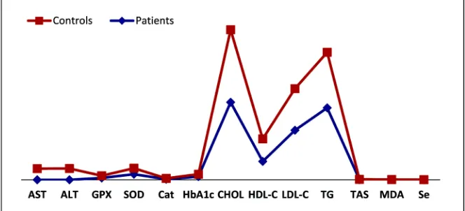
The mean percentage changes of the oxidant and antioxidant parameters in patients as compared to the controls are shown in (Figure 2). In comparison with control group, the level of MDA was increased significantly (P < 0.001) from 0.066 ± 0.048 to 0.52 ± 0.32 nmol/mgP by 213% and a significant increase in HbA1c levels (P < 0.001) from 5.27% ± 0.40% to 6.88 ± 1.74% by 30.55% was observed in atheroserotic patients with respect to normal healthy control sub-jects (Table 1 and Figure 2). A significant decrease in Cat activity (P < 0.002) by 3.98% (from 1.885 ± 0.84 U/mgP to 1.81 ± 0.90 U/mg P) was found in patients as compared with control groups (Table 1 and Figure 2).
Table 1 shows the distribution of serum selenium concentration in patients and controls. The mean concentration of serum selenium was marked decrease
Values are expressed as mean ± SD for n subjects, HbA1c: Glycated haemoglobin A1C; SOD:
Supe-roxide dismutase; GPx: Glutathione peroxidase; MDA: Malondialdehyde; TAS: Total Antioxidant status.
Figure 2. Percentage change of mean oxidant and antioxidant parameters in
DOI: 10.4236/jbm.2019.77005 56 Journal of Biosciences and Medicines (P < 0.00) by 56.91% (from 0.094 ± 0.0114 to 0.0405 ± 0.0039 ppm for controls and atherosclerotic subjects) (Table 1 and Figure 2).
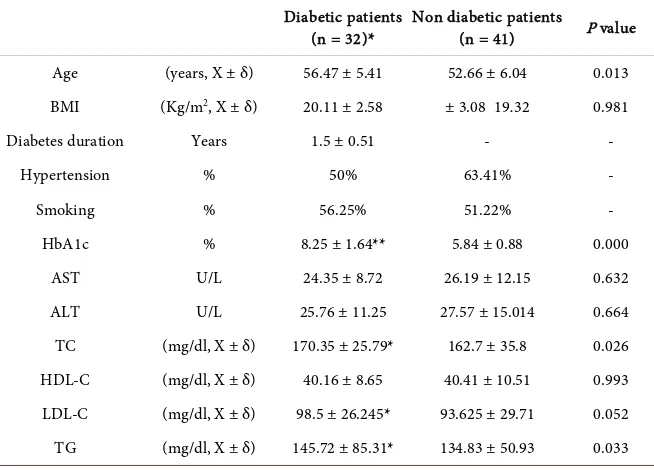
Thirty-two atherosclerotic type-II diabetes mellitus and forty-one atheroscle-rotic non-diabetic age matched normal blood pressure controls were included in the study. Baseline characteristics of both groups are shown in Table 2. In type-II diabetes mellitus group, HbA1c was significantly higher than the non-diabetic group (P < 0.000) (Table 2). Serum cholesterol, TG and LDL con-centrations in diabetic subjects were significantly raised as compared to non-diabetic group (TC: 170.35 ± 25.79 mg/dl vs. 162.7 ± 35.8 mg/dl, TG: 145.72 ± 85.31 mg/dl vs. 134.83 ± 50.93 mg/dl, LDL: 98.5 ± 26.245 mg/dl vs. 93.625 ± 29.71 mg/dl. However, there was no significant change was observed in the level of HDL AST, ALT in diabetic group with respect to the control group (P > 0.05).
[image:6.595.208.539.324.660.2]Figure 3 shows the values of MDA and anti-oxidative parameters in atheros-clerotic diabetic patients and atherosatheros-clerotic none diabetic subjects. The results
Table 1. Characteristic laboratory variables and the values of anti-oxidative parameters
atherosclerotic and control subjects.
Measure Indices (unit)
Patients with atherosclerosis (n = 73)
Controls
(n = 30) P value
Age (Years, X ± δ) 54.33 ± 6.04 48.52 ± 5.03 0.000
BMI (Kg/m2, X ± δ) 19.66 ± 2.88 16.96 ± 2.71 0.009
Diabetes n (%) 43.84% -
Hypertension n (%) 57.53% -
Smoking n (%) 53.42% -
AST (U/l, X ± δ) 25.38 ± 10.75 24.29 ± 4.18 0.664
ALT (U/l, X ± δ) 26.77 ± 13.44 24.74 ± 11.83 0.632
GPx (U/mg P, X ± δ) 4.02 ± 1.95 4.9 ± 2.73 0.137
SOD (U/mg P, X ± δ) 12.09 ± 5.5 13.32 ± 6.18 0.537
Cat (U/mg P, X ± δ) 1.18 ± 0.90 1.885 ± 0.84 0.002
HbA1c % 6.88 ± 1.74 5.27 ± 0.40 0.000
TC (mg/dl, X ± δ) 170.21 ± 45.01 159.6 ± 27.37 0.015
HDL-C (mg/dl, X ± δ) 40.30 ± 9.7 49.59 ± 10.33 0.000
LDL-C (mg/dl, X ± δ) 108.7 ± 28.65 91.75 ± 24.24 0.047
TG (mg/dl, X ± δ) 158.15 ± 97.14 122 ± 52.2 0.111
TAS (nmol/mgP, X ± δ) 0.34 ± 0.22 0.39 ± 0.20 0.597
MDA (nmol/mgP, X ± δ) 0.52 ± 0.32 0.166 ± 0.048 0.000
Se (ppm, X ± δ) 0.0405 ± 0.0039 0.094 ± 0.0114 0.000
Values are expressed as mean ± SD for n subjects, BMI, Body mass index, TC: Total cholesterol; TG: Trig-lyceride; LDL-C: Low-density lipoprotein cholesterol; HDL-C: High-density lipoprotein cholesterol, HbA1C: Glycated haemoglobin A1C; SOD: Superoxide dismutase; GPx: Glutathione peroxidase; MDA:
DOI: 10.4236/jbm.2019.77005 57 Journal of Biosciences and Medicines
Table 2. Characteristic laboratory variables and the values of anti-oxidative parameters
atherosclerotic patients with and without type 2 diabetes.
[image:7.595.214.532.400.590.2]Diabetic patients
(n = 32)* Non diabetic patients (n = 41) P value
Age (years, X ± δ) 56.47 ± 5.41 52.66 ± 6.04 0.013
BMI (Kg/m2, X ± δ) 20.11 ± 2.58 ± 3.08 19.32 0.981
Diabetes duration Years 1.5 ± 0.51 - -
Hypertension % 50% 63.41% -
Smoking % 56.25% 51.22% -
HbA1c % 8.25 ± 1.64** 5.84 ± 0.88 0.000
AST U/L 24.35 ± 8.72 26.19 ± 12.15 0.632
ALT U/L 25.76 ± 11.25 27.57 ± 15.014 0.664
TC (mg/dl, X ± δ) 170.35 ± 25.79* 162.7 ± 35.8 0.026
HDL-C (mg/dl, X ± δ) 40.16 ± 8.65 40.41 ± 10.51 0.993
LDL-C (mg/dl, X ± δ) 98.5 ± 26.245* 93.625 ± 29.71 0.052
TG (mg/dl, X ± δ) 145.72 ± 85.31* 134.83 ± 50.93 0.033
Values are expressed as mean ± SD for n subjects, BMI, Body mass index, *: non-insulin dependent (Type-2) diabetes mellitus, TC: Total cholesterol; TG: Triglyceride; LDL-C: Low-density lipoprotein cho-lesterol; HDL C: High density lipoprotein chocho-lesterol; HbA1C: Glycosylated hemoglobin. One-way ANOVA
followed by Tuckey test, n = 3. Non-significant groups: P ≥ 0.05; *P < 0.05; **P < 0.001 considered statisti-cally significant as compared to atherosclerotic non-diabetic group.
Values are expressed as mean ± SD for n subjects, Diabitcs: non-insulin dependent (Type-II) di-abetes mellitus, MDA: Malondialdehyde; One-way ANOVA followed by Tuceky test, n = 3. Non-significant groups: P ≥ 0.05; *P < 0.05; **P < 0.001 considered statistically significant as com-pared to atherosclerotic non-diabetic group.
Figure 3. Level of MDA in atherosclerotic patients with and without type 2 diabetes.
DOI: 10.4236/jbm.2019.77005 58 Journal of Biosciences and Medicines to the non-diabetics group (TAS: 0.35 ± 0.22 mmol/l vs. 0.33 ± 0.23 mmol/l, GPx: U/mgP 4.27 ± 1.76 vs. 3.81 ± 2.08 U/mgP, SOD: 11.64 ± 4.61 U/mgP vs. 12.433 ± 6.11 U/mgP, Cat: 1.089 ± 0.79 U/mgP vs. 1.25 ± 0.99 U/mgP). Athe-rosclerotic patients with type II diabetes had slightly higher values of Se in rela-tion to its level in non-diabetic patients (0.039 ± 0.0032 ppm vs. 0.042 ± 0.0042 ppm) (Figure 4).
[image:8.595.158.537.325.655.2]3.2. Correlations
Table 3 shows the correlations between TAS activity and some clinical and la-boratory variables in atherosclerotic diabetic patients as well as atherosclerotic none diabetic subjects. Pearson’s correlation coefficient in atherosclerotic di-abetic patients was observed a significant positive correlation between TAS and HbA1c (r = 0.134, P < 0.049) while a negative correlation was found between TAS and MDA (r = −0.061, P < 0.039) (Table 3). In atherosclerotic non-diabetic patients, TAS showed significant negative correlation with the TG (r = −0.423, P < 0.006), MDA (r = −0.200, P < 0.02) (Table 3).
Values are expressed as mean ± SD for n subjects, SOD: Superoxide dismutase; GPx: Glutathione peroxidase; Cat: Catalase. One-way ANOVA followed by Tuckey test, n = 3. Non-significant groups: P ≥ 0.05; *P < 0.05; **P < 0.001 considered statistically significant as compared to atherosclerotic non-diabetic group.
Figure 4. Values of antioxidative parameters in atherosclerotic patients with and without type 2
DOI: 10.4236/jbm.2019.77005 59 Journal of Biosciences and Medicines
Table 3. Correlations of Total Antioxidants (TAS) activity with some clinical and
labora-tory variables in atherosclerotic patients with and without type 2 diabetes.
Parameters Diabetic patients (n = 32) Non diabetic patients (n = 41)
r P r P
Age −0.220 0.227 0.089 0.584
BMI −0.172 0.354 −0.215 0.182
HbA1C 0.134 0.049 −0.143 0.419
GPX 0.068 0.711 0.004 0.983
SOD −0.065 0.729 0.176 0.278
Cat −0.014 0.937 −0.042 0.797
HbA1c −0.052 0.793 −0.010 0.956
HDL-C 0.042 0.820 0.051 0.753
LDL-C −0.090 0.626 −0.197 0.224
TG 0.074 0.686 −0.423** 0.006
TC −0.025 −0.891 −0.261 0.108
MDA −0.061 0.039 −0.200 0.021
Se 0.468 0.172 0.230 0.522
r = correlation coefficient. BMI, Body mass index, *: non-insulin dependent (Type-2) diabetes mellitus, TC: Total cholesterol; TG: Triglyceride; LDL-C: Low-density lipoprotein cholesterol; HDL-C: High-density li-poprotein cholesterol, HbA1C: Glycosylated hemoglobin, SOD: Superoxide dismutase; GPx: Glutathione
peroxidase; Cat: Catalase; MDA: Malondialdehyde. Non-significant groups: P ≥ 0.05; *P < 0.05; **P < 0.001 considered statistically significant as compared to atherosclerotic non-diabetic group.
There was a correlation of GPx activity with some clinical and laboratory va-riables in atherosclerotic diabetic patients as well as atherosclerotic none diabetic subjects (Table 4). A significant correlation was found between GPx and SOD (r = 0.359, P < 0.047), GPx and Cat (r = 0.488, P < 0.005), GPx and MDA (r = 0.575, P < 0.001) in atherosclerotic diabetic patients.
Atherosclerotic diabetic group, atherosclerotic non-diabetic group showed highly significant correlations between GPx and SOD (r = 0.684, P < 0.000), GPx and Cat (r = 0.488, P < 0.001) (Table 4).
In addition, Pearson’s correlation reveals a positive significant correlation between MDA and GPx (r = 0.575, P < 0.001), MDA and Cat (r = 0.673, P < 0.00), MDA and SOD (r = 0.449, P < 0.011) in atherosclerotic diabetic groups and in atherosclerotic non-diabetic patients, a highly significant positive correla-tion between MDA and Cat (r = −0.604, P < 0.00) was obtained (Table 5).
Moreover, a significant positive correlations was obtained between SOD-GPx (r = 0.359, P < 0.047), and SOD-MDA (r = 0.449, P < 0.011) in atherosclerotic diabetics while in atherosclerotic non-diabetics as marked positive correlations were observed between SOD-GPx (r = 0.684, P < 0.00), and GPx-Cat (r = 0.421, P < 0.006) (Table 6).
DOI: 10.4236/jbm.2019.77005 60 Journal of Biosciences and Medicines
Table 4. Correlations of Glutathione peroxidase (GPx) activity with some clinical and
la-boratory variables in atherosclerotic patients with and without type 2 diabetes.
Parameters Diabetic patients (n = 32) Non diabetic patients (n = 41)
r P r P
Age −0.321 0.073 −0.089 0.579
BMI −0.321 0.078 0.040 0.802
CBC 0.194 0.323 −0.221 0.201
TAS 0.068 0.711 0.004 0.983
SOD 0.359* 0.047 0.684** 0.000
Cat 0.488** 0.005 0.489** 0.001
HbA1c −0.047 0.814 −0.021 0.901
HDL-C −0.030 0.870 −0.168 0.294
LDL-C −0.028 0.879 −0.127 0.430
TG 0.163 0.374 −0.074 0.644
TC 0.011 0.952 0.004 0.983
MDA 0.575** 0.001 0.275 0.086
Se 0.412 0.236 0.430 0.215
r = correlation coefficient. BMI, Body mass index, *: non-insulin dependent (Type-2) diabetes mellitus, TC: Total cholesterol; TG: Triglyceride; LDL-C: Low-density lipoprotein cholesterol; HDL-C: High-density li-poprotein cholesterol, HbA1C: Glycosylated hemoglobin, SOD: Superoxide dismutase; Cat: Catalase; MDA:
Malondialdehyde; TAS: Total Antioxidants. Non-significant groups: P ≥ 0.05; *P < 0.05; **P < 0.001 consi-dered statistically significant as compared to atherosclerotic non-diabetic group.
Table 5. Correlations of Malondialdehyde (MDA) activity with some clinical and laboratory
variables in atherosclerotic patients with and without type 2 diabetes.
Parameters Diabetic patients (n = 32) Non diabetic patients (n = 41)
r P r P
Age −0.320 0.074 0.206 0.202
BMI −0.235 0.203 −0.015 0.928
GPx 0.575** 0.001 0.275 0.086
TAS −0.061 0.739 −0.200 0.216
SOD 0.449* 0.011 0.155 0.341
Cat 0.673** 0.000 0.604** 0.000
HbA1c 0.027 0.894 0.043 0.805
HDL-C −0.070 0.704 −0.204 0.207
LDL-C −0.077 0.677 −0.056 0.733
TG −0.107 0.561 0.000 0.999
TC −0.126 0.493 −0.143 0.385
Se 0.309 0.385 −0.174 0.630
r = correlation coefficient. BMI, Body mass index, *: non-insulin dependent (Type-II) diabetes mellitus, TC: Total cholesterol; TG: Triglyceride; LDL-C: Low-density lipoprotein cholesterol; HDL-C: High-density li-poprotein cholesterol, HbA1C: Glycated haemoglobin A1C; GPx: Glutathione peroxidase; Cat: Catalase;
[image:10.595.207.538.440.666.2]DOI: 10.4236/jbm.2019.77005 61 Journal of Biosciences and Medicines
Table 6. Correlations of Superoxide dismutase (SOD) activity with some clinical and
la-boratory variables in atherosclerotic patients with and without type 2 diabetes.
Parameters Diabetic patients (n = 32) Non diabetic patients (n = 41)
r P r P
Age −0.099 0.598 −0.147 0.360
BMI −0.043 0.822 −0.197 0.218
CBC −0.114 0.564 −0.242 0.162
GPx 0.359* 0.047 0.684** .000
TAS −0.065 0.729 0.176 0.278
MDA 0.449* 0.011 0.155 0.341
Cat 0.066 0.726 0.421** 0.006
HbA1c −0.020 0.918 −0.050 0.771
HDL-C 0.082 0.659 0.061 0.706
LDL-C −0.230 0.213 −0.101 0.529
TG −0.153 0.410 −0.077 0.632
TC −0.236 0.202 −0.106 0.514
Se 0.623 0.054 0.314 0.378
r = correlation coefficient. BMI, Body mass index, *: non-insulin dependent (Type-2) diabetes mellitus, TC: Total cholesterol; TG: Triglyceride; LDL-C: Low-density lipoprotein cholesterol; HDL-C: High-density li-poprotein cholesterol; HbA1C: Glycosylated hemoglobin; GPx: Glutathione peroxidase; Cat: Catalase; MDA:
Malondialdehyde; TAS: Total Antioxidants. Non-significant groups: P ≥ 0.05; *P < 0.05; **P < 0.001 consi-dered statistically significant as compared to atherosclerotic non-diabetic group.
clinical and laboratory variables in atherosclerotic diabetics as well as atheros-clerotic non-diabetics. In atherosatheros-clerotic diabetic patients, Pearson’s correlation coefficient shows significant positive correlation between Cat and GPx (r = 0.488, P < 0.005) and Cat-MDA (r = 0.673, P < 0.00). In atherosclerotic non-diabetic patients Pearson’s correlation observes a significant positive corre-lation between Cat-GPx (r = 0.489, P < 0.001), Cat-MDA (r = 0.604, P < 0.00) and Cat-SOD (r = 0.421, P < 0.000) (Table 7).
4. Discussion
DOI: 10.4236/jbm.2019.77005 62 Journal of Biosciences and Medicines
Table 7. Correlations of Catalase (Cat) activity with some clinical and laboratory
va-riables in atherosclerotic patients with and without type 2 diabetes.
Parameters Diabetics patients (n = 32) Non diabetics patients (n = 41)
r P r P
Age −0.224 0.217 −0.058 0.720
BMI −0.155 0.404 −0.178 0.265
GPx 0.488** 0.005 0.489** 0.001
TAS −0.014 0.937 −0.042 0.797
MDA 0.673** 0.000 0.604** 0.000
SOD 0.066 0.726 0.421** 0.006
HbA1c −0.132 0.502 −0.009 0.957
HDL-C −0.001 0.997 0.004 0.980
LDL-C 0.063 0.732 0.006 0.968
TG 0.148 0.420 0.079 0.625
TC 0.079 0.668 0.012 0.940
Se 0.197 0.586 0.029 0.937
r = correlation coefficient. BMI, Body mass index, *: non-insulin dependent (Type-2) diabetes mellitus, TC: Total cholesterol; TG: Triglyceride; LDL-C: Low-density lipoprotein cholesterol; HDL-C: High-density li-poprotein cholesterol, HbA1C: Glycosylated hemoglobin, SOD: Superoxide dismutase; GPx: Glutathione
peroxidase; MDA: Malondialdehyde; TAS: Total Antioxidants. Non-significant groups: P ≥ 0.05; *P < 0.05; **P < 0.001 considered statistically significant as compared to atherosclerotic non-diabetic group.
patients in relation to healthy subjects (Table 1).
Various studies have demonstrated that diabetes mellitus, smoking, and hypertension, consider the main risk factors of atherosclerosis, which are corre-lated with an increased production of ROS by the endothelium [21] [22] [23]. It has been recently found a good correlation between elevated HbA1c values and the atherosclerosis [24] [25] [26] which result from oxidative stress induced by various glucose oxidation, change in antioxidant enzymes status and impaired glutathione metabolism [27] [28].
DOI: 10.4236/jbm.2019.77005 63 Journal of Biosciences and Medicines plasma antioxidants and increases lipid peroxidation [8].
ROS and reactive nitrogen species (RNS) produced by the endothelium pro-mote oxidative modification of LDL (low-density lipoprotein). Oxidized LDL has been implicated in several facets of the inflammatory process, including the invasion of monocytes and macrophages, their differentiation, and the induction of injury to endothelial cells [31]. It has been proven that oxidized LDL involved in plaque formation in atherosclerosis as it is naturally being considered to play the most important role in lipid storage and inflammation [32] [33]. Previous studies have found that atheroscloresis is related to increase in lipid peroxidation [34] [35] [36]. An increase in lipid peroxidation product especially malondial-dehyde level (MDA) may also related to the higher level of oxidized LDL. Present results correlate with previous findings [37] [38] [39] which illustrated that elevated levels of MDA indicate increase in the level of production of oxy-gen free radicals, suggesting their possible role in atherooxy-genesis.
Antioxidants are a diverse group of compounds within which play important role in inhibiting oxidant formation, intercepting oxidants once they have formed, and repairing oxidant-induced injury. Although It is well documented that exposure to ROS increases the expression of antioxidant enzymes [40] [41], many studies have revealed that a decrease in the level of antioxidant defense as an expression of increase in free radical production [42] [43] [44]. Various en-zyme systems including superoxide dismutase (SOD), glutathione peroxidases (GPx) and catalase (Cat) are involved in regulating ROS production and degra-dation in vascular cells [45] [46].
SOD, Cat and GPx play association role in the elimination of superoxide rad-icals, in which SOD transforms them to H2O2 while GPx and Cat convert H2O2 to water [47] [48]. Therefore, prevent the formation of hydroxyl radicals, which is considered highly toxic molecule [49] [50]. In this study, results demonstrated lower levels of SOD, GPx, and catalase in serum of atherosclerotic patients with and without diabetes as compared to controls (Figure 3 and Figure 4), that was in agreement with the outcomes of other studies [51] [52] [53]. In atherosclerot-ic with diabetes group, there was found a very good positive correlation between elevated MDA and decreased GPx level (r = 0.575., P < 0.001) (Table 5). In ad-dition, Significant positive correlations were noted between serum MDA level and SOD activity (r = 0.449, P = 0.011) and serum MDA level and Cat activity (r = 0.673, P = 0.000) (Table 5) which was in consistent with previous study [54]. The declined in the activity of SOD was feasibly due to the accumulation of H2O2, and glycation of these enzymes, which was in concord with previous find-ings [55] [56]. In addition, [57] were reported that the lower activity of GPx ac-tivity in atherosclerotic with diabetes group was related to inactivation of the enzyme because of severe oxidative stress condition.
dem-DOI: 10.4236/jbm.2019.77005 64 Journal of Biosciences and Medicines onstrated that the selenium concentrations were lower in the atherosclerotic pa-tients in relation to its content in controls which was in agreement with the out-comes of other studies [58] [59]. However, the Figure 4 shows that the distribu-tion of patients with and without diabetes was similar at all selenium concentra-tions, indicating that a serum selenium is not related to the regulation and sever-ity of the disease [17] [60].
5. Conclusion
An imbalance between increased oxidative stress and impaired antioxidant de-fense may play a role in the progression of atherosclerotic disease. Aforemen-tioned observations indicate that oxidative stress was increased in atherosclerotic diabetic patients as approved by the significant changes in the levels of oxidative stress parameters. The relationship between oxidative stress parameters and im-balance antioxidants suggests their strong involvement in atherosclerosis. Fur-ther investigations with large clinical trials are recommended.
Acknowledgements
The authors would like to thank the medical record in charge and staff of Ta-joura National Heart Center, Tripoli, Libya, for their cooperation and support during the study and patients who participate in this study.
Conflicts of Interest
The authors declare no conflicts of interest regarding the publication of this paper.
Authors’ Contributions
This work was carried out in collaboration between all authors. Author RA de-signed the study, wrote the protocol and wrote the first draft of the manuscript. Authors SE and EA preformed the experiments, ZA collected the data and sam-ples, author NA interpreted the analyses of the study and contributed important intellectual content, author ME performed the statistical analysis, and author AR managed the literature searches and critically revised the manuscript. All au-thors read and approved the final manuscript.
Ethics Approval and Consent to Participate
The Ethics committee of Biotechnology Research Center (Tripoli, Libya) ap-proved the study (BEC-BTRC 03-2016). All patients provided a written in-formed consent prior to start of the study procedures.
References
DOI: 10.4236/jbm.2019.77005 65 Journal of Biosciences and Medicines 3rd Edition. Oxford University Press, New York, 10-121.
[3] Bonomini, F., Tengattini, S., Fabiano, A., Bianchi, R. and Rezzani, R. (2008) Athe-rosclerosis and Oxidative Stress. Histology and Histopathology Cellular and
Mole-cular Biology, 23, 381-390.
[4] Witztum, J. (1997) The Oxidation Hypothesis of Atherosclerosis. The Lancet, 344, 793-795.https://doi.org/10.1016/S0140-6736(94)92346-9
[5] Witztum, J. and Steinberg, D. (2001) The Oxidative Modification Hypothesis of Atherosclerosis Does It Hold for Humans? Trends in Cardiovascular Medicine, 11, 93-102.https://doi.org/10.1016/S1050-1738(01)00111-6
[6] Stocker, R. and Keaney, J.F. (2004) Role of Oxidative Modifications in Atheroscle-rosis. Physiological Reviews, 84, 1381-1478.
[7] Steinberg, D., Parthasarathy S., Carew, T.E., Khoo, J.C. and Witztum, J.L. (1989) Beyond Cholesterol. Modifications of Low-Density Lipoprotein that Increase Its Atherogenicity. TheNew England Journal of Medicine, 320, 915-924.
https://doi.org/10.1056/NEJM198904063201407
[8] Liaudet, L., Rosenblatt-Velin, N. and Pacher, P. (2013) Role of Peroxynitrite in the Cardiovascular Dysfunction of Septic Shock. Current Vascular Pharmacology, 11, 196-207.
[9] Incalza, M.A., D’Oria, R., Natalicchio, A., Perrini, S., Laviola, L. and Giorgino, F. (2018) Oxidative Stress and Reactive Oxygen Species in Endothelial Dysfunction Associated with Cardiovascular and Metabolic Diseases. Vascular Pharmacology, 100, 1-19.https://doi.org/10.1016/j.vph.2017.05.005
[10] Victor, V., Rocha, M., Sola, E., Banuls, C., Garcia-Malpartida, K. and Hernan-dez-Mijares, A. (2009) Oxidative Stress, Endothelial Dysfunction and Atherosclero-sis. Current Pharmaceutical Design, 15, 2988-3002.
[11] Chen, J.Y., Ye, Z.X., Wang, X.F., Chang, J., Yang, M.W., Zhong, H.H., Hong, F.F. and Yang, S.L. (2018) Nitric Oxide Bioavailability Dysfunction Involves in Atheros-clerosis. Biomedicine and Pharmacotherapy, 97, 423-428.
https://doi.org/10.1016/j.biopha.2017.10.122
[12] Katakami, N. (2017) Mechanism of Development of Atherosclerosis and Car-dio-Vascular Disease in Diabetes Mellitus. Journal of Atherosclerosis and Throm-bosis, 25, 27-39.
[13] Severino, P., D’Amato, A., Netti, L., Pucci, M., De Marchis, M., Palmirotta, R., Vol-terrani, M., Mancone, M. and Fedele, F. (2018) Diabetes Mellitus and Ischemic Heart Disease: The Role of Ion Channels. International Journal of Molecular
Sciences, 19, 802.https://doi.org/10.3390/ijms19030802
[14] Pai, J.K., Cahill, L.E., Hu, F.B., Rexrode, K.M., Manson, J.E. and Rimm, E.B. (2013) Hemoglobin A1c Is Associated with Increased Risk of Incident Coronary Heart Dis-ease among Apparently Healthy, Nondiabetic Men and Women. Journal of the
American Heart Association, 2, e000077.https://doi.org/10.1161/JAHA.112.000077
[15] Hjalmarsson, C., Manhem, K., Bokemark, L. and Andersson, B. (2014) The Role of Prestroke Glycemic Control on Severity and Outcome of Acute Ischemic Stroke.
Stroke Research and Treatment,2014, Article ID: 694569.
https://doi.org/10.1155/2014/694569
DOI: 10.4236/jbm.2019.77005 66 Journal of Biosciences and Medicines [17] Kornhauser, C., Garcia-Ramirez, J.R., Wrobe, K., Perez-Luque, E.L., Garay-Sevilla,
M.E. and Wrobe, K. (2008) Serum Selenium and Glutathione Peroxidase Concen-trations in Type 2 Diabetes Mellitus Patients. Primary Care Diabetes, 2, 81-85.
https://doi.org/10.1016/j.pcd.2008.02.003
[18] Zhang, Y.-T., Zheng, Q.-S., Pan, J. and Zheng, R.-L. (2004) Oxidative Damage of Biomolecules in Mouse Liver Induced by Morphine and Protected by Antioxidants.
Basic & Clinical Pharmacology & Toxicology, 95, 53-58.
https://doi.org/10.1111/j.1742-7843.2004.950202.x
[19] Hadwan, M.H. (2016) New Method for Assessment of Serum Catalase Activity.
In-dian Journal of Science and Technology, 9, 1-5.
https://doi.org/10.17485/ijst/2016/v9i4/80499
[20] Hafeman, D.G., Sunde, R.A. and Hoekstra, W.G. (1974). Effect of Dietary Selenium on Erythrocyte and Liver Glutathione Peroxidase in the Rat. TheJournal of Nutri-tion, 104, 580-587.https://doi.org/10.1093/jn/104.5.580
[21] Forbes, J.M. and Cooper, M.E. (2013) Mechanisms of Diabetic Complications.
Phy-siological Reviews, 93, 137-188.
[22] Fan, L.M., Douglas, G., Bendall, J.K., McNeill, E., Crabtree, M.J., Hale, A.B., Jian-Mei Li, A.M., McAteer, M.A., Schneider, J.E., Choudhury, R.P. and Channon, K.M. (2014) Endothelial Cell-Specific Reactive Oxygen Species Production Increas-es Susceptibility to Aortic Dissection. Circulation,129, 2661-2672.
https://doi.org/10.1161/CIRCULATIONAHA.113.005062
[23] Yuan, T., Yang, T., Chen, H., Fu, D., Hu, Y., Wang, J., Yuan, Q., Yu, H., Xu, W. and Xie, X. (2019) New Insights into Oxidative Stress and Inflammation during Di-abetes Mellitus-Accelerated Atherosclerosis. Redox Biology, 20, 247-260.
https://doi.org/10.1016/j.redox.2018.09.025
[24] Xing, F.Y., Neeland, I.J., Gore, M.O., Ayers, C.R., Paixao, A.R., Turer, A.T., Berry, J.D., Khera, A., de Lemos, J.A. and McGuire, D.K. (2014) Association of Prediabetes By Fasting Glucose and/or Haemoglobin A1c Levels with Subclinical Atherosclerosis and Impaired Renal Function: Observations from the Dallas Heart Study. Diabetes
and Vascular Disease Research,11, 11-18.
https://doi.org/10.1177/1479164113514239
[25] Fernández-Friera, L., Fuster, V., López-Melgar, B., Oliva, B., García-Ruiz, J.M., Mendiguren, J., Bueno, H., Pocock, S., Ibáñez, B., Fernández-Ortiz, A. and Sanz, J. (2017) Normal LDL-Cholesterol Levels Are Associated with Subclinical Atheros-clerosis in the Absence of Risk Factors. Journal of the American College of
Cardi-ology, 70, 2979-2991.https://doi.org/10.1016/j.jacc.2017.10.024
[26] Nazish, S., Zafar, A., Shahid, R., Albakr, A., Alkhamis, F.A., Aljaafari, D., Alabdali, M., Alsulaiman, A. and Al-Mulla, F.A. (2018) Relationship between Glycated Hae-moglobin and Carotid Atherosclerotic Disease among Patients with Acute Ischae-mic Stroke. Sultan Qaboos University Medical Journal, 18, 311-317.
[27] Butterfield, D.A., Domenico, F. and Di.and Barone, E. (2014) Elevated Risk of Type 2 Diabetes for Development of Alzheimer Disease: A Key Role for Oxidative Stress in Brain. Biochimica et Biophysica Acta (BBA)—Molecular Basis of Disease, 1842, 1693-1706.https://doi.org/10.1016/j.bbadis.2014.06.010
[28] Nguyen, D., Hsu, J.W., Jahoor, F. and Sekhar, R.V. (2014) Effect of Increasing Glu-tathione with Cysteine and Glycine Supplementation on Mitochondrial Fuel Oxida-tion, Insulin Sensitivity, and Body Composition in Older HIV Infected Patients. The
Journal of Clinical Endocrinology and Metabolism, 99, 169-177.
DOI: 10.4236/jbm.2019.77005 67 Journal of Biosciences and Medicines [29] Chen, Q., Wang, Q., Zhu, J., Xiao, Q. and Zhang, L. (2017) Reactive Oxygen Spe-cies: Key Regulators in Vascular Health and Diseases. British Journal of
Pharma-cology, 175, 1279-1292.https://doi.org/10.1111/bph.13828
[30] Heymes C, Bendall, J.K., Ratajczak, P., Cave, A.C., Samuel, J.L., Hasenfuss, G. and Shah, A.M. (2003) Increased Myocardial NADPH Oxidase Activity in Human Heart Failure. Journal of the American College of Cardiology, 41, 2164-2171.
https://doi.org/10.1016/S0735-1097(03)00471-6
[31] Yamada, N. (2001) Atherosclerosis and Oxidative Stress. Japan Medical Association
Journal, 44, 529-534.
[32] Uchida, K. (2015) Aldehyde Adducts Generated during Lipid Peroxidation Modifi-cation of Proteins. Free Radical Research, 49, 896-904.
https://doi.org/10.3109/10715762.2015.1036052
[33] Orekhov, A.N. and Myasoedova, V.A. (2019) Low Density Lipoprotein-Induced Li-pid Accumulation Is a Key Phenomenon of Atherogenesis at the Arterial Cell Level.
Vessel Plus,3, 14.https://doi.org/10.20517/2574-1209.2018.80
[34] Amaki, T., Suzuki, T., Nakamura, F., Hayashi, D., Imai, Y., Morita, H., Fukino, K., Nojiri, T., Kitano, S., Hibi, N., Yamazaki, T. and Nagai, R. (2004) Circulating Ma-londialdehyde Modified LDL Is a Biochemical Risk Marker for Coronary Artery Disease. Heart, 90, 1211-1213.https://doi.org/10.1136/hrt.2003.018226
[35] Polidori, M.C., Pratico, D., Parente, B., Mariani, E., Cecchetti, R., Yao, Y., Sies, H., Cao, P., Mecocci, P. and Stahl, W. (2007) Elevated Lipid Peroxidation Biomarkers and Low Antioxidant Status in Atherosclerotic Patients with Increased Carotid or Iliofemoral Intima Media Thickness. Journal of Investigative Medicine, 55, 163-167.
https://doi.org/10.2310/6650.2007.06043
[36] Vogiatzi, G., Tousoulis, D. and Christodoulos, S. (2009) The Role of Oxidative Stress in Atherosclerosis Oxidative Stress and Atherosclerosis. Hellenic Journal of
Cardiology, 50, 402-409.
[37] Yang, R.L., Shi, Y.H., Hao, G., Li, W. and Le, G.W. (2008) Increasing Oxidative Stress with Progressive Hyperlipidemia in Human: Relation between Ma-lon-Dialdehyde and Atherogenic Index. Journal of Clinical Biochemistry and Nutri-tion, 43, 154-158.https://doi.org/10.3164/jcbn.2008044
[38] Viigimaa, M., Abina, J., Zemtsovskaya, G., Tikhaze, A., Konovalova, G., Kumskova, E. and Lankin, V. (2010) Malondialdehyde-Modified Low-Density Lipoproteins as Biomarker for Atherosclerosis. Blood Pressure,19, 164-168.
https://doi.org/10.3109/08037051.2010.484158
[39] Stanek, A., Cholewka, A., Wielkoszyński, T., Romuk, E., Sieroń, K. and Sieroń, A. (2017) Increased Levels of Oxidative Stress Markers, Soluble CD40 Ligand, and Ca-rotid Intima-Media Thickness Reflect Acceleration of Atherosclerosis in Male Pa-tients with Ankylosing Spondylitis in Active Phase and without the Classical Cardi-ovascular Risk Factors. Oxidative Medicine and Cellular Longevity, 2017, Article ID: 9712536. https://doi.org/10.1155/2017/9712536
[40] Worthington Enzyme Manual (2009) Worthington Biochemical Corporation.
http://www.worthington-biochem.com/index/manual.html
[41] Ray, P.D. Huang, B.W. and Tsuji, Y. (2012) Reactive Oxygen Species (ROS) Ho-meostasis and Redox Regulation in Cellular Signaling. Cellular Signalling, 24, 981-990.https://doi.org/10.1016/j.cellsig.2012.01.008
DOI: 10.4236/jbm.2019.77005 68 Journal of Biosciences and Medicines [43] Milanlioglu, A., Aslan, M., Ozkol, H., Çilingir, V., Nuri Aydın, M. and Karadas, S.
(2016) Serum Antioxidant Enzymes Activities and Oxidative Stress Levels in Pa-tients with Acute Ischemic Stroke: Influence on Neurological Status and Outcome.
Wiener Klinische Wochenschrift, 128, 169-174.
https://doi.org/10.1007/s00508-015-0742-6
[44] Ziaadini, F., Aminae, M., Rastegar, M.M., Abbasian, S. and Memari, A.H. (2017) Melatonin Supplementation Decreases Aerobic Exercise Training Induced-Lipid Peroxidation and Malondialdehyde in Sedentary Young Women. Polish Journal of
Food and Nutrition Sciences, 67, 225-232.https://doi.org/10.1515/pjfns-2017-0001
[45] Papaharalambus, M.C.A. and Griendling, K.K. (2007) Basic Mechanisms of Oxida-tive Stress and ReacOxida-tive Oxygen Species in Cardiovascular Injury. Trends in
Cardi-ovascular Medicine, 17, 48-54.https://doi.org/10.1016/j.tcm.2006.11.005
[46] Lü, J.M., Lin, P.H., Yao, Q. and Chen, C. (2010) Chemical and Molecular Mechan-isms of Antioxidants: Experimental Approaches and Model Systems. Journal of
Cellular and Molecular Medicine, 14, 840-860.
https://doi.org/10.1111/j.1582-4934.2009.00897.x
[47] Dal, S. and Sigrist, S. (2016) The Protective Effect of Antioxidants Consumption on Diabetes and Vascular Complications. Diseases,4, 24.
https://doi.org/10.3390/diseases4030024
[48] Kwon, I., Jang, Y., Song, W., Roltsch, M.H. and Lee, Y. (2018) Mitochondrial Anti-oxidant Enzymes and Endurance Exercise-Induced Cardioprotection against Ischemia-Reperfusion Injury. Sports and Exercise Medicine Open Journal, 4, 9-15.
https://doi.org/10.17140/SEMOJ-4-155
[49] Styskal, J., van Remmen, H., Richardson, A. and Salmon, A.B. (2012) Oxidative Stress and Diabetes: What Can We Learn about Insulin Resistance from Antioxi-dant Mutant Mouse Models? Free Radical Biology and Medicine, 52, 46-58.
https://doi.org/10.1016/j.freeradbiomed.2011.10.441
[50] Li, H., Horke, S. and Förstermann, U. (2013) Oxidative Stress in Vascular Disease and Its Pharmacological Prevention. Trends in Pharmacological Sciences, 34, 313-319.https://doi.org/10.1016/j.tips.2013.03.007
[51] Zawadzka-Bartczak, E. (2005) Activities of Red Blood Cell Anti-Oxidative Enzymes (SOD, GPx) and Total Anti-Oxidative Capacity of Serum (TAS) in Men with Coro-nary Atherosclerosis and in Healthy Pilots. Medical Science Monitor, 11, 440-444. [52] Čolak, E., Dimitrijević-Srećković, V., Djordjević, R.B., Stanković, S., Glišić1, B.,
koSrećković, B. and Majkic-Singh, N. (2008) Biomarkers of Enzymatic and Nonen-zymatic Antioxidative Defense in Type 2 Diabetes Mellitus-Comparative Analysis.
Biochemia Medica, 18, 42-51.https://doi.org/10.11613/BM.2008.006
[53] Chehaibi, K., Trabelsi, I., Mahdouani, K. and Slimane, M.N. (2016) Correlation of Oxidative Stress Parameters and Inflammatory Markers in Ischemic Stroke Patients.
Journal of Stroke and Cerebrovascular Diseases, 25, 2585-2593.
https://doi.org/10.1016/j.jstrokecerebrovasdis.2016.06.042
[54] Abou-Seif, M.A. and Youssef, A.A. (2004) Evaluation of Some Biochemical Changes in Diabetic Patients. Clinica Chimica Acta, 346, 161-170.
https://doi.org/10.1016/j.cccn.2004.03.030
[55] Matough, F.A., Budin, S.B., Hamid, Z.A., Alwahaibi, N. and Mohamed, J. (2012) The Role of Oxidative Stress and Antioxidants in Diabetic Complications. Sultan
Qaboos University Medical Journal, 12, 5-18.
DOI: 10.4236/jbm.2019.77005 69 Journal of Biosciences and Medicines Standing Type 2 Diabetes in Port Harcourt, Rivers State, Nigeria. International
Journal of Science and Research,5, 1282-1288.
https://doi.org/10.21275/v4i5.NOV162149
[57] Condell, R.A. and Tapell, A.L. (1983) Evidence for Suitability of Glutathione Pe-roxidase as a Protective Enzyme: Studies of Oxidative Damage, Restoration and Proteolysis. Archives of Biochemistry and Biophysics,223, 407-416.
[58] Sanmartin, C., Plano, D., Font, M. and Palop, J.A. (2011) Selenium and Clinical Tri-als: New Therapeutic Evidence for Multiple Diseases. Current Medicinal Chemistry, 18, 4635-4650.https://doi.org/10.2174/092986711797379249
[59] Liang, J., Gao, Y., Tang, S., Feng, S., Han, J. and Hu, Q. (2018) Serum Levels of Trace Elements and Vitamins in Coronary Artery Disease and Their Predictive Values. International Journal of Clinical and Experimental Medicine, 11, 922-931. [60] Kljai, K. and Runje, R. (2001) Selenium and Glycogen Levels in Diabetic Patients.
Biological Trace Element Research, 83, 223-229.