ULTRASOUND IN ADNEXAL TUMOR: NEW CONCEPT
1, *
Dr. Nagham Muhammed Utba,
1
Department of Radiology, Baghdad Teaching Hospital, Medical City, Iraq
2
Department of Gynecology, Baghdad Teaching Hospital, Medical City, Iraq
ARTICLE INFO ABSTRACT
Background
Analysis (IOTA) ADNEX model can be used by medical doctors to diagnose ovarian cancer in women who have at least one persistent adnexal and estimates the pro
benign or malignant.
in differentiation between malignant and benign ovarian tumor.
patients presented with suspicious adnexal mass to the radiological department in Baghdad teaching hospital in medical city complex in the period from February 2016 till the end of December 2016. U/S was performed to confirm & characterize the a
patients underwent surgery to remove the mass and the masses were sent for histopathological study. Results
the deter
specificity was 98% in detection the malignant at cut off point 49.25%. (ADNEX model) was highly sensitive (92%) and highly specific (98%) in d malignant at score of 49.25.
Copyright©2019, Nagham Muhammed Utba et al. This
unrestricted use, distribution, and reproduction in any medium,
INTRODUCTION
Ovarian cancer has the highest fatality-to-case ratio (70.3%) and is the most challenging of the gynecological cancers
et al., 2011).
Typesofovariantumors: Approximately90%
are epithelial ovariancarcinomas. Epithelial
adenocarcinomas. In this type of cancer, a malignant tumor originates in the surface epithelium tissue, which is the lining on the outside of the ovary (Seidman et al., 2003).
Clinical features: The suspicion of malignancy must be
particularly high in a postmenopausal female with a symptomatic and palpable pelvic mass. Most women with early ovarian cancer are asymptomatic. In very advanced
patients may have symptoms related to the presence of ascites or metastatic disease, which may include abdominal distention, abdominal pain, bloating, constipation, nausea, emesis, weight loss, anorexia, reflux, or early satiety (Goff et al.,
Imaging diagnosis:For pre-surgical assessment of an adnexal
mass, transvaginal ultrasonography (US) combined with Doppler techniques is the first-line and best imaging technique (Rosemarie et al., 2016). When a lesion is large or extends
ISSN: 0975-833X
Article History:
Received 17th November, 2018
Received in revised form
03rd December, 2018
Accepted 24th January, 2019
Published online 28th February, 2019
Citation: Dr. Nagham Muhammed Utba, Dr. Farah Fawzi Marzook, Dr. Atheer Adnan Fadhil and Dr. Nawfal Azzo
Tumor: New Concept”, International Journal of Current
Key Words:
Ultrasound, IOTA, ADNEX, Ovarian tumor.
*Corresponding author:
Dr. Nagham Muhammed Utba
RESEARCH ARTICLE
ULTRASOUND IN ADNEXAL TUMOR: NEW CONCEPT
Dr. Nagham Muhammed Utba,
1Dr. Farah Fawzi Marzook,
1Dr. Atheer Adnan Fadhil and
2Dr. Nawfal Azzo
Department of Radiology, Baghdad Teaching Hospital, Medical City, Iraq
Department of Gynecology, Baghdad Teaching Hospital, Medical City, Iraq
ABSTRACT
Background: For pre-surgical assessment of an adnexal mass, the International Ovarian Tumor Analysis (IOTA) ADNEX model can be used by medical doctors to diagnose ovarian cancer in women who have at least one persistent adnexal and estimates the pro
benign or malignant. Objective: To study the value of the international ovarian tumor analysis (IOTA) in differentiation between malignant and benign ovarian tumor.
patients presented with suspicious adnexal mass to the radiological department in Baghdad teaching hospital in medical city complex in the period from February 2016 till the end of December 2016. U/S was performed to confirm & characterize the adnexal mass. CA-125 tumor marker was measured. All patients underwent surgery to remove the mass and the masses were sent for histopathological study. Results: the IOTA score was calculated and compared, and shows that IOTA score was efficient in the determination of benign or malignant. The sensitivity of the IOTA score system was 92% and specificity was 98% in detection the malignant at cut off point 49.25%.
(ADNEX model) was highly sensitive (92%) and highly specific (98%) in d malignant at score of 49.25.
This is an open access article distributed under the Creative Commons medium, provided the original work is properly cited.
case ratio (70.3%) and is the most challenging of the gynecological cancers (Jemal
90%ofovariancancers Epithelial malignancies are adenocarcinomas. In this type of cancer, a malignant tumor originates in the surface epithelium tissue, which is the lining
2003).
The suspicion of malignancy must be particularly high in a postmenopausal female with a symptomatic and palpable pelvic mass. Most women with early ovarian cancer are asymptomatic. In very advanced disease, patients may have symptoms related to the presence of ascites or metastatic disease, which may include abdominal distention, abdominal pain, bloating, constipation, nausea, emesis, weight
et al., 2004).
surgical assessment of an adnexal mass, transvaginal ultrasonography (US) combined with line and best imaging technique When a lesion is large or extends
beyond the field of view of transvaginal US, complementary transabdominal US should be performed according to the 2013 American College of Radiology appropriateness criteria. MRI is usually considered as complementary problem
modality (Spencer et al., 2010).
clinical features (e.g., menopausal status) and serum biomarkers (CA-125) allow further risk stratification, e.g., as in the widely used risk of malignancy index (RMI). The value of gray scale and color Doppler US has been extens
by the International Ovarian Tumor Analysis (IOTA) group. The pattern recognition of specific ultrasound findings and assignment into categories of diagnostic certainty of malignancy is well established
IOTA Scoring system: In 2008, the International Ovarian
Tumor Analysis (IOTA) group proposed simple ultrasound based rules for the diagnosis of ovarian malignancy. These rules are based on the simple identification of certain findings on ultrasound examination, some o
malignant lesions (malignant or M
benign lesions (benign or B-features). A prospective internal validation from the IOTA group showed that these rules can be applied to 77% of adnexal masses and that, w
diagnostic performance is high
examiner analyzed the mass according to the IOTA simple rules to determine whether there were malignant (M) features
International Journal of Current Research Vol. 11, Issue, 02, pp.1620-1624, February, 2019
DOI: https://doi.org/10.24941/ijcr.34333.02.2019
Dr. Nagham Muhammed Utba, Dr. Farah Fawzi Marzook, Dr. Atheer Adnan Fadhil and Dr. Nawfal Azzo
International Journal of Current Research, 11, (02), 1620-1624.
ULTRASOUND IN ADNEXAL TUMOR: NEW CONCEPT
Dr. Atheer Adnan Fadhil and
Department of Radiology, Baghdad Teaching Hospital, Medical City, Iraq
Department of Gynecology, Baghdad Teaching Hospital, Medical City, Iraq
surgical assessment of an adnexal mass, the International Ovarian Tumor Analysis (IOTA) ADNEX model can be used by medical doctors to diagnose ovarian cancer in women who have at least one persistent adnexal and estimates the probability that an adnexal tumor is To study the value of the international ovarian tumor analysis (IOTA) in differentiation between malignant and benign ovarian tumor. Patients and methods: Fifty one patients presented with suspicious adnexal mass to the radiological department in Baghdad teaching hospital in medical city complex in the period from February 2016 till the end of December 2016. U/S 125 tumor marker was measured. All patients underwent surgery to remove the mass and the masses were sent for histopathological study. he IOTA score was calculated and compared, and shows that IOTA score was efficient in mination of benign or malignant. The sensitivity of the IOTA score system was 92% and specificity was 98% in detection the malignant at cut off point 49.25%. Conclusion: the IOTA score (ADNEX model) was highly sensitive (92%) and highly specific (98%) in detection of benign or
Commons Attribution License, which permits
field of view of transvaginal US, complementary transabdominal US should be performed according to the 2013 American College of Radiology appropriateness criteria. MRI is usually considered as complementary problem-solving 2010). Integration of additional clinical features (e.g., menopausal status) and serum 125) allow further risk stratification, e.g., as in the widely used risk of malignancy index (RMI). The value of gray scale and color Doppler US has been extensively analyzed by the International Ovarian Tumor Analysis (IOTA) group. The pattern recognition of specific ultrasound findings and assignment into categories of diagnostic certainty of malignancy is well established (Valentin et al., 2011).
In 2008, the International Ovarian Tumor Analysis (IOTA) group proposed simple ultrasound-based rules for the diagnosis of ovarian malignancy. These rules are based on the simple identification of certain findings on ultrasound examination, some of which are characteristic of malignant lesions (malignant or M-features) and others of features). A prospective internal validation from the IOTA group showed that these rules can be applied to 77% of adnexal masses and that, when applied, the diagnostic performance is high (AlcaZar et al., 2013). The examiner analyzed the mass according to the IOTA simple rules to determine whether there were malignant (M) features
INTERNATIONAL JOURNAL OF CURRENT RESEARCH
(M1, irregular solid tumor; M2, presence of ascites; M3, at least four papillary projections; M4, irregular multilocular solid tumor with largest diameter ≥100 mm; M5, very strong blood flow (IOTA color score 4)) or benign (B) features (B1, unilocular tumor; B2, presence of solid components with solid component largest diameter <7 mm; B3, presence of acoustic shadows; B4, smooth multilocular tumor with largest diameter <100 mm; B5, no blood flow (IOTA color score 1)) (Timmerman et al., 2008). At the end of the examination, a diagnosis of benign, malignant or inconclusive was provided. The mass was classified as benign if one or more B-features were present in the absence of M-features. The mass was classified as malignant if one or more M-features were present in the absence of B-features. If both B- and M-features were present, or none was present, the mass was classified as inconclusive (Timmerman et al., 2010). The ADNEX risk model can be used by medical doctors to diagnose ovarian cancer in women who have at least one persistent adnexal (ovarian, para-ovarian, and tubal) tumor and are considered to require surgery. ADNEX estimates the probability that an adnexal tumor is benign, borderline, stage I cancer, stage II-IV cancer, or secondary metastatic cancer (i.e. metastasis of non-adnexal cancer to the ovary). The model was developed by clinicians and statisticians from the International Ovarian Tumor Analysis (IOTA) group, and is based on clinical and ultrasound data from almost 6000 women recruited at 24 centers in 10 countries (Italy, Belgium, Sweden, Czech Republic, Poland, France, UK, China, Spain, and Canada) (Epstein et al., 2016). The ADNEX model uses nine predictors. There are three clinical variables, age, serum CA-125 level, and type of center (oncology referral center vs other), and six ultrasound variables, maximal diameter of lesion, proportion of solid tissue, more than 10 cyst locules, number of papillary projections, acoustic shadows, and ascites. All patients included required surgery as judged by a local clinician. As with all current diagnostic models for adnexal tumors (e.g. IOTA models, RMI, ROMA) it implies that patients selected for expectant management were excluded when creating the model. As a consequence ADNEX cannot be applied to conservatively treated adnexal tumors (Epstein et al., 2016).
Objectives: To study the value of the international ovarian
tumor analysis (IOTA) in differentiation between malignant and benign ovarian tumor.
MATERIALS AND METHODS
This is a prospective study performed in radiological department in Baghdad teaching hospital in medical city complex in the period from February 2016 till the end of December 2016. Fifty one adult women patients with clinical suspecion of adnexal masses were enrolled in this study. Informed consent was obtained from the patients to participate in this study,
Exclusion criteria:
1. Patients who don't have definite histopathological results 2. Any patient with non ovarian lesion (e.g: pendunculated
fibroid, paratubal cyst).
A grey scale and color Doppler ultrasound examination was performed (Transabdominal evaluation using a curved array 3 – 5 MHZ transducer) and (Transvaginal high frequency 5 – 10 MHZ transducer) to obtain morphological and blood flow
variables to characterize each adnexal mass. A hospital type ultrasound equipment (Voluson, GE Healthcare Austria) or (Philips HD11 XE Netherland) were used. Then the mass was stated likely to be malignant or benign on the basis of subjective evaluation of ultrasonic findings (“subjective assessment”). The risk of malignancy was calculated using the IOTA logistic regression models ADNEX. The IOTA model uses nine predictors. There are three clinical variables, age, serum CA-125 level, and type of center, and six ultrasound variables, maximal diameter of lesion, proportion of solid tissue, more than 10 cyst locules, number of papillary projections, acoustic shadows, and ascites (Epstein et al.,
2016). Results of CA 125 marker was recorded. All patients underwent surgery, and the mass was removed and sent for histopathological study. The IOTA score was calculated by application by entering the data as shown in Figure 3.
Statistical analysis
Analysis of data was carried out using the available statistical package of SPSS-22 (Statistical Packages for Social Sciences- version 22).
The significance of difference of different means (quantitative data) were tested using Students-t-test for difference between two independent means.
The significance of difference of different percentages (qualitative data) were tested using Pearson Chi-square test (2-test) with application of Yate's correction or Fisher Exact test whenever applicable. Statistical significance was considered whenever the P value was equal or less than 0.05.
Reliability tests (Sensitivity and specificity) and critical cut-off points were calculated using receiver operator curve (ROC).
RESULTS
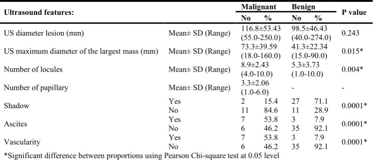
Table 1. Ultrasound features in the study sample
Ultrasound features: Malignant Benign P value
No % No %
US diameter lesion (mm) Mean± SD (Range) 116.8±53.43
(55.0-250.0)
98.5±46.43
(40.0-274.0) 0.243
US maximum diameter of the largest mass (mm) Mean± SD (Range) 73.3±39.59
(18.0-160.0)
41.3±22.34
(15.0-90.0) 0.015*
Number of locules Mean± SD (Range) 8.9±2.43
(4.0-10.0)
5.3±3.73
(1.0-10.0) 0.004*
Number of papillary Mean± SD (Range) 3.3±2.06
(1.0-6.0) - -
Shadow Yes 2 15.4 27 71.1 0.0001*
No 11 84.6 11 28.9
Ascites Yes 7 53.8 3 7.9 0.0001*
No 6 46.2 35 92.1
Vascularity Yes 7 53.8 3 7.9 0.0001*
No 6 46.2 35 92.1
*Significant difference between proportions using Pearson Chi-square test at 0.05 level
Table 2. IOTA score between the malignant and benign patients
Type of tumor IOTA score P value
Benign 8.44 + 9.04 (0.3 -38.5)
0.0001*
Malignancy 78.68±14.8
(60.0-97.1)
[image:3.595.109.489.307.798.2]*Significant difference between two independent means using Students-t-test at 0.05 level
Table 3. Histopathology in the study groups
Histopathology No %
Benign
Functional Cyst 2 5.26%
Dermoid 9 23.68%
Endometrioma 3 7.89%
Hemorrhagic corpus luteal cyst 7 18.42%
Mucinous cyst 3 7.89%
Papillary serrouscystadenofibroma 2 5.26%
Seromucinous tumor 1 2.63%
Serous cystadenoma 8 21.05%
Serrouscystadenofibroma 2 5.26%
Sex cord stroma 1 2.63%
Fibromatosis 1 7.69%
Malignant
Immature cystic teratoma 2 15.38%
Metastasis 2 15.38%
Mucinous cystadenocarcinoma 4 30.77%
Papillary serrous adenocarcinoma 1 7.69%
Serrous and mucinous tumor 1 7.69%
Serrouscystadenocarcinoma 1 7.69%
Transitional cell carcinoma 1 7.69%
Figure2.Acousticshadowinanovarianmass,suggestingdermoid, benign score IOTA 8.64
Figure 3. Multilocular complex ovarian cystic lesion with solid component, malignant IOTA score 76.1 indicating malignancy
In analysis of the CA 125 marker, the mean CA 125 marker seen in the malignancy was 69.6 u/ml, while the mean CA 125 marker seen in the benign was 30.6 u/ml. The IOTA score was calculated and analyzed, and shows that IOTA score was efficient in defferentiation of benign from malignant ovarian tumors, The IOTA score in histopathologically proved malignant cases was 78.7 while in benign cases, it was 8.2 with a p value was 0.0001 which was statistically significant. There were 38 cases with benign according to histopathology (37 of them was benign according to the IOTA score and 1 was malignant), and there was 13 patients with malignant according to histopathology (12 of them was malignant according to IOTA score and 1 was benign). The sensitivity of the IOTA score system was 92% and specificity of the IOTA was 98% in detection of malignancy.The most common benign tumor was dermoid (9 cases (23% of the benign cases)), while the most common malignant tumor was mucinous cystadenocarcinoma (4cases(30%ofthemalignantpatients),theotherhistopathology seen in the study groups were listed in the Table 3.
DISCUSSION
The clinical impact of defining whether an adnexal mass is benign or malignant is enormous. Thus, predictive models have been developed to triage women presenting with adnexal masses to an appropriate treatment regimen (Rosemarie Forstner et al., 2016).In analysis of our study data the patients grouped into two groups according to their final diagnosis whether benign or malignant depending on the histopathological results .There were no statistical significant difference had been noticed between the two groups in relevant to the age at time of presentation, which means that the age was not considered to be a predictor in determining the mass to be benign or malignant.This finding, when compared with other study shows that the incidence of the cancer increased with age but not the type of the tumor, and this was mentioned in Sassone et al (USA 1991) (Margherita Sassone et al., 1991) while in the Brown et al (USA 2010) (Douglas et al., 2010). In this study, IOTA score when calculated depend on the ultrasound parameter with ca 125 marker and recorded, the percentage of the malignancy and the benign was calculated, and found that the IOTA score in detecting the malignancy was 92% sensitive and 98% specific if the score was 49.2%. Abbas et al (Egypt 2014) (Ahmed Mohamed Abbas et al., 2014) shows that the benign was more than the malignant and the main benign tumor was endometriotic cyst 27% followed by simple cyst 16% while the malignant tumor was mainly mucinous cystadenocarcinoma 4.4% but they use only 3d ultrasound and they found it 3D ultrasound MSV can be helpful in the morphological assessment of adnexal masses especially in detection of papillary projections in adnexal cysts, and this was compatible with our study.In brown et al (Douglas et al.,
2010) they showed that the overwhelming majority of adnexal masses are benign and most can be recognized on the basis of characteristic US features. Malignancy, while infrequent, is likewise usually identifiable by a different set of distinguishing US features. Accordingly, in most cases the report should reflect a reasonably confident diagnosis of a benign or malignant entity. Clear communication of the US results will assist in proper patient care and should include a sufficient description and/or conclusion regarding the most likely diagnosis. Fischerova, Zikan, Dundr et al. (Czech 2012) (Daniela Fischerova et al., 2012) showed that ovarian tumors represent a wide spectrum of tumors with different biological potential and uncertain malignant potential. No precise prognostic or predictive markers exist to clearly distinguish between tumors of purely benign behavior and those with risk of malignant transformation into carcinomas. Therefore, the oncologic safety must be always balanced again less radical treatment, and this was incompatible to our results because they did not depend on the IOTA score as we did. Forstner et al
[image:4.595.35.293.326.548.2]to be able to interact with the assisting physician with the purpose of proposing strategies that may best guide the specific therapy for the patient, and this support our study results.
Conclusion
1. The IOTA scoring system (ADNEX model) was sensitive and specific for the detection of the malignancy before surgery.
2. The age was not a predictor for the type of ovarian tumor (benign or malignant).
Recommendation
Installation of the mobile phone application (IOTA score calculator) is recommended to assist in evaluation of ovarian masses by ultrasound and calculated according to IOTA score (ADNEX model) to exclude malignancy
Abbreviation list
CT computed tomography
HU Hounsfield unit
IOTA International Ovarian Tumor Analysis
IUP Intrauterine pregnancy
LH luteinizing hormone
Mm Millimeter
MRI Magnetic resonance imaging
MSV MultiSlice View
P value Predicted value
PMP Postmenopausal
U Unit
U/S Ultrasound
REFERENCES
Ahmed Mohamed Abbas, Kamal M. Zahran, Ahmed Nasr et al. 2014. Evaluation of Adnexal Masses by Three-Dimensional Ultrasound Multi-slice View: Do we really need it?. Thai Journal of Obstetrics and Gynaecology, Vol. 22; pp. 150-155.
Alca Zar J. L., M. A. Pascual, B. Olartecoechea, 2013. IOTA simple rules for discriminating between benign and malignant adnexal masses: prospective external validation.
Ultrasound Obstet Gynecol., 42: 467–471
Andrade Neto F, Palma-Dias R, Costa FS. 2011. Ultrasonography of adnexal masses: imaging findings.
Radiol Bras, 441:59–67.
Daniela Fischerova, Michal Zikan, Pavel Dundr, David Cibula, 2012. Diagnosis, Treatment, and Follow-Up of Borderline Ovarian Tumors, The Oncologist, 17:1515–1533
Douglas L. Brown, Kika M. Dudiak, Faye C. Laing, 2010. Adnexal Masses: US Characterization and Reporting.
Radiology, 254: 342-354
Epstein E, et al. 2016. Subjective ultrasound assessment, the ADNEX model and ultrasound-guided true cut biopsy to differentiate disseminated primary ovarian cancer from metastatic non-ovarian cancer. Ultrasound Obstet Gynecol., 47:110-6
Goff BA, Mandel LS, Melancon CH, Muntz HG. 2004. Frequency of symptoms of ovarian cancer in women presenting to primary care clinics. JAMA, 291:2705–2712. Jemal A, Bray F, Center MM, et al. 2011. Global cancer
statistics. CA: A Cancer Journal for Clinicians, 61:69–90. Margherita Sassone A., Ilan E, Timor Tritsch et al. 1991.
Transvaginal sonographic characteristization of ovarian disease. Obst and gyncol., 78 p:70 -76.
Rosemarie Forstner, Matthias Meissnitzer, Teresa Margarida Cunha, 2016. Update on Imaging of Ovarian Cancer. Curr Radiol Rep., 4:31
Rosemarie Forstner, Matthias Meissnitzer, Teresa Margarida Cunha, 2016. Update on Imaging of Ovarian Cancer. Curr Radiol Rep., 4:31-37
Seidman JD, Kurman RJ. 2003. Pathology of ovarian carcinoma. Hematol Oncol Clin North Am., 17:909–925. Spencer JA, Forstner R, Cunha TM, Kinkel K. 2010. On behalf
of the ESUR Female Imaging Sub-Committee. ESUR guidelines for MR imaging of the sonographically indeterminate adnexal mass: an algorithmic approach. Eur Radiol., 20:25–35
Timmerman D, Ameye L, Fischerova D, Epstein E, Melis GB, Guerriero S, Van Holsbeke C, Savelli L, Fruscio R, Lissoni AA, Testa AC, Veldman J, Vergote I, Van Huffel S, Bourne T, Valentin L. 2010. Simple ultrasound rules to distinguish between benign and malignant adnexal masses before surgery: prospective validation by IOTA group.
BMJ, 341: 1-8.
Timmerman D, Testa AC, Bourne T, Ameye L, Jurkovic D, Van Holsbeke C, Paladini D, Van Calster B, Vergote I, Van Huffel S, Valentin L. 2008. Simple ultrasound-based rules for the diagnosis of ovarian cancer. Ultrasound Obstet Gynecol., 31: 681–690.
Valentin L, Ameye L, Savelli L, et al. 2011. Adnexal masses difficult to classify as benign or malignant using subjective assessment of gray-scale ad Doppler ultrasound findings. Logistic regression models do not help. Ultrasound Obstet Gynecol., 38:456–65.