LEVEL 1 EBM
EXPEDITED
PUBLICATION
SPINE
A Randomized Trial Comparing Balloon Kyphoplasty and
Vertebroplasty for Vertebral Compression Fractures due to
Osteoporosis
M. Dohm, C.M. Black, A. Dacre, J.B. Tillman, G. Fueredi, on behalf of the KAVIAR investigators
EBM 1
ABSTRACT
BACKGROUND AND PURPOSE: Several trials have compared vertebral augmentation with nonsurgical treatment for vertebral compres-sion fractures. This trial compares the efficacy and safety of balloon kyphoplasty and vertebroplasty.
MATERIALS AND METHODS: Patients with osteoporosis with 1–3 acute fractures (T5–L5) were randomized and treated with kyphoplasty (n⫽191) or vertebroplasty (n⫽190) and were not blinded to the treatment assignment. Twelve- and 24-month subsequent radiographic fracture incidence was the primary end point. Due to low enrollment and early withdrawals, the study was terminated with 404/1234 (32.7%) patients enrolled.
RESULTS:The average age of patients was 75.6 years (77.4% female). Mean procedure duration was longer for kyphoplasty (40.0 versus 31.8 minutes,P⬍.001). At 12 months, 7.8% fewer patients with kyphoplasty (50/140 versus 57/131) had subsequent radiographic fracture, and there were 8.6% fewer at 24 months (54/110 versus 64/111). The results were not statistically significant (P⬎.21). When we used time to event for new clinical fractures, kyphoplasty approached statistical significance in longer fracture-free survival (Wilcoxon,P⫽.0596). Similar pain and function improvements were observed. CT demonstrated lower cement extravasation for kyphoplasty (157/214 versus 164/201 levels treated,P⫽.047). For kyphoplasty versus vertebroplasty, common adverse events within 30 postoperative days were procedural pain (12/191, 9/190), back pain (14/191, 28/190), and new vertebral fractures (9/191, 17/190); similar 2-year occurrence of device-related cement embolism (1/191, 1/190), procedural pain (3/191, 3/190), back pain (2/191, 3/190), and new vertebral fracture (2/191, 2/190) was observed.
CONCLUSIONS: Kyphoplasty and vertebroplasty had similar long-term improvement in pain and disability with similar safety profiles and few device-related complications. Procedure duration was shorter with vertebroplasty. Kyphoplasty had fewer cement leakages and a trend toward longer fracture-free survival.
ABBREVIATIONS:AE⫽adverse event; BKP⫽balloon kyphoplasty; EQ-5D⫽EuroQol-5-Domain; KAVIAR⫽Kyphoplasty and Vertebroplasty In the Augmentation and Restoration of vertebral body compression fractures; MedDRA⫽Medical Dictionary for Regulatory Activities; ODI⫽Oswestry Disability Index; RCT⫽randomized controlled trial; VCF⫽vertebral compression fracture; VP⫽vertebroplasty
V
ertebral compression fractures (VCFs) are clinically recog-nized in 1.4 million individuals worldwide annually,1oftenresulting in pain, disability, vertebral deformity,2and
consider-able negative economic impact.3Balloon kyphoplasty (BKP) and
vertebroplasty (VP) are percutaneous procedures aimed at reduc-ing pain and providreduc-ing fracture stability. Balloon kyphoplasty uses orthopedic inflatable bone tamps before bone cement injec-tion in an attempt to correct vertebral deformity and control ce-ment distribution.4-6Vertebroplasty is similar, using needles to
deliver bone cement without orthopedic balloons.7When
Ky-phoplasty and Vertebroplasty In the Augmentation and Restora-tion of vertebral body compression fractures (KAVIAR) was ini-tiated, no comparative randomized controlled trials (RCTs) existed, and evidence remains limited.8Several RCTs
demon-strated better clinical outcomes for kyphoplasty and vertebro-plasty compared with nonsurgical management.4,7,9-11 The
KAVIAR study objectives were to document and compare BKP and VP safety and effectiveness in patients with osteoporosis with VCF. The primary end point, subsequent radiographic
Received June 15, 2014; accepted after revision August 25.
From the Department of Orthopaedic Surgery, University of Arizona College of Medicine (M.D.), Tucson, Arizona; Utah Valley Interventional Associates and Utah Valley Regional Medical Center (C.M.B.), Provo, Utah; OrthoMontana (A.D.), Billings, Montana; Medtronic Spine (J.B.T.), Sunnyvale, California; and Aurora Memorial Hos-pital of Burlington (G.F.), Burlington, Wisconsin.
This work was sponsored and funded by Medtronic Spine.
Please address correspondence to Michael Dohm, MD, Department of Orthopae-dic Surgery, University of Arizona College of MeOrthopae-dicine, 2800 E Ajo Way, Suite 200, PO Box 245005, Tucson, AZ 85724; e-mail: mdohm@ortho.arizona.edu
Indicates open access to non-subscribers at www.ajnr.org
Indicates article with supplemental on-line table. EBM
VCF incidence, was selected because stabilization and defor-mity correction may have an effect on new VCF occurrence.12
Secondary end points included pain, disability, and quality-of-life assessments.
MATERIALS AND METHODS
Patients
A protocol steering committee, established before study enroll-ment (see the “Acknowledgenroll-ments”), comprised kyphoplasty and vertebroplasty experts, to reduce the potential for bias and pro-vide critical input to study design elements.
Participants had 1–3 acute, painful, VCFs from T5 to L5 due to osteoporosis and had correlative clinical findings with edema on MR imaging, uptake on radionuclide bone scans, or acute verte-bral height loss within 6 months by CT, MR imaging, or x-ray. Patients were excluded if back pain was not attributable to VCF, they had⬎3 acute fractures, had VCFs⬎6 months old, had frac-tures due to cancer or high-energy trauma, required procedures other than BKP or VP for fracture stabilization, they had contra-indications such as irreversible coagulopathy or known allergies to bone cement or contrast, or had evidence of local or systemic infection. In the absence of cancer or trauma (see exclusions above), the presence of VCF is a hallmark of osteoporosis13;
there-fore, the decision to perform diagnostic bone mineral attenuation examinations was determined by treating physicians and was not a study requirement. Before enrollment, participants gave written informed consent, which included risks for both procedures. The protocol and consent forms were approved by the institutional review board for enrolling sites.
Research Design
Patients were randomized to kyphoplasty (n⫽199) or vertebro-plasty (n⫽205) by computer by using a dynamic minimization technique stratified by the number of prevalent VCFs, etiology, and study center; patients were not blinded—that is, on randomization, they were aware of the treatment assignment.
Investigator requirements were 50 lifetime procedures or 20 in the last year for each procedure. Investigators qualified for only 1 procedure could participate as a team—that is, they could partner with an investigator qualified in the other technique to treat pa-tients randomized to the alternative treatment. Treating physi-cians partnering as a team were to consult on patient screening (before enrollment and randomization) to ensure agreement that the patient could be treated with either VP or BKP. Tools and polymethylmethacrylate bone cement used were approved or cleared by the FDA for treating VCFs by using BKP and VP, re-spectively. BKP was performed by using a bilateral approach as previously described (Kyphon Osteo Introducer Systems, Inflat-able Bone Tamps, HV-R Bone Cement, Bone Filler Devices, and other BKP devices were manufactured by Medtronic Spine, Sunnyvale, California).4-6,14VP was performed according to local
practices.
The primary end points were 12- and 24-month new radio-graphic VCF (including any new or worsening index fracture) incidence by using the method of Genant et al.15Originally, the
1234 enrollment goal stemmed from an 8.7% difference in subse-quent radiographic fracture (40% in VP, 31.3% in BKP), 20%
withdrawal, 80% power, and 5%␣. Due to high unanticipated withdrawal (38%) and limited enrollment, the sponsor, unaware of outcomes, terminated the study with only 404/1234 patients enrolled. This decision was discussed with the protocol steering committee and data safety monitoring board members (see the “Acknowledgments”) with subsequent investigator notification. Enrolled patients were terminated without additional follow-up except that any not reaching the 1-month visit were followed to collect 30-day safety data. Investigators reviewed and signed case report forms in an Electronic Data Capture system (Outcome, Cambridge, Massachusetts), and data were 100% source-verified. All adverse events (AEs) were collected, reported, and evalu-ated by investigators for device and procedure relationship. AEs were systematically classified into preferred terms and system or-gan class according to the Medical Dictionary for Regulatory Ac-tivities (MedDRA)16by using the verbatim language reported by
investigators into Electronic Data Capture. An independent data safety monitoring board reviewed safety data and associated MedDRA coding for the trial. New clinical fractures were defined as subsequent, painful vertebral fractures coming to clinical atten-tion. Data were derived from specific, subsequent fracture and adverse event data entered by sites in the Electronic Data Capture. Secondary outcomes at 1, 3, 12, and 24 months included the SF-36 Physical Component Summary,17the EuroQol-5-Domain
(EQ-5D) questionnaire,18a numeric rating scale for back pain,19
and the Oswestry Disability Index (ODI) (Section 8, regarding sexual activity, was removed from the ODI questionnaire and therefore not asked of these typically elderly patients).20Back pain
was also assessed 7 days postoperatively. For EQ-5D, US utilities were applied.21
Standing lateral spine radiographs were obtained at baseline, postoperatively, and at 3, 12, and 24 months. Standing lateral images were used for determining new radiographic fracture by using the method of Genant et al15; and vertebral kyphotic
angu-lation, by using quantitative morphometry.22The angle formed
by lines drawn parallel to the caudal and cranial fractured verte-bral body endplates determined the kyphotic angulation. A post-procedural CT scan was obtained through treated levels and was used for determining cement volume and leakage. All im-ages were read centrally (Synarc, Newark, California) by a blinded radiologist. Cement volume was determined by ce-ment injected intraoperatively as reported by investigators, and an independent radiologist used postoperative CTs and computer-assisted segmentation of vertebrae, with cement de-fined as voxels⬎850. Image segments were inspected, and vox-els⬎850 were removed if clearly part of native bone.
Statistical Methods
variables, between-group comparisons were assessed by using the
2or Fisher exact test. For cement leakage, cement volume, and
kyphotic angulation, analyses were based on treated levels.Pⱕ.05 was statistically significant.
Funding Source
Medtronic Spine sponsored this study and contributed to study design, data monitoring, statistical analysis, and reporting of
re-sults and paid for independent core laboratory and data safety-monitoring board services.
RESULTS
Patient Disposition and Demographics
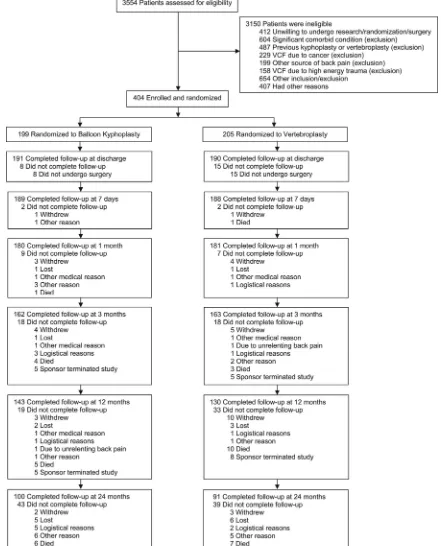
Patients were enrolled and randomized between October 2006 and May 2011. Figure 1 shows patient disposition.
[image:3.594.70.511.53.599.2]and 78.5% had single fractures treated (On-line Table). The base-line proportion of patients using bisphosphonates, calcium, and vi-tamin D is shown in the On-line Table; 151/261 (57.9%), 212/261 (81.2%), and 214/261 (82.0%) were using bisphosphonates, calcium, and vitamin D at 12 months respectively; and 93/191 (48.7%), 149/ 191 (78.0%), and 151/191 (79.1%), at 24 months. Use of these med-ications was not statistically significant between groups.
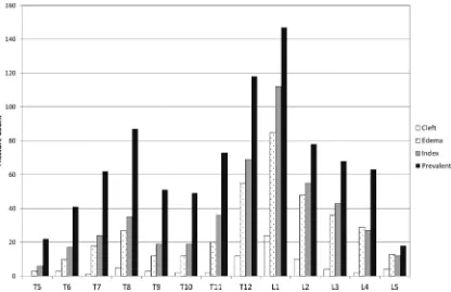
There was a higher radiographic fracture prevalence than identified clinically, and most treated fractures had active lesions confirmed on MR imaging (Fig 2). Most fractures occurred at T12 and L1.
Procedure Characteristics
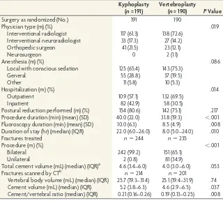
Most patients (70.3%) had local anesthesia with conscious seda-tion (Table 1). Fewer radiologists performed kyphoplasty (150/ 191, 78.5%) compared with vertebroplasty (165/190, 86.8%). Vertebroplasty had a shorter mean procedure duration (BKP, 40.0 minutes; VP, 31.8 minutes;P⬍.001) and median hospital-ization duration (BKP, 22 hours; VP, 8 hours;P ⫽.010). For vertebroplasty, 152/189 (80.4%) procedures were performed by using an injector kit. Kyphoplasty had statistically significantly higher cement volumes assessed by CT (Table 1).
Subsequent Vertebral Fracture
Regarding the primary end points (Table 2), 7.8% fewer patients with BKP had subsequent radiographic fractures compared with those who had VP at 12 months (P⫽.21), and 8.6% fewer, at 24 months (P⫽.23); but results were not statistically significant. Because subsequent radiographic fractures included any worsen-ing index fractures, we analyzed those separately; 4 of 140 (2.9%)
subjects with BKP and 10 of 131 (7.6%) subjects with VP had worsening index fractures (P⫽.10) at 12 months. No additional worsening index fractures were detected at 24 months.
Kaplan-Meier analysis of new clinically identified fractures (Fig 3) approached statistical significance for longer fracture-free survival in the BKP group (Wilcoxon test,P⫽.0596). For the 88 subjects with new clinically recognized fractures (Fig 3), 85 of 88 (96.6%) were specifically associated with new-onset pain. Sixty-three (71.6%) subjects had a VCF confirmed by MR imaging; 15 (17.0%) had VCF confirmed by using x-ray only, which consisted of a change of at least 1 Genant grade. Six (6.8%) had CT, 2 (2.2%) had bone scans, and 2 (2.2%) had an imaging technique not spec-ified. With regard to subsequent treatment, 70 (79.5%) had ver-tebral augmentation for at least 1 VCF, 16 (18.1%) had nonsurgi-cal care alone, 1 (1.1%) had subsequent fusion surgery with instrumentation, and 1 (1.1%) had therapy not identified.
Pain, Disability, and Quality of Life
Kyphoplasty and vertebroplasty groups had similar baseline back pain, SF-36 Physical Component Summary, and EQ-5D quality-of-life and ODI scores. For each outcome, statistically significant improvements from baseline were observed for each group, but differences between treatment groups were not significant (Fig 4). Concomitant with pain relief, use of opioid medications dropped from 73.9% (122/165) of patients at baseline (On-line Table) to 17.6% (25/142) for BKP and from 74.6% (126/169) to 23.9% (34/142) for VP at 6 months (P⫽.24). Results were 17.6% (16/91) for BKP and 25.6% (21/82) for VP at 24 months (P⫽.26).
[image:4.594.86.502.45.314.2]Secondary Radiographic End Points
Investigators were to attempt vertebral deformity correction, regardless of treatment; 154/191 (80.6%) patients with BKP and 142/189 (75.1%) with VP had perioperative postural re-duction. Compared with the preoperative condition, average postoperative kyphotic correction was statistically significant at each time point for both the BKP and VP groups (Table 3). Postoperatively, the ANCOVA estimate of mean difference be-tween groups was 0.21° (95% CI,⫺0.73°–1.14°) and was not statistically different (P⫽.663). At 24 months, kyphosis cor-rection was better in the BKP group with a mean difference between groups of 1.42° (95% CI, 0.10°–2.74°), which was sta-tistically significant (P⫽.036).
Safety
The most common AEs (classified ac-cording to MedDRA) within 30 days of surgery were procedural pain (BKP: 12/ 191, VP: 9/190), back pain (BKP: 14/ 191, VP: 28/190), and new symptomatic fracture (BKP: 9/191, VP: 17/190). Common during 2 years were bronchitis (BKP: 10/191, VP: 10/190), pneumonia (BKP: 15/191, VP: 12/190), urinary tract infection (BKP: 11/191, VP: 19/190), falls (BKP: 47/191, VP: 46/190), proce-dural pain (BKP: 12/191, VP: 9/190), ar-thralgia (BKP: 18/191, VP: 12/190), back pain (BKP: 49/191, VP: 59/190), lumbar vertebral facture (BKP: 9/191, VP: 13/ 190), and thoracic vertebral fracture (BKP: 20/191, VP: 21/190). Device- and procedure-related AEs during 2 years are detailed in Table 4; most were ob-served within 30 days postsurgery. All AEs are posted on www.clinicaltrials. gov (NCT00323609). A few AEs, includ-ing procedural pain (BKP: 3/191, VP: 3/190), back pain (BKP: 2/191, VP: 3/ 190), new fractures (BKP: 2/191, VP: 2/190), cement embolism (BKP: 1/191, VP: 1/190), muscle spasm (BKP: 1/191, VP: 0/190), arthralgia (BKP: 1/191, VP: 0/190), bone marrow edema (BKP: 0/191, VP: 1/190), and implant-site ex-travasation (BKP: 0/191, VP: 1/190), were specifically considered to be device-related (Table 4). Five BKP AEs (constipation, pro-cedural nausea, propro-cedural hypotension, hallucination, exacer-bated chronic obstructive pulmonary disease) and 5 VP AEs (hy-persensitivity, mental status changes, hypoxia, respiratory failure, hematoma) were considered anesthesia- or procedure-related (Table 4). No deaths were noted as device- or procedure-related. Overall cement extravasation, assessed by postoperative CT (Fig 5), was lower (P⫽.047) for BKP (157/214 levels treated) compared with VP (164/201 levels treated). There was lower in-travascular extravasation for BKP (59/214 levels treated) com-pared with VP (76/201 levels treated,P⫽.028). As indicated in Table 4, 1 patient with BKP and 1 with VP each presented with symptoms and were found to have cement embolism. One patient with VP had a new symptomatic fracture occur within 2 days postoperatively (inferior to the index level), with inferior cement leakage that was considered possibly bone cement–related.
DISCUSSION
This was the first large-scale RCT of BKP and VP with long-term follow-up. Both treatments provided similar sustained improve-ment from baseline in pain, disability, and quality of life that lasted for 2 years. These improvements were statistically signifi-cant and clinically relevant but were not statistically different be-tween groups. Safety data support the safe use of both BKP and VP. Kyphoplasty trended toward fewer fractures, had lower ce-Table 1: Procedure characteristics
Kyphoplasty (n= 191)
Vertebroplasty
(n= 190) PValue
Surgery as randomized (No.) 191 190
Physician type (m) (%) .019
Interventional radiologist 117 (61.3) 138 (72.6) Interventional neuroradiologist 33 (17.3) 27 (14.2)
Orthopedic surgeon 41 (21.5) 23 (12.1)
Neurosurgeon 0 2 (1.1)
Anesthesia (m) (%) .086
Local with conscious sedation 125 (65.4) 143 (75.3)
General 55 (28.8) 37 (19.5)
Other 11 (5.8) 10 (5.3)
Hospitalization (m) (%) .014
Outpatient 109 (57.1) 132 (69.5)
Inpatient 82 (42.9) 58 (30.5)
Postural reduction performed (m) (%) 154 (80.6) 142 (75.1) .217 Procedure duration (min) (mean) (SD) 40.0 (22.0) 31.8 (19.3) ⬍.001 Fluoroscopy duration (min) (mean) (SD) 10.0 (6.3) 8.5 (4.9) .008 Duration of stay (hr) (median) (IQR) 22.0 (6.0–26.0) 8.0 (5.0–24.0) .010
Fractures treated n⫽244 n⫽235
Procedure (m) (%) ⬍.001
Bilateral 242 (99.2) 151 (65.1)
Unilateral 2 (0.8) 81 (34.9)
Total cement volume (mL) (median) (IQR)a 4.6 (3.4–6.0) 4.0 (3.0–6.0) .053
Fractures scanned by CTb n⫽214 n⫽201
Vertebral body volume (mL) (median) (IQR) 25.7 (19.3–31.4) 25.1 (19.4–31.9) .74 Cement volume (mL) (median) (IQR) 5.2 (3.8–6.3) 4.6 (2.9–6.5) .037 Cement/vertebral ratio (median) (IQR) 0.21 (0.16–0.26) 0.19 (0.13–0.25) .008 Note:—(m) indicates numerator (No.) in category; IQR, interquartile range.
a
For BKP, all patients were treated with HV-R bone cement (Kyphon; Medtronic Spine, Sunnyvale, California). For VP, 46 (24.3%) patients were treated with Spineplex (Stryker, Kalamazoo, Michigan); 43 (22.8%), with Parallax (ArthroCare, Austin, Texas); 34 (18.0%), with AVAtex (CareFusion, San Diego, California); 14 (7.4%), with Confidence (DePuy Spine, Raynham, Massachusetts); 11 (5.8%), with HV-R (Kyphon); 10 (5.3%), with Visioplast (Tecres, Verona, Italy); and 10 (5.3%), with Vertefix (Cook, Bloomington, Indiana). The remaining 21 (11.1%) were treated with other bone cements.
b
[image:5.594.55.375.56.344.2]CT assessments were made by the core radiographic laboratory.
Table 2: Patients with new radiographic fracturesa
Kyphoplasty Vertebroplasty PValue 0–3 Mo
None 115 (76.7%) 106 (72.6%) .43
All subsequent 35 (23.3%) 40 (27.4%) 0–12 Mob
None 90 (64.3%) 74 (56.5%) .21
All subsequent 50 (35.7%) 57 (43.5%) 0–24 Mob
None 56 (50.9%) 47 (42.3%) .23
All subsequent 54 (49.1%) 64 (57.7%) a
Radiographic fractures identified by a core laboratory.
[image:5.594.52.287.435.540.2]ment extravasation, and more kyphotic deformity correction at 24 months. With vertebroplasty, there was slightly more local anesthesia use, more outpatient procedures, and a shorter dura-tion of stay; it is likely that these findings are due to the different physician profiles performing the procedures (Table 1) and asso-ciated practice patterns.23,24Differences in physician profile likely
stem from the study design allowing investigators to participate as a team (see Material and Methods). Shorter procedure and fluo-roscopy duration for VP may be related to more unipedicular vertebral access during VP treatment (Table 1).
Minimally clinically important differences are commonly used thresholds to estimate the clinical significance of out-comes.25 Improvements from baseline in the SF-36 Physical
Component Summary were⬎7.5 points at 12 and 24 months for both groups, exceeding the estimated minimally clinically impor-tant difference of 3.5– 4.3 points.25Similarly, improvements of ⱖ4 points exceeded the 1- to 2.5-point threshold for back pain,19,25improvements of⬎25 points exceeded the 10- to
15-point threshold for ODI,20,25and improvements ofⱖ0.28 points
exceeded the 0.08 threshold for EQ-5D.26
Cumulative evidence demonstrates that kyphoplasty and verte-broplasty provide better outcome than nonsurgical management in RCTs and meta-analyses4,5,7,9-11,27,28and acceptable
cost-effective-ness ratios.11,29-31Several large retrospective studies using claims
data, investigating BKP, VP, and nonsurgical management, provide additional evidence.32-35Although 2 blinded RCTs concluded that
vertebroplasty was similar to a local anesthetic “sham” interven-tion,36,37these trials have several important limitations, including
atypically broad inclusion criteria, allowance of chronic frac-tures, small sample size, and, in 1 study, high crossover, all of
which preclude definitive conclusions.28,38,39For example, in 1
study, higher crossover in the sham group compared with verte-broplasty (51% versus 13%,P⬍.001) at 3 months suggests that any short-term effects of the sham intervention are not long-last-ing.36Here, and in several other RCTs, kyphoplasty and
vertebro-plasty had statistically significant and sustained clinical improve-ment from baseline in pain, disability, and quality-of-life outcomes for 1 and 2 years; and compared with nonsurgical man-agement, benefits persisted throughout 1 and 2 years for several outcomes.7,9-11Such results are inconsistent with placebo effects.
Currently, there is 1 small randomized study showing similar pain outcomes for BKP and VP but statistically significantly better height restoration for BKP.8Nonrandomized comparisons show
similar results.40,41Here, postsurgery kyphotic angulation
tion was similar between groups. There was some loss of correc-tion in both groups at 3 months; however, for VP, the trend was more loss, with a statistically significant 24-month difference be-tween groups (Table 3). Our postoperative results of approxi-mately 3° of kyphosis correction are consistent with 2 other BKP RCTs5,6; one with long-term follow-up reported minimal
correc-tion loss during 2 years.5Postural reduction has been shown to
provide deformity correction for vertebroplasty,42,43achievable
in 35%–50% of acute fractures that have dynamic mobility.6,43
Likewise, in several BKP studies, substantial postural reduction with additional inflatable bone tamp contributions of 27%–100% has been documented.6,44,45With KAVIAR, 75% of patients with VP
had postural reduction, accounting for deformity correction in the VP group, and this finding may explain less postoperative correction in VP (compared to BKP) reported in other studies.8,40,41
[image:6.594.56.531.42.344.2]another RCT5,10and is likely due to the high number of prevalent
fractures at baseline, a potent risk factor.13This finding
under-scores the importance of additional therapeutic measures such as pharmacologics to help reduce fracture risk. Because randomiza-tion was stratified by baseline fracture prevalence, BKP and VP groups were well-balanced. Although not statistically significant, likely due to lack of statistical power, the BKP group was 7.8%– 8.6% lower in 1- and 2-year radiographic fracture rates, consistent with the prespecified protocol originally powered to detect an 8.7% difference. Furthermore, the Kaplan-Meier analysis of new clinically recognized fractures approached statistical significance for a longer time to first clinical fracture within the BKP group. These results are consistent with several meta-analyses showing fewer new VCFs in BKP.46-48
Kyphoplasty and vertebroplasty were found to be safe in this
pop-ulation, having similar safety profiles (Table 4). A similar number of patients in each group had device-related cement embolism (1 BKP, 1 VP), back pain (2 BKPs, 3 VPs), procedural pain (2 BKPs, 3 VPs), and new fractures considered to be related to the procedure (2 BKPs, 2 VPs). Several meta-analyses suggested that while both procedures have a low complication rate, BKP may have a lower rate of serious and symptomatic complica-tions.47-49The similar safety profile observed here may be due
to highly experienced physicians required by the protocol. CT assessment showed significantly less cement extravasation in BKP- (73%) versus VP-treated (82%) vertebrae (P⫽.047). These rates are higher than those in most reports, likely relating to use of CT, but are consistent with leakage being lower in BKP.46-49
[image:7.594.58.529.46.361.2]Most other studies reported cement leakage on the basis of inves-tigator assessment, by using reviews of conventional x-ray images, FIG 4. Quality-of-life, disability, and pain assessments at baseline and after balloon kyphoplasty or vertebroplasty. Means and 95% confidence intervals are shown for balloon kyphoplasty (solid lines) and vertebroplasty (dashed lines) for the SF-36 Physical Component Summary (scale 0 –100) (A); the total EQ-5D scores (scale 0 –1) (B); back pain (scale 0 –10) (C); and the Oswestry Disability Index (scale 0 –100) (D). The treatmentPvalue in each panel indicates the comparison between groups. Below each panel, thenfor each group is shown for baseline, 3, 12, and 24 months and the group average for change from baseline and 95% CI for 3, 12, and 24 months. TheasteriskindicatesP⬍.001 for all change from baseline scores.
Table 3: Index vertebral body kyphotic angulation correction
Levels (No.)
Raw Data Change from Baseline in Degrees
(Mean) (95% CI)a
ANCOVA Change from Baseline in Degrees
(LS Mean) (95% CI) ANCOVA Estimate ofMean Difference
(95% CI)
ANCOVA PValue
BKP VP BKP VP BKP VP
Post-op 213 195 3.10 (2.39–3.80) 3.41 (2.61–4.21) 3.34 (2.70–3.99) 3.14 (2.47–3.81) 0.21 (⫺0.73–1.14) .663 3 Mo 168 155 1.78 (0.98–2.58) 2.28 (1.37–3.19) 2.00 (1.27–2.72) 2.04 (1.28–2.80) ⫺0.04 (⫺1.10–1.01) .933 12 Mo 154 127 1.97 (1.11–2.82) 1.51 (0.58–2.44) 2.18 (1.47–2.89) 1.26 (0.48–2.04) 0.92 (⫺0.14–1.98) .089 24 Mo 99 92 2.09 (0.90–3.28) 1.43 (0.39–2.47) 2.46 (1.55–3.37) 1.04 (0.09–1.98) 1.42 (0.10–2.74) .036 Note:—Post-op indicates postoperative; LS, least squares.
a
[image:7.594.53.536.435.523.2]which are prone to interpretation bias and are less sensitive. These results are similar to those in another study using CT, also report-ing less leakage in BKP (49%) versus VP (87%).50Leakage into the
perispinal vasculature was significantly less (P⫽.028) for BKP. Meta-analyses of complications suggest that BKP results in fewer
symptomatic cement leakages, which include embolism and spi-nal cord compression.46-48
[image:8.594.55.532.56.283.2]The primary limitations in this study were the lack of patient blinding, substantial loss to follow-up, and early termination, which resulted in lack of statistical power for the primary end point. Verte-FIG 5. Cement extravasation. The percentage of treated vertebrae in each treatment group having cement extravasation, measured by using postoperative CT, is shown; results are based on evaluable CT data for 168/191 patients with BKP and 160/190 with VP, accounting for 214/244 and 201/233 levels, respectively. Fischer exactPvalues comparing the 2 treatment groups for each category are shown.
Table 4: Device/procedure/anesthesia–related adverse events during 2 years
No. of Patientsa
Kyphoplasty (n= 191)
Vertebroplasty (n= 190)
With procedure/device/anesthesia-related (or possibly) AEs 12 11
Blood and lymphatic disorders Bone marrow edema 0 1b
Gastrointestinal disorders Constipation 1c 0
Immune system disorders Hypersensitivity 0 1c
Injury or procedural complications Cement embolism 1d 1d
Implant site extravasation 0 1e
Mental status changes postoperative 0 1c
Procedural hypotension 1c 0
Procedural nausea/vomiting 1c 0
Procedural pain 3f 3f
Spinal fracture 1g 0
Musculoskeletal disorders Arthralgia 1g 0
Back pain 2g 3g
Muscle spasm 1g 0
Symptomatic vertebral fracture 1g 2g
Psychiatric disorders Hallucination 1c 0
Respiratory disorders COPD 1c 0
Hypoxia 0 1c
Respiratory failure 0 1c
Vascular disorders Hematoma 0 1h
Note:—COPD indicates chronic obstructive pulmonary disease.
a
Patients may have had multiple AEs; all system organ class categories are listed when there was an event considered related (or possibly related) to device/procedure/anesthesia.
b
In 1 patient, 1 event was considered nonserious and possibly related to bone cement.
c
In 1 patient, 1 event was considered serious and related (or possibly related) to anesthesia and was resolved with medical intervention.
d
In 1 patient, 1 event each was considered serious and bone cement–related (or possibly related). In each case, the event occurred after the surgical treatment of a subsequent fracture; the BKP event was resolved with oxygen, and the VP event was ongoing at final follow-up.
e
In 1 patient, 1 event was considered bone cement–related and nonserious. A spinal canal leak was detected intraoperatively; CT was immediately performed without significant canal stenosis observed with no medical intervention given.
f
Three patients in each group with 3 (2 serious) and 4 (all serious) events in the BKP and VP groups, respectively, were considered device-related (or possibly related) but were resolved.
g
The number of patients reflected in the Table had events that were considered serious and related (or possibly related) to the device.
h
[image:8.594.111.476.409.625.2]broplasty treatment was not standardized among centers. This lack of standardization may be viewed as a limitation and likely accounts for observed differences between groups in bilateral treatment and may, at least partially, account for differences in cement volumes and leak-age. Differences in bilateral treatment, in turn, may have had an effect on the statistically significant changes between groups in kyphotic angulation at 24 months. However, because there is no established standard for vertebroplasty, for generalizability, every study center was asked to provide care consistent with local practices. Nonethe-less, the strengths are the randomized, multicenter design, a relatively large sample size, and long-term follow-up. The results of this trial confirm the effectiveness of vertebral body cement augmentation for patients with osteoporosis with ongoing pain at index levels corre-lated by physical examination and imaging. Our results are consistent with recently updated guidelines published by the National Institute for Health and Care Excellence in the United Kingdom.51
CONCLUSIONS
Kyphoplasty and vertebroplasty had similar statistically signifi-cant sustained clinical improvement in pain and disability with similar AE profiles. Procedure duration and hospitalization were shorter with vertebroplasty. Kyphoplasty had fewer cement leak-ages, a trend of longer fracture-free survival, and less loss of ky-photic-deformity correction during 2 years.
ACKNOWLEDGMENTS
The authors are indebted to all participating staff at the investiga-tional centers and the patients who consented to participate in the KAVIAR trial, and they thank Li Ni, PhD, and Feng Tang, PhD, (Medtronic Spine) for contributions to statistical analysis. The authors gratefully acknowledge the KAVIAR protocol steering committee members, Drs Jacques Dion, Michael Ford, Joshua Hirsch, Mary Jensen, Reginald Knight, Isador Lieberman, James Lindley, Mark Myers, Wayne Olan, Frank Phillips, and Gregg Zoarski for contributions to the study design and the data safety monitoring board members, Drs Gunnar Andersson, Harry Cloft, Deborah Kado, Kern Singh, and Joel Verter, for contributions in monitoring patient safety throughout the trial.
APPENDIX
KAVIAR Investigators
Principal investigator, city, state (number of patients enrolled), for enrolling centers:
United States. G. Fueredi, Burlington, Wisconsin (42); G. So, Torrance, California (39); M. Dohm, Grand Junction, Colorado (36); L. Haikal, Huntington, West Virginia (28); C. Black, Provo, Utah (26); J. Milburn, New Orleans, Louisiana (22); R. DiSalle, Toledo, Ohio (21); A. Dacre, Billings, Montana (20); J. Neil, Scottsdale, Arizona (19); N. Cooper, Atlanta, Georgia (19); P. Chesis, Kansas City, Missouri (17); D. Sacks, Reading, Pennsylva-nia (17); S. Pledger, Middletown, Ohio (17); J. Small, Temple Terrace, Florida (16); P. Minor, Milwaukee, Wisconsin (12); H. Haykal, Houston, Texas (9); M. Montgomery, Temple, Texas (5); C. Kazmierczak, Royal Oak, Michigan (5); S. Bukata, Rochester, New York (4); A. Padidar, San Jose, California (4); P. Schloesser, Murray, Utah (4); B. Ward, Bend, Oregon (3); V. Lewis, Roanoke,
Virginia (3); D. Beall, Oklahoma City, Oklahoma (3); C. Graham, Columbia, South Carolina (1).
Canada. C. Guest, Barrie, Ontario (12).
Disclosures: Michael Dohm—RELATED:Fees for Participation in Review Activities such as Data Monitoring Boards, Statistical Analysis, Endpoint Committees, and the Like: Medtronic Spine,*Comments: The Western Slope Study Group was com-pensated for study-specific data collection related to this study only. I received no money because the Western Slope Study Group is a quality-improvement organiza-tion, 501 (c) 3, and I am not a paid employee. I received no compensation at any time, in any form from Medtronic. Carl M. Black—RELATED:Grant: Kyphon/Medtronic,* Comments: Utah Valley Regional Medical Center received remuneration for admin-istrative support during the research trial;Support for Travel to Meetings for the Study or Other Purposes: Kyphon/Medtronic,Comments: for inservice and training on research protocol;Provision of Writing Assistance, Medicines, Equipment, or Administrative Support: Kyphon/Medtronic*,Comments: data collection and sta-tistical support;Other: As a study investigator, my institution received compensa-tion from Medtronic Spine for study-specific data colleccompensa-tion. Alan Dacre—RELATED: Provision of Writing Assistance, Medicines, Equipment, or Administrative Support: Kyphon/Medtronic,*Comments: to support the collection of data for the study; Other: As a study investigator, my institution received compensation from Medtronic Spine for study-specific data collection;UNRELATED:Consultancy: Medtronic,Comments: I have an agreement but have not done any remunerable work. John B. Tillman—RELATED:Other: Medtronic,Comments: employed as a Clin-ical Program Director;UNRELATED:Stock/Stock Options: Medtronic. George Fu-eredi—RELATED:Grant: Aurora Medical Group*;Other: As a study investigator, my institution received compensation from Medtronic Spine for study-specific data collection and has received compensation for consulting for Medtronic Spine. *Money paid to the institution.
REFERENCES
1. Johnell O, Kanis JA.An estimate of the worldwide prevalence and disability associated with osteoporotic fractures.Osteoporos Int 2006;17:1726 –33
2. Silverman S.The clinical consequences of vertebral compression fracture.Bone1992;13:S27–31
3. Lindsay R, Burge RT, Strauss DM.One year outcomes and costs following a vertebral fracture.Osteoporos Int2005;16:78 – 85 4. Berenson J, Pflugmacher R, Jarzem P, et al.Balloon kyphoplasty
versus non-surgical fracture management for treatment of painful vertebral body compression fractures in patients with cancer: a multicentre, randomised controlled trial. Lancet Oncol 2011;12:225–35
5. Van Meirhaeghe J, Bastian L, Boonen S, et al.A randomized trial of balloon kyphoplasty and non-surgical management for treating acute vertebral compression fractures: vertebral body kyphosis correction and surgical parameters. Spine (Phila Pa 1976) 2013;38:971– 83
6. Bastian L, Schils F, Tillman JB, et al.A randomized trial comparing 2 techniques of balloon kyphoplasty and curette use for obtaining vertebral body height restoration and angular-deformity correc-tion in vertebral compression fractures due to osteoporosis.AJNR Am J Neuroradiol2013;34:666 –75
7. Farrokhi MR, Alibai E, Maghami Z.Randomized controlled trial of percutaneous vertebroplasty versus optimal medical management for the relief of pain and disability in acute osteoporotic vertebral compression fractures.J Neurosurg Spine2011;14:561– 69 8. Liu JT, Liao WJ, Tan WC, et al.Balloon kyphoplasty versus
verte-broplasty for treatment of osteoporotic vertebral compression fracture: a prospective, comparative, and randomized clinical study.Osteoporos Int2010;21:359 – 64
9. Wardlaw D, Cummings SR, Van Meirhaeghe J, et al.Efficacy and safety of balloon kyphoplasty compared with non-surgical care for vertebral compression fracture (FREE): a randomised controlled trial.Lancet2009;373:1016 –24
11. Klazen CA, Lohle PN, de Vries J, et al.Vertebroplasty versus conservative treatment in acute osteoporotic vertebral compres-sion fractures (Vertos II): an open-label randomised trial.Lancet 2010;376:1085–92
12. Briggs AM, Wrigley TV, van Dieen JH, et al.The effect of osteopo-rotic vertebral fracture on predicted spinal loads in vivo.Eur Spine J 2006;15:1785–95
13. Lindsay R, Silverman SL, Cooper C, et al.Risk of new vertebral frac-ture in the year following a fracfrac-ture.JAMA2001;285:320 –23 14. Wardlaw D, Van Meirhaeghe J, Ranstam J, et al.Balloon kyphoplasty
in patients with osteoporotic vertebral compression fractures. Ex-pert Rev Med Devices2012;9:423–36
15. Genant HK, Wu CY, van Kuijk C, et al.Vertebral fracture assess-ment using a semiquantitative technique. J Bone Miner Res 1993;8:1137– 48
16. MedDRA term selection: points to consider. ICH endorsed guide for MedDRA users. Release 4.2, based on MedDRA Version 14.1, October 2011. http://www.meddra.org/sites/default/files/guidance/file/9491-1410_termselptc_r4_2_sep2011.pdf. Accessed July 2, 2014
17. Ware J, Kosinski M, Dewey J.How to Score Version 2 of the SF-36 Health Survey.Lincoln: QualityMetric; 2000
18. EQ-5D User Guide. Rotterdam, the Netherlands: EuroQol Group; 1996
19. Farrar JT, Young JP Jr, LaMoreaux L, et al.Clinical importance of changes in chronic pain intensity measured on an 11-point numer-ical pain rating scale.Pain2001;94:149 –58
20. Fairbank JC, Pynsent PB.The Oswestry Disability Index. Spine 2000;25:2940 –52
21. Agency for Healthcare Research and Quality. U.S. Valuation of the Eu-roQol EQ-5D Health States. January 2012. http://www.ahrq.gov/ professionals/clinicians-providers/resources/rice/EQ5Dproj.html. Ac-cessed June 7, 2013
22. Eastell R, Cedel SL, Wahner HW, et al.Classification of vertebral fractures.J Bone Miner Res1991;6:207–15
23. Goz V, Koehler SM, Egorova NN, et al. Kyphoplasty and vertebroplasty: trends in use in ambulatory and inpatient settings.
Spine J2011;11:737– 44
24. Mehio AK, Lerner JH, Engelhart LM, et al.Comparative hospital economics and patient presentation: vertebroplasty and kyphop-lasty for the treatment of vertebral compression fracture.AJNR Am J Neuroradiol2011;32:1290 –94
25. Copay AG, Glassman SD, Subach BR, et al.The minimum clinically important difference in lumbar spine surgery patients: a choice of methods using the Oswestry Disability Index, Medical Outcomes Study Questionnaire Short Form 36, and Pain Scales. Spine J 2008;8:968 –74
26. Walters SJ, Brazier JE.Comparison of the minimally important dif-ference for two health state utility measures: EQ-5D and SF-6D.
Qual Life Res2005;14:1523–32
27. Papanastassiou ID, Phillips FM, Van Meirhaeghe J, et al.Comparing effects of kyphoplasty, vertebroplasty, and non-surgical manage-ment in a systematic review of randomized and non-randomized controlled studies.Eur Spine J2012;21:1826 – 43
28. Anderson PA, Froyshteter AB, Tontz WL Jr.Meta-analysis of verte-bral augmentation compared to conservative treatment for osteo-porotic spinal fractures.J Bone Miner Res2013;28:372– 82 29. Strom O, Leonard C, Marsh D, et al.Cost-effectiveness of balloon
kyphoplasty in patients with symptomatic vertebral compression fractures in a UK setting.Osteoporos Int2010;21:1599 – 608 30. Svedbom A, Alvares L, Cooper C, et al.Balloon kyphoplasty
com-pared to vertebroplasty and nonsurgical management in pa-tients hospitalised with acute osteoporotic vertebral compres-sion fracture: a UK cost-effectiveness analysis.Osteoporos Int 2013;24:355– 67
31. Edidin AA, Ong KL, Lau E, et al.Cost-effectiveness analysis of treat-ments for vertebral compression fractures.Appl Health Econ Health Policy2012;10:273– 84
32. Edidin AA, Ong KL, Lau E, et al.Mortality risk for operated and nonoperated vertebral fracture patients in the Medicare popula-tion.J Bone Miner Res2011;26:1617–26
33. McCullough BJ, Comstock BA, Deyo RA, et al.Major medical out-comes with spinal augmentation vs conservative therapy.JAMA In-tern Med2013;173:1514 –21
34. Lange A, Kasperk CP, Alvares L, et al.Survival and cost comparison of kyphoplasty and percutaneous vertebroplasty using German claims data.Spine (Phila Pa 1976)2014;39:318 –26
35. Chen AT, Cohen DB, Skolasky RL.Impact of nonoperative treat-ment, vertebroplasty, and kyphoplasty on survival and morbidity after vertebral compression fracture in the Medicare population.
J Bone Joint Surg Am2013;95:1729 –36
36. Kallmes DF, Comstock BA, Heagerty PJ, et al.A randomized trial of vertebroplasty for osteoporotic spinal fractures. N Engl J Med 2009;361:569 –79
37. Buchbinder R, Osborne RH, Ebeling PR, et al.A randomized trial of vertebroplasty for painful osteoporotic vertebral fractures.N Engl J Med2009;361:557– 68
38. Bono CM, Heggeness M, Mick C, et al.North American Spine Society: newly released vertebroplasty randomized controlled tri-als—a tale of two trials.Spine J2010;10:238 – 40
39. Clark W, Lyon S, Burnes J, et al.Trials of vertebroplasty for vertebral fractures.N Engl J Med2009;361:2097–98
40. Li X, Yang H, Tang T, et al.Comparison of kyphoplasty and verte-broplasty for treatment of painful osteoporotic vertebral compres-sion fractures: twelve-month follow-up in a prospective nonran-domized comparative study. J Spinal Disord Tech2012;25:142– 49 41. Grohs JG, Matzner M, Trieb K, et al.Minimal invasive stabilization
of osteoporotic vertebral fractures: a prospective nonrandomized comparison of vertebroplasty and balloon kyphoplasty.J Spinal Disord Tech2005;18:238 – 42
42. Chin DK, Kim YS, Cho YE, et al.Efficacy of postural reduction in osteoporotic vertebral compression fractures followed by percuta-neous vertebroplasty. Neurosurgery 2006;58:695–700; discussion 695–700
43. McKiernan F, Jensen R, Faciszewski T.The dynamic mobility of vertebral compression fractures.J Bone Miner Res2003;18:24 –29 44. Voggenreiter G.Balloon kyphoplasty is effective in deformity cor-rection of osteoporotic vertebral compression fractures. Spine 2005;30:2806 –12
45. Shindle MK, Gardner MJ, Koob J, et al.Vertebral height restora-tion in osteoporotic compression fractures: kyphoplasty bal-loon tamp is superior to postural correction alone.Osteoporos Int 2006;17:1815–19
46. Eck JC, Nachtigall D, Humphreys SC, et al.Comparison of verte-broplasty and balloon kyphoplasty for treatment of vertebral compression fractures: a meta-analysis of the literature.Spine J 2008;8:488 –97
47. Taylor RS, Taylor RJ, Fritzell P.Balloon kyphoplasty and verte-broplasty for vertebral compression fractures: a comparative systematic review of efficacy and safety.Spine2006;31:2747–55 48. Lee MJ, Dumonski M, Cahill P, et al.Percutaneous treatment of
vertebral compression fractures: a meta-analysis of complications.
Spine2009;34:1228 –32
49. Hulme PA, Krebs J, Ferguson SJ, et al.Vertebroplasty and kyphoplasty: a systematic review of 69 clinical studies.Spine2006;31:1983–2001 50. Lee IJ, Choi AL, Yie MY, et al.CT evaluation of local leakage of bone
cement after percutaneous kyphoplasty and vertebroplasty.Acta Radiol2010;51:649 –54