TOWARDS A BETTER
UNDERSTANDING OF
COCHLEAR MECHANICS:
A
New Cochlear Model
A Thesis presented for the Degree of Doctor of Philosophy
in
Electrical and Electronic Engineering a.t the
University of Canterbury, Christchurch, New Zealand.
by
Paul
J
ohannes
~olstonBE (Hons 1)
I
Abstract
Improving our understanding of cochlear mechanics requires the analysis of cochlear mod-els. In the formulation of such models, it is necessary to make assumptions as to the relative importance of the many structures which comprise the cochlear partition. Data on the relative motion of these structures are virtually non-existent, and so the accuracy of many of the assumptions made is questionable~ The bulk of the content of this thesis relates to the formulation and analysis of a new type of linear mechanical cochlear model. The new assumptions made in the formulation of the model are justified on the basis of the structure of the cochlear partition. The apparent realism of the model (both its struc-ture and certain feastruc-tures of its response) seriously questions the hypothesis that response tuning and changes in response resulting from trauma prove the existence of active pro-cesses in the cochlea. Furthermore, arguments are presented that question the validity of using the presence of spontaneous otoacoustic emissions to justify the existence of active processes in cochlear tuning. The new model suggests that the complicated structural geometry of the cochlear partition (particularly the organ of Corti) must be incorporated in a model before conclusions relating to real cochlear behaviour can be drawn from it. In particular, the model suggests that a mechanical second filter exists in the cochlea, from rather broad tuning in the pectinate zone of the basilar membrane to sharper, neural-like tuning in the arcuate zone. It is concluded that the only way to properly check the validity of cochlear models is to obtain more experimental data pertaining to the relative motions of the various components that constitute the cochlear partition. Before this is done, we should not place too much faith in our present (alleged) understanding of cochlear mechanics. Also presented in this thesis are new modelling techniques for improving the realism of electrical transmission line cochlear models: the ability to include longitudinal and radial mechanical coupling, multidimensional fluid motion and stick-slip friction. It is shown that the inclusion of mechanical coupling and two-dimensional fluid motion in the new cochlear model has a predictable (and trivial) effect on-its response. The use of the stick-slip friction modelling technique is illustrated by means of two simple examples.
Acknowledgements
I am very grateful to Dr. Peter T. Gough for allowing me the freedom to pursue this field of research. I am indebted to Prof.dr. Egbert de Boer and Prof.dr.ir. Max A. Viergever for showing an interest in my work and for enabling me to undertake part of my studies in The Netherlands.
Thanks are due to those members of staff and postgraduate students in the Department of Electrical and Electronic Engineering, University of Canterbury, and in the Vakgroep Toegepaste Analyse, Faculteit Technische Wiskunde en Informatica, Technische Univer-siteit Delft, who provided me with technical help, companionship and amusement.
This work was financially supported by the Vakgroep Toegepaste Analyse, Faculteit Tech-nische Wiskunde en Informatica, TechTech-nische Universiteit Delft, and by a Postgraduate Scholarship from the New Zealand University Grants Committee.
reface
For at least the last five years virtually all literature on cochlear mechanics has made mention of an "active process" being necessary within the cochlea in order for it to produce the sharply-tuned mechanical response curves that have been recently observed experimentally. I believ(:! that the evidence cited (to date) in support of the existence of such an energy-producing process is not convincing. In support of this conclusion, I have developed a passive cochlear model that exhibits certain features of cochlear response which previously required the incorporation of active processes in cochlear models.
Considering the anatomy of the cochlear partition (CP), it seems odd that few descriptions of cochlear mechanics have taken into account the gross radial structural differences of the CPo I have found that the division of the CP radially into two zones, combined with a few other assumptions, results in a passive cochlear model with. realistic parameters being able to exhibit a sharply-tuned mechanical response, a realistic phase response, and show realistic response changes as a result of trauma. Even so, I do not believe that the new cochlear model described in this thesis is an accurate representation of the cochlea, since it ignores most of the structural features of the organ of Corti. However, all other cochlear models are (at best) equally deficient and so I believe that we must be more sceptical in our interpretations of modelling studies.
This thesis does not present an overview of cochlear mechanics. Topics such as non-linearities, electrophysiology, transduction, and mechanics of non-mammalian cochleae are completely ignored, since they do not contribute to the conclusions that can be drawn from the work described herein. A chapter-by-chapter description of the thesis contents now follows.
III The Introduction presents justifications for pursuing this field of research. A few
definitions and abbreviations are also provided.
1/ In Chapter 2 the anatomy of the ear is briefly summarized. Obvious features of cochlear anatomy that seem to have been largely overlooked by other modellers are described in more detail. In particular, the gross structural differences between the arcuate and pectinate zones of the CP are emphasized .
.. Chapter 3 includes a non-mathematical description ofthe formation of the travelling wave. Also presented in Chapter 3 is an overview of recent measurements of the mechanical response of the cochlea.
viii
• Chapter 4 includes a critical review of the assumptions made in the formulation of cochlear models. Considering these assumptions in the light of cochlear anatomy and response (as described in Chapters 2 and 3), I conclude that all cochlear models may not represent those features of cochlear mechanics that are responsible for the cochlea's exquisite tuning. Active models are briefly reviewed and the formulation of transmission line cochlear models is described in detail.
• The new type of cochlear model that I have proposed is described in Chapter 5. This model has been the subject of a number of papers: Kolston (1988a,c), Kolston and Viergever (1989a), Kolston et al. (1989). In the formulation of this model, the CP is divided radially into its two zones, and the presence of the outer hair cells greatly alters the mechanics of the arcuate zone. The model simulates realistic selectivity and phase response, and realistic changes in response as a result of trauma, without needing to incorporate active processes. The model's response is evaluated on the basis of recent experimental mechanical response data.
• Electrical transmission line (ETL) cochlear models have a number of unique ad-vantages over other modelling techniques. The most obvious is that when an ETL model is realized as a physical circuit, its response is available in real time to real inputs (e.g. speech), thereby enabling direct observations of its operation and re-sponse. Until now, such models have been deficient in that they did not include mechanical coupling in the CP nor, more importantly, multi-dimensional fluid mo-tion. Chapter 6 describes two new modeliing techniques whlch enable incorporation of these two features in an ETL model. The effect of adding mechanical coupling and two-dimensional fluid motion to the new cochlear model (described in Chapter 5) is assessed.
• The Discussion presents an appraisal of the new model, and the Conclusion com-ments on the model's significance and makes suggestions on how best to further improve our understanding of cochlear mechanics.
• The Appendix presents a new technique for modelling stick-slip friction, a form of friction that may play a role in cochlear micromechanical fluid motion. The technique enables incorporation of stick-slip friction in an ETL cochlear model, but this has not been attempted. Instead, two test examples are used to illustrate the validity of the technique.
Some of the work contained in this thesis has appeared (or will appear) in published form:
Koiston, P.J. (1988a). Sharp mechanical tuning in a cochlear model without negative damping. Journal of the Acoustical Society of America, Vol. 83, pp 1481-1487.
ix
Kolston, P.J. (1988c). Micromechanics remove the need for active processes in cochlear tuning. In Basic Issues in Hearing, edited by H. Duiibuis, J.W. Horst
and H.P. Wit, (Academic Press, London), pp 74-79.
Koiston, P.J. and Viergever, M.A. (1989a). Realistic basilar membrane tuning does not require active processes. In Cochlear Mechanisms - Structure, Function and Models, edited by J.P. Wilson and D.T. Kemp, (Plenum Press, London), pp
415-424.
Koiston, P.J. and Viergever, M.A. (1989b). How do the outer hair cells influence cochlear mechanics? Submitted for publication.
Koiston, P.J., Viergever, M.A., de Boer, E., and Diependaal, R.J. (1989). Realistic mechanical tuning in a micromechanical cochlear model. Journal of the Acoustical Society of America, Vol. 86 (In Press).
I was fortunate to have been able to attend (and present papers at) the following confer-ences:
8th International Conference on Hearing (Paterswolde, The Netherlands, 5-9 April, 1988). Mechanics of Hearing 1988 (Keele, England, 4-8 July, 1988).
During the periods June 1985 to May 1987, and August 1988 to February 1989, the work for this thesis was undertaken in the Department of.Electrical and Electronic Engineering, University of Canterbury, Christchurch, New Zealand. From June 1987 to July 1988 the work was undertaken in the Department of Technical Mathematics and Informatics, Delft· University of Technology, Delft, The Netherlands.
Contents
Abstract
Acknowledgements
Preface
1 Introduction
1.1 Aim of the Thesis.
1.2 Why Cochlear Research? .
1.3 Why Cochlear Modelling?
1.4 Definitions and Symbols
2 Anatomy
2.1 Outer and Middle Ear
2.2 Cochlear Apatomy . .
2.2.1 Cross-sectional structure.
2.2.2 Longitudinal structure . .
3 Response
3.1 Formation of the Travelling Wave.
3.1.1 Historical . . . .
3.1.2 A qualitative description.
3.1.3 Fluid wavelength
3.2 Measured Responses . .
3.2.1 Measurement techniques.
3.2.2 Phase response . . . .
xi III V Vll 1 1 1
2-..
3xii
3.2.3 Magnitude response . . . .
4 Models
4.1 Mathematical and Physical Models
4.2 Assumptions . , . . . . . . .
4.2.1 Fluid Dimensionality .
4.2.2 Degrees of Freedom
4.3 Active models . . . .
4.4 Transmission Line Models
4.4.1
4.4.2
4.4:3
Mechanical Transmission Line.
Electrical Transmission Line.
Parameters
5 The OHCAP Model
5.1 Assumptions
5.2 Formulation.
5.3 Operation..
5.4 Models for the Outer Hair Cells .
5.4.1 FOHC model
5.4.2 DOHC model
5.5 Response . . . . ..
5.5.1 Analysis techniques
5.5.2 Parameters . . .
5.5.3 Typical response
5.5.4 Comparisons with experimental data .
6 Extensions to ETL Cochlear Models
6.1 Mechanical Coupling .
6.1.1 Description . .
6.1.2 Effect of radial coupling on OHCAP model response
6.2 Multi-dimensional Fluid Modelling
6.2.1 Description . . . .
6.2.2 Two-dimensional fluid motion.
23 27 27 28 30 30 31 32 32 34 36 39 39
40
42 43 43 45 48 48 49 49 52 65 66 66 69 71 71 736.2.3 Effect of 2-D fluid on OHCAP model response
7 Discussion
7.1 Anatomical support for the OHCAP model
7.2 Advantages of a passive model . . .
7.3 Arguments supporting active models
7.3.1 Acoustic emissions . . . .
7.3.2 Experimentally-measured tuning data
8 Conclusion
An Electrical Model of Stick-slip Friction
A.1 Description . .
A.2 Test examples .
References
xiii
76
81
81
82
82 82 83
85
87 87
91
List of Figure
201
Drawing of the human ear2.2
Schematic of the ear...
2.3
Diagram of a cross-section of the cochlea.2.4
Diagram of a cross-section of the cochlear canal .2.5
Diagram of the cochlear partition (CP)2.6
Structure of the basilar membrane (BM) .2.7
BM fibre geometry in arcuate and pectinate zones2.8
Diagram of the longitudinal structure of the organ of Corti3.1 Simplified diagram of the cochlea . . . .
3.2
Variation of fluid wavelength within the cochlea.6
6
8
910
12
13
14
19 203.3 The mechanical response of the cochlea . . . 22
3.4
Response in the arcuate and pectinate zones of the BM 244.1
Schematic representation of one section of the cochlea . . . . .33
\4.2 Schematic of one section of an electrical transmission line (ETL) cochlear model . . .
35
4.3
Schematic of a short length of an ETL cochlear model ,4.4
Appearance of an ETL model for one stimulus frequency.5.1
Schematic of the CP . . . ' ..I
5.2
Lumped component representati~n of the CP5.3
One section of the OHCAP model, in ETL form5.4
FOHC model in electrical 'circuit formxvi
5.5 Schematic of the DOHC model . . . 46
5.6 . Typical response of the FOHCAP model. 50
5.7 Impedances in the OHCAP model . . . . 51
5.8 Comparison of FOHCAP model response with the experimental data of Khanna and Leonard (1982). . . . . 56
5.9 Comparison of FOHCAP model response with the experimental data of Khanna and Leonard (1986). . . . . 57
5.10 Comparison of FOHCAP model response with the experimental data of Sellick et al. (1982 - animal 29) . . . .. 58
5.11 Comparison of FOHCAP model response with the experimental data of Sellick et al. (1982 - animal 35) . . . .. 59
5.12 Comparison of FOHCAP model response with the experimental data of Sellick et al. (1983a) . . . .. 60
5.13 Comparison of DOHCAP model.response with the experimental data of Sellick et al. (1983a) . . . . . 61
5.14 Comparison of FOHCAP model response with the experimental data of Robles et al. (1986) . . . 62
5.15 Comparison of DOHCAP model response with the experimental data of Robles .et at. (1986) . . . 63
6.1 Transmission line model with longitudinal mechanical coupling 67
6.2 ETL model with longitudinal mechanical coupling . . . 68
6.3 Effect of adding radial mechanical coupling to the FOHCAP model. 70
6.4 Fluid particle.motion on a streamline.
6.5 2-D approximatioIi of an inviscid fluid
6.6 2-D fluid motion in an ETL cochlear model
6.7 2-D fluid motion in an ETL cochlear model with high scalae .
6.8 2-D fluid motion in an ETL cochlear model with low scalae
6.9 Effect of 2-D fluid on the FOHCAP model.
I
A.1 A typical stick-slip friction characteristic .
A.2 The electrical stick-slip friction model. .
A.3 How the transition region characteristic is obtained .
, I
AA Some possible friction characteristics
A.5 Simple spring-mass system. . . . . .
A.6 Response of the simple spring-mass system
A.7 System with interacting stick-slip forces . .
A.S Response of the system with interacting stick-slip forces
xvii
90
92 92
93
The shaking air rattled Lord Edward's
membrana
tympani;
the interlocked malleus, incus
and
stirrup bones were set in motion
so as to
agitate the
membrane
of the oval window
and
raise
an
infinitesimal storm in the fluid of the labyrinth.
The hairy endings of the auditory nerve
shuddered like weeds in
a
rough sea;
a
vast number of obscure miracles were performed in the brain,
and
Lord Edward ecstatically whispered
'Bach!'
from
Ohapter 1
Introduction
This chapter outlines some of the reasons for investigating the operation of the cochlea; including an explanation of the role that the formulation and analysis of cochlear models plays in improving our understanding of cochlear mechanics. Also included are definitions for a number of technical terms used throughout the thesis. It should be noted that only the mammalian cochlea is considered in this thesis.
1.1
Aim of the Thesis
A proper understanding of cochlear mechanics can only be obtained by combining information gained from three quite different areas of research: observations of cochlear anatomy; psychophysical, electrophysiological and mechanical measurements of cochlear response; and analysis of cochlear models. The main aim of the work undertaken for this thesis is to improve our understanding of cochlear mechanics by the formulation and analysis of cochlear models. The most valuable contribution of this work is the description of a new model of the linear mechanical motion of the cochlea. a model that has prompted the author to question the present state of our understanding of cochlear mechanics.
1.2 Why Cochlear Research?
The air pressure variations generated by, for example, a voice, a musical instrument, or an environmental disturbance, enter our ears and undergo complicated signal-processing procedures before we can perceive them as sound. However, what we hear is largely determined by what we listen to, in that the last part of this analysis process occurs at a conscious level within the higher centres of the brain. The ear, or in more formal language the peripheral auditory system, consists of the first pa.rt of the hearing chain
2 CHAPTER 1. INTRODUCTION
that takes the air pressure variations and converts them into nerve pulses which travel to the brain. Irrespective of what we choose t9 listen to, the ear always senses the air pressure variations and sends the processed information to the brain.
Although a large amount of the signal processing in our sense of hearing takes place after the ear, the ear performs important initial processing. Most of this initial processing , occurs within the cochlea. The cochlea is that part of the ear that functions as a
micro-phone, converting mechanical motion into nerve pulses. It acts as an interface between the acoustic environment and the neural environment, and constitutes one of the more accessible stages of our hearing process. Furthermore, hearing disorders are often the result of a malfunctioning cochlea and so a greater understanding of its operation should improve our ability to alleviate the suffering of the deaf and hearing-impaired.
To better understand the operation of the human cochlea, it is necessary to know both its structure and its response. Humans are able to prevent destructive irrevers.ible experi-ments being performed on their bodies and therefore much of our understanding of human cochlea function is derived from experiments performed on animal cochleae. Fortunately, the basic structure of all mammalian cochleae is very similar and so it is assumed that the response measured in animal cochleae, reflects that of human cochleae.
1.3 Why Cochlear Modelling?
The purpose of modelling cochlear mechanics is to gain a better understanding of the operation of the real cochlea by the formulation and analysis of models that reflect the structure and mechanical response (see Section 1.4) of the real cochlea. When a model is found that conforms with these criteria, it can be assumed that the model is an accurate representation of the real thing. Observing the effects of changing the parameter values or altering the structure of the model can be helpful in determining the important (abnormal) features characterizing damaged cochleae. This information can be used to minimize damage caused by harmful influences on normal cochleae.
1.4. DEFINITIONS AND SYMBOLS 3
check if these assumptions are valid.
In the formulation of a cochlear model certain structures can be disregarded, enabling study of only those variables which govern the particular phenomenon of interest. For example, the first variable considered essential to the response of cochlea was the elasticity variation observed along the length of the cochlear partition. Therefore, this was the first structural parameter measured in the human cochlea and was the main feature of the first cochlear models investigated by von Bekesy. Cochlear modellers are very reliant on experimental studies to determine which variables should be included and which can be disregarded. Unfortunately, all cochlear models require considerable assumptions to be made in their formulation and so they seldom reflect the microstructure of the cochlea to any realistic extent.
The importance of following an iterative process cannot be overemphasized: using structural observations to formulate a model, analysing its response, comparing this with that observed in the real cochlea, and using this comparison to improve the model and to suggest new observations. An accurate cochlear model must be consistent with the structure and the response of the cochlea. It is the author's opinion that there is no current
model which satisfies both these criteria.
1.4
Definitions and Symbols
As is usual in highly-specialized fields of research, cochlear mechanics nomenclature plays an impbrtant role in communicating specific ideas. To ensure that the nomenclature used in this thesis has the same meaning to all readers, it is appropriate to include here a list of terms which have possible ambiguities in their meanings.
Motile/active processes
In the author's opinion, the term active process is applied incorrectly and inconsistently by many investigators in the field of cochlear mechanics. It is the current trend to use this term to describe the metabolically-sensitive process that exerts forces on the structures within the cochlea. In an engineering sense, the word "active" implies that these forces involve the injection of mechanical energy into the cochlea. However, many elements within a mechanical system generate forces without adding energy to the system. The direction (or phase) in which an element exerts a force, in relation to the controlling signal, determines whether the element adds energy or dissipates energy. For example, a dashpot (an element which exerts resistive forces in a mechanical system) can be replaced by a force generator whose output is directly proportional to an input velocity (with the correct phase relationship). Both the dashpot and the generator have exactly the same effect on the system, so to call one "passive" (the dashpot) and the other "active" (the generator) is misleading.
metabolically-4 CHAPTER 1. INTRODUCTION
sensitive process present in the cochlea. The convention adopted here is to describe such a process as a motile process, a convention tying in nicely with the description of hair cell motility, a likely source of such a process within. the cochlea. The term active process is reserved for a motile process that injects mechanical energy into the cochlea.
Symbols and other detlnitions
The cochlear partition (CP) is defined as encompassing all of the flexible structure that mechanically divides the cochlea into two scalae; the basilar membrane (BM) is just one component (albeit a very important one). The mechanical response of the cochlea refers to the motion of the BM for a certain stapes (or eardrum) excitation. The BM m~tion is influenced by the mechanics of the CPo With reference to the latter, the terms stiffness and compliance are used interchangeably (one is the inverse of the other).
With reference to a cross-section of the cochlea, that part of the CP extending from the edge of the spiral lamina to the edge of the outer pillar of Corti is defined as the arcuate
zone. The remainder of the CP, extending out to the spiral ligament, is defined as the pectinate zone.
Selectivity is defined as the shape of the tuning curve (the relative amplitude of motion
as a function of frequency or place), and sensitivity is defined as the absolute position of the tuning curve (the actual magnitude of vibration of the BM for a certain magnitude of stapes (or eardrum) excitation).
Chapter
2
Anatomy
This chapter presents a summary of those aspects of cochlear anatomy that are directly relevant to the formulation of a linear mechanical model of the cochlea. The chapter begins with a brief description of outer ear and middle ear anatomy. It is concluded that the arcuate and pectinate zones of the CP have gross (and mechanically important) structural differences which must be taken into account when considering basic aspects of cochlear mechanics.
Much of the material in this chapter comes from the reviews of Yost and Nielsen (1977) and Lim (1980, 1986); extra references are added where appropriate. The dimensions quoted here are approximate. Where there is considerable variation of dimensions (e.g. in different animal species) an average value is quoted. Most of the dimensions and descriptions relate to the human ear.
2.1
Outer and Middle Ear
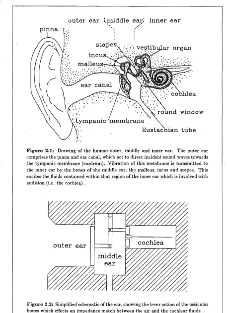
Figures 2.1 and 2.2 show, respectively, a drawing and a schematic of the human ear. The ear is nominally divided into three functional units: outer, middle, and inner ear. The most obvious component of this chain, the outer ear, consists of the pinna, the external auditory meatus (ear canal), and the tympanic membrane (eardrum). The main purposes of the outer ear are the "collection" of sound (to maximize sensitivity) and the improvement of spatial localization by causing the spectral content of the sound entering the ear canal to be a function of source position. In humans, localization performance is further enhanced by the effects of reflections from shoulders and diffraction around the head.
On the other side of the eardrum is the middle ear, an air-filled cavity that contains the ossicular bones; the malleus, incus and stapes. Attached to the middle of the eardrum is one end of the malleus bone. The malleus is connected to the incus which is, in turn, connected to the stapes. The stapes is located in the oval window of the inner ear. The only other opening to the inner ear is the round window, which is covered by a thin, flexible membrane. Through this combination of bones, a vibration of the eardrum (caused by air pressure vibrations in the ear canal) results in piston-like motion of the stapes, and
6
,,-'
(
outer ear
,
.
:'
..
,.
'lmiddle
, ,')
I
I
ear)
,,"
,,"
II
I !
CHAPTER 2, ANATOMY
inner ear
organ
,.
,
~..!.
membrane
Eustac
tube
Figure 2.1: Drawing of the human outer, middle and inner ear. The outer ear comprises the pinna and ear canal, which act to direct incident sound waves towards the tympanic membrane (eardrum). Vibration of this membrane is transmitted to tbe inner ear by the bones of the middle ear; the malleus, incus and stapes. This excites the fluids contained within that region of the inner ear which is involved with audition (i.e. the cochlea) ,
outer ear
0 [image:24.566.37.495.70.701.2]2.2. COCHLEAR ANATOMY 7
hence excites the fluids of the inner ear. The inner ear comprises the vestibular organ (the organ of balance) and the cochlea (the organ of hearing). Although they contain the ,same fluid, the two organs operate quite independently. Only the cochlea is considered
in this thesis.
The area of the stapes footplate is 17 times less than that of the eardrum. This feature, combined with a 1.3-fold ratio in the malleus-incus lever system, effects an impedance transformation between the (low impedance) air and the (high impedance) fluids of the cochlea. The resulting impedance match effects an efficient transfer of energy (50-75 % from 0.3-3 kHz) from sound waves to fluid waves. Sound can also enter the cochlea by bone conduction, but the thresh?ld for this is approximately 80 dB higher than that for
eardrum vibration.
The middle ear protects the cochlea from damage by loud sounds by the contraction of muscles which impedes the transmission of sound from the eardrum to the cochlea. The amount of contraction increases with sound intensity, thereby preventing the ossicles from bouncing out of contact and causing excessive distortion at high levels. The Eustachian tube, connecting the middle ear cavity to the throat, ensures a constant operating position for the eardrum by equalizing the pressure in the middle ear cavity with the environmental pressure. This is achieved by the tube opening (e.g. by swallowing or yawning).
The transmission of sound through the middle ear is frequency dependent. The middle ear acts as a lowpass filter, with a corner frequency of 1-2 kHz. However, this outer ear-to-cochlea transfer function is insignificant compared to the filtering performed by
th~ cochlea, and so the middle ear is ignored in this thesis (except when comparisons are made between cochlear model responses and experimental data; for the latter a reference of eardrum pressure is often used).
2.2
Cochlear Anatomy
Most of the signal analysis performed by the ear occurs within the cochlea. The cochlea acts as a transducer, converting stapes motion into nerve pulses that are sent to the brain.
2.2.1
Cross-sectional structure
A diagram showing a cross-section of the cochlea appears in Fig. 2.3. The cochlea consists of three turns of an approximately circular canal, wound in a very similar manner to that of a snail shell (hence the name, from the Greek word kochlias). When stretched out, the cochlea is 25-35 mm long and has a cross-sectional area of 2 mm2 •
8 CHAPTER 2. ANATOMY
apical turn
turn
Figure 2.3: Diagram of a cross-section of the cochlea, showing its spiral shape embedded in the temporal bone. The stapes is positioned in the basal turn. The acoustic nerve projects down the centre of the cochlea which, together with the vestibular nerve bundle, forms the VIn nerve to the brain.
[image:26.566.44.486.57.535.2]2.2. COCHLEAR ANATOMY
Reissner's
cochlear partition
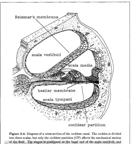
Figure 2.4: Diagram of a cross-section of the cochlear canal. The cochlea is divided
into three scalae, but only the cochlear partition (CP) affects the mechanical motion
~. ~f \~jt d.
l!j)}I~~.Witr~~~ ..•..
·fii
·,ari~~t~iekn~s;·alpngt
e.cocfor a large stiffness gradient in the CP This results in the formation of a transverse travelling wave on the BM, that propagates from base to apex.
9
T.~~~t~~~~t~eia(~~Ma4~a~IQtili~t~fthe
[image:27.566.85.515.78.546.2]10
CHAPTER 2. ANATOMYlamina
pectinate zone
BM
Figure 2.5: Diagram of the CPo The BM is divided into its two zones. The sensory hair cells, pillars of Corti, and Deiters', Hensen's and Claudius' cells constitute the organ of Corti. The hair cells are divided into two distinct groups: inner hair cells (IHCs) and outer hair cells (OHCs). Vertical motion of the arcuate zone results in shear motion in the sub tectorial space, bending the hair cell stereocilia and thereby causing excitation of the hair cells.
an important role in the development of the new cochlear model described in Chapter 5. In particular, the need for a clear differentiation of the CP into its arcuate and pectinate zones can be justified on the bases of anatomical structure and experimental observations.
Pi BM,
e are attached at different points, so that vertical motion of the BM arcuate zone results
ii{
shear motion in the subtectorial space. It is likely that this directly stimulates the sensory hair cells, by the bending of their stereocilia. This is known as the forward transduction process, with information concerning the form of the stimulus (at the BM and hence at the(Deiters" Rensen's and Claudius') cells. The sensory hair cells are divided into two distinct groups: inner hair cells (IRCs) and outer hair cells (ORCs). The IRCs are supported by the inner pillar of Corti. Projections from the top of the outer pillars extend out in a radial direction. These projections, along with the ends of phalangeal processes ~~~~~~~~:~~l~~t~R~:$~retli~~~~~~m~~c~~_~~~~~~~~t~~~~t~_,~opof
[image:28.566.60.491.70.464.2]The most obvious anatomical distinction between the two types of hair cells is that there are three rows of OHCs but only one row of IHCs at each position along the cochlear length. The IHCs are innervated by 90-95% of the afferent nerve fibres (Spoendlin, 1969), and so are largely responsible for the information that is sent to the brain (Le. the forward transduction process). The OHCs, on the other hand, are mainly innervated by efferent nerve fibres (from the brain). The response of the cochlea is altered when the efferent fibres are stimulated electrically (Mountain, 1985). Furthermore, the selectivity and sensitivity of the cochlear response decrease when the condition of the OHCs deteriorates (Khanna and Leonard, 1986). These observations suggest that a reverse transduction process exists within in the cochlea, in which forces generated by the OHCs affect the mechanics of the BM. The OHCs are located in the arcuate zone of the BM (see Fig. 2.5), and so it is assumed that such a reverse transduction process will directly affect the arcuate zone only; the pectinate zone will be affected via its coupling to the arcuate zone provided by the BM fibres and the cochlear fluid.
The tops of the OHC stereocilia are embedded in the underside of the tectorial membrane.
It is uncertain whether or not the IHC stereocilia are embedded (Lim, 1980; Steel, 1983). If they are not, then it is likely that the IHCs would respond to the velocity of the subtectorial fluid motion, and hence to the velocity of the BM arcuate zone. This is supported by experimental measurements (Sellick et al., 1983a - see Section 3.2.3).
In each hair cell the stereocilia are arranged into three distinct rows. In each row the stereocilia are approximately the same height, but the heights are graded between the three . rows. In the IHe the rows run parallel to the longitudinal axis of the cochlea, whereas in the OHCs the rows form a very distinct "V" shape, pointing away from the IHC. The tallest stereocilia of each hair cell are longest for those cells near the apex, and it has been shown that this height gradation along the cochlear length could result in a stereocilia stiffness gradient which closely matches that of the BM (Wright, 1984). Strelioff and Flock (1984) measured a smaller variation of stereocilia stiffness than proposed by Wright. However, allowing for changes in the dimensions of the tectorial membrane and the organ of Corti, they concluded that OHC stereocilia stiffness could significantly influence BM mechanics. Since the actual means by which the OHCs alter BM mechanics is still uncertain (e.g. Zenner et al., 1988), it is merely assumed in this thesis that the presence of the OHCs does indeed alter BM (arcuate) mechanics.
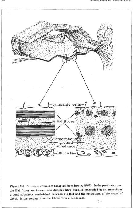
The width and thickness of the BM vary along the cochlear length, both increasing towards the apex. The associated stiffness gradient (from 109 dyn'cm-3 to 105 dyn'cm-3 - these are specific acoustic stiffness parameters (see Section 4.4,1» gives rise to a travelling wave on the BM (see Chapter 3), The structure of the BM is shown in Fig. 2.6. The two cross-sectional diagrams in Fig. 2.6 show that in the pectinate zone the BM consists of three layers: a supporting layer of BM cells, an intracellular layer and the covering epithelium layer of tympanic cells (Iurato, 1962). The cell layers do not affect the mechanical properties of the BM. The intracellular layer, having neither cells nor vessels, consists of an amorphous ground substance in which small proteinaceous fibres (approximately 10 nm in diameter) are embedded. These fibres, which run in a radial direction spanning the full width of the cochlea, determine the mechanical properties of the BM.
along the
c~chlear
length, a number that closely matches the number of hair cell rows.12 CHAPTER 2. ANATOMY
2.2. COCHLEAR ANATOMY
arcuate zone
section
A-A
pectinate zone
lffi>
\W1
lffi>
'lli1
[image:31.566.69.504.57.565.2]section
B-B
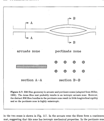
Figure 2.7: BM fibre geometry in arcuate and pectinate zones (adapted from Miller, 1985). The dense fibre mat probably results in an isotropic arcuate zone. However, the distinct BM fibre bundles in the pectinate zone result in little longitudinal rigidity and so the pectinate zone is highly anisotropic.
13
in the two zones is shown in Fig. 2.1. In the arcuate zone the fibres form a continuous mat, suggesting that this zone has isotropic mechanical properties. In the pectinate zone the fibres are grouped into bundles which lie in two distinct layers. The ground substance in which the fibre bundles are embedded has a very low shear modulus and so it provides very little longitudinal coupling between adjacent bundles. There is a significant change in fibre geometry at the transition point between the arcuate and pectinate zones. This geometry change would allow for significantly different motion in the two zones.
2.2.2 Longitudinal structure
14 CHAPTER 2. ANATOMY
pillar cells
arcuate zone
---fBM
pectinate zone
Deiters' cell
phalangeal process
Figure 2.8: Diagram of the organ of Corti's longitudinal structure, showing the
geometric arrangement of the ORCs, the Deiters cells, their phalangeal processes, and the pillars of Corti (adapted from Voldrich, 1983).
2.2. COCHLEAR ANATOMY
15
The accepted amount of anisotropy in the BM has varied over the years, with more re-cent studies having lead to the general acceptance of a highly anisotropic BM. However, the greatly differing structure of the BM in its two zones can be helpful in providing an explanation for the contradictory observations of its mechanical properties. Vpn Bekesy (1960) observed that the BM displayed isotropic elastic properties under light pressures and anisotropic properties under much stronger forces. yoldrich (1978) found that freshly prepared samples exhibited anisotropic properties, whereas physically or chemically dam-aged (or post mortem) preparations were always isotropic. He attributed von Bekesy's
observations to the fact that the earlier experiments were conducted on post mortem
Chapter 3
Response
This chapter describes the mechanical response of the cochlea. The formation of the trav-elling wave is qualitatively described, and the form of the mechanical response measured experimentally is outlined. It is concluded that the response of the BM is probably quite different in its arcuate and pectinate zones.
3.1
Formation of the Travelling Wave
Some important aspects of cochlear anatomy are outlined in Chapter 2. The most im-portant feature of this, in terms of the gross mechanical (macromechanical) response of the cochlea, is that the cochlea is a canal divided lengthwise into two scalae by a flexible partition that possesses a substantial stiffness gradient. For all forms of stapes stimulus, this anatomical feature results in the formation of a travelling wave on the BM.
3.1.1 Historical
The formation of the travelling wave, which travels from the base to the apex of the cochlea, is one of the (few) aspects of cochlear response that is universally accepted today. However, at the time of von Bekesy's (1928) observations there was considerable controversy concerning the form of BM motion. One of the more famous theories was the resonance theory, proposed by Helmholtz (1863). He assumed that the CP was comprised of a series of resonators tuned to different frequencies, with the cochlear fluid acting only to lower the resonant frequency of these resonators. Therefore, when the sound stimulus contained several frequencies, each would produce a separate vibration maximum at a different location on the BM, corresponding to a crude (spatial) Fourier analysis. The problem with this theory was the paradox it generated. For a resonator to be effective in fine frequency analysis, as our pitch perception abilities require, it must have very low damping. However, if the cochlea was equipped with such underdamped resonators we would have difficulty comprehending the rapidly-changing events in music and speech (since the resonators would exhibit prolonged ringing during the transients). The actual
18 CHAPTER 3. RESPONSE
analysis performed by the cochlea is still uncertain, but it has been suggested that the response to sine signals approaches a pure Fourier analysis, the response to transients approaches a pure waveform (time) analysis.
Von Bekesy's observations were initially in a physical model of the cochlea, and later in post mortem (and a few live) specimens. When fine metallic particles were added to the cochlea fluid, they were seen (using a light microscope) to move from base to apex, indicating the presence of a travelling wave. Eddies of fluid motion were also seen in the region of maximum response. The presence of a travelling wave was later confirmed in the cochleae of various animals in a fresh state and in the living guinea pig.
3.1.2 A
qualitative description
A diagram of the unrolled cochlea is shown in Fig. 3.1. It is generally considered that the coiled nature of the cochlea has little affect on its mechanical response (as suggested by the analyses of Viergever (1978a), Loh (1983), and Steele and Zais (1985». Consider a pulse-type stimulus to the cochlea, consisting of a force applied to the stapes tending to cause it to move inwards. This produces a compressional wave that propagates away from the stapes travelling at the speed of sound in the fluid (approximately 1500 m· 8-1 ). For such a compressional wave the BM appears transparent and so the wave invades both scalae. The only other opening to the cochlea is the round window, which normally has the middle ear air pressure keeping it at an equilibrium position. The compressional wave causes an unequal pressure to be exerted on the round window, which tends to make it bulge outwards. The (potential) movement of the round window results in the formation of a pressure differential across the BM.
Conservation of fluid mass dictates that the fluid displacement of the stapes and the round window must equal the integrated volume displacement of the BM. The BM constitutes the membrane with the largest impedance, and hence neither the stapes nor the round window will bulge outwards until the response time of the BM has passed. The response time is defined as the time taken for the BM to begin moving after the application of a force. It can be estimated by considering the BM to be an assemblage of independent masses and springs, each tuned to a frequency determined by the point values of mass and stiffness at that location. The response time of any such spring-mass system is governed by the system's bandwidth (defined as the frequency range over which the system can absorb energy). If the bandwidth is large, this implies that the system is capable of absorbing energy from the stimulus over a wide frequency range and hence will tend to respond to that stimulus more rapidly (and vice versa for a system with a small bandwidth).
PhD
Thesis Revisions11. (page 18, para. 3 - page 20, para. 1) "Author's
explanation" :
10
3.1. FORMATION OF THE TRAVELLING WAVE
basilar membrane
oval
window
( stapes)
~:tJ:J:j~~:r::t:
round
window
helicotrema
Figure 3.1: Diagram of longitudinal section of the cochlea. When a force is applied to the stapes it will not move until the response time of the BM has passed. Instead, a compressional wave invades both scalae (since the BM is transparent for such waves). The unequal pressure on the round window causes it to bulge outwards. Conservation of fluid mass means that the stapes moves inwards and the BM bulges downwards with the same volume displacement. The most basal regipn of the BM deflects first, since it has the largest bandwidth and hence the smallest response time. The BM deflection heralds tJ:te beginning a transverse wave which travels towards the apex. The travelling wave propagates as far as the (lessening) BM bandwidth will allow, at which point the wave reaches a peak, thereafter being rapidly attenuated. The point of resonance is near the base for high frequency stimuli and near the apex for low frequency stimuli.
19
frequency, it effectively stops and the energy contained in the wave is absorbed by the local BM resistance. Points beyond this position receive no excitation from the travelling wave; instead they are excited (at a very low level) by the initial pressure in the scala vestibuli, resulting in the characteristic phase plateau (at a multiple of 1r radians) for all
positions beyond the point of resonance.
20 CHAPTER 3. RESPONSE
fluid moving
longitudinally
envelope of EM motion
Figure 3.2: Variation of fluid wavelength within the c.ochlea (only the scala vestibuli
is shown - the fluid motion is equal and opposite in the scala tympani). For regions far basal of the characteristic place all the fluid participates in the longitudinal flow (the "long-wave" region). Near the characteristic place, however, the wavelength becomes comparable to the scala height and so the amount of fluid moving longitudinally is reduced (the "short-wave" region).
frequency, the position of the peak is termed the "characteristic place" .
3.1.3 Fluid wavelength
For regions far basal of the characteristic place, the wavelength of the travelling wave is much larger than the dimensions of the scalae. Hence the fluid motion can be considered to be essentially one-dimensional in a longitudinal direction. This is known as the "long-wave" region. Nearer the characteristic place the wavelength becomes comparable to the scalae dimensions, and hence the longitudinal movement of fluid is confined to a smaller distance above the BM. This corresponds to "short-wave" fluid behaviour. The transition from long-wave to short-wave region 1s shown graphically in Fig. 3.2.
3.2. MEASURED RESPONSES 21
(owing to the fluid effectively being "squeezed" through a smaller cross-sectional area) and the reduced BM impedance (owing to a reduction in the amount of fluid "riding" with the BM), result in a larger peak at the characteristic place. Both of these features are beneficial in that they maximize the energy transfer from the stapes to the sensory hair cells.
3.2
Measured Responses
This section summarizes the available response data obtained from experimental inves-tigations on animals. The magnitude response data illustrate the remarkable sensitivity and selectivity of the mechanical response of the cochlea, whereas the phase response data confirm the presence of a travelling wave. The changes in the mechanical response, as a result of trauma, are also outlined in this section. The data referred to here are for the response to pure tone stimuli. It should be remembered that the response of the BM exhibits the same degree of selectivity for single frequency and noise stimuli (de Boer, 1973).
3.2.1 Measurement techniques
There are three main techniques used for measuring the very small mechanical movements of the BM (e.g. de Boer, 1980; Khanna, 1986): Mossbauer, Laser Interferometry, and Capacitive Probe. In the Mossbauer technique, a small gamma-ray source containing
52 Co is placed on the BM. An absorber (enriched in 52Fe) is positioned so that the rays
penetrating are detected on a scintillation counter. The rays reaching the counter are minimized when there is no relative motion between the source and the absorber, since the resonance frequencies of the two nuclides are then the same. When the source vibrates, the emitted rays are frequency modulated (owing to the Doppler effect) and therefore undergo varying absorption. The absorption-time functions so obtained are used to calculate the magnitude and phase of the source motion. In the Capacitive Probe technique, a small probe that is part of an electrical resonant circuit is brought into close proximity with the BM. Motion of the BM results in variations of capacitance between the BM and the probe (the scala tympani is drained), and hence causes changes in the circuit's resonant frequency. In Interferometric techniques a small mirror is placed on the BM and is illuminated by a coherent laser beam. Frequency and phase variations of the reflected beam, caused by motion of the mirror, are detected using interferometry. This information is used to determine the BM motion.
22
CHAPTER 3. RESPONSE100~---~
-..
~ ... 4)
"'t1 ~
--
Q2 Q2Q)
e
"'t1 t:lt
::;
I
~
...
d
tll)
([J 4)
:::s
...o,---~
-4~----~--~--~.-~~~----~
1000
20000
Frequency (Hz)
PhD Thesis Revisions
13. (page 23, para .. 2) "Author's explanation":
24 CHAPTER 3. RESPONSE
100,---~
""
\ x'\
f
I
20,---.-.-.-~~.---~--~
3000
30000
Frequency (Hz)
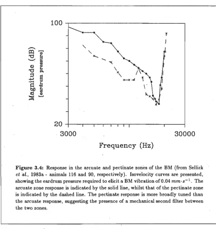
Figure 3.4: Response in the arcuat~ and pectinate zones of the BM (from Sellick
et ·al., 1983a - animals 116 and 90, respectively). Isovelocity curves are presented, showing the eardrum pressure required to elicit a BM vibration of 0.04 mm·s-1 . The arcuate zone response is indicated by the solid line, whilst that of the pectinate zone is indicated by the dashed line. The pectinate response is more broadly tuned than the arcuate response, suggesting the presence of a mechanical second filter between the two zones.
susceptible to trauma, and that it is only when exceptional care is taken to minimize cochlea damage that high selectivity and sensitivity is observed. The data of Sellick et ai.
(1983a,b) and Robles et ai. (1986) further support this hypothesis.
The selective vulnerability of the BM response peak (i.e. the ability to remove it, leaving the remainder of the tuning curve, including high frequency rolloff, essentially intact) suggests that its underlying mechanisms are separate from those governing the formation of the travelling wave. The likely site for this extra, metabolically-sensitive process is in the organ of Corti (see Section 2.2). If this is the case, then anatomical considerations (as outlined in Chapter 2) suggest that the response in the arcuate and pectinate zones should exhibit significant differences. In the response measurements of Sellick et al. (l983a,b),
[image:43.566.57.479.74.521.2]3.2. MEASURED RESPONSES
25
zone compared to that measured with a source on the arcuate zone. The measured neural response resembles more closely the arcuate mechanical response than it does the pectinate response.
Evans and Wilson (1975) made concurrent BM and nerve fibre response measurements. In their data the neural response always had a large peak, whereas the BM response, measured using a capacitive probe, did not. This observation is in conflict with those of other investigators, since the large neural peak implies a cochlea free from trauma, whereas the broad mechanical response implies a traumatized cochlea. This could be a result of one ofthe inherent disadvantages of the Capacitive Probe technique (for a review see Khanna, 1986)~ The spatial resolution of this technique is severely limited by the size of the probe used. The diameter of their capacitive probe was of the same order as the width of the BM at the position of measurement (approximately 0.2 mm). Therefore, they were measuring the average BM velocity. Their results suggest that the mechanical response of the BM is different across its width·, with the average response exhibiting low selectivity while the neural response is determined by a small section of the BM that exhibits a high selectivity. This hypothesis is also supported by the broad tuning curves measured (using the Capacitive Probe technique) by Le Page (1987).
Chapter
4
Models
This chapter presents a brief description of a number of different types of cochlear model, and outlines the contribution they have made to the improvement of our understanding of cochlear mechanics. The formulation of electrical transmission line (ETL) cochlear models is described in detail, since the cochlear model described in Chapter 5 takes the form of an ETL. It is concluded that some of the fundamental assumptions made in the formulation of cochlear models are probably inaccurate ..
4.1
Mathematical and Physical Models
On a microscopic (or micromechanical) scale, the cochlea is a very complicated system with many unusual structural features. In the formulation of cochlear models, assump-tions must be made concerning the relative importance of these features. If the assump-tions made are accurate, the resulting model can provide new insight into the mechanics of the cochlea and can be used as a basis for determining the experimental investigations that would further improve our understanding of cochlear mechanics. A useful cochlear model must satisfy three criteria (Viergever, 1980): it must be the simplest possible, without neglecting structural features that are mechanically important; it must exhibit a realistic response; and, through its analysis, it must be capable of elucidating the impor-tant aspects of cochlear mechanics.
Different modelling techniques allure themselves to investigations of the cochlea. The most obvious distinction to be made is that between mathematical and physical models. The present trend is to formulate mathematical cochlear models and then analyse them analytically or numerically. Analytical investigations have the advantage that the solution method itself can elucidate physical properties of the model. Numerical investigations have the advantage that, in general, fewer assumptions are required in the formulation of the model. Both analysis techniques contribute significantly to our understanding of cochlear mechanics. However, in mathematical analyses there can be a tendency to regard the solution as accurately describing real cochlear operation, by forgetting the gross assumptions that are always required in the formulation of the model.
28 CHAPTER 4. MODELS
Most of what we know "for sure" about the basic operation of the cochlea - for instance, the formation of the travelling wave - is the result of investigations performed on physical hydrodynamic models by von Bekesy (1960). However, since then the (few) such models that have appeared have had serious shortcomings. For example, the model of Cancelli
et ai. (1985) did not exhibit a realistic response, despite the incorporation of many of the
cochlear partition structures as well as allowing for fully three-dimensional (3-D) fluid flow.
Another type of cochlear model, the physical ETL (e.g. Bogert, 1951; Zwicker, 1986) can be considered in the same way as physical hydrodynamic models, in that the response and stimulus appear simultaneously. This represents an important advantage over computer or analytical solutions. However, all such models to date have the disadvantages of a very simple fluid representation and an inability to incorporate mechanical coupling in the CP (techniques for overcoming these shortcomings are presented in Chapter 6).
Assumptions
The need to make assumptions in the formulation of a cochlear model is clear (for a review of the common assumptions made, see Neely (1981) or Viergever (1980, 1986». Those . assumptions which, in the opinion of the author, are considered to be accurate are:
Isolated cavity: The cochlea is considered to communicate (mechanically) with the out-side world only via its two windows. The cochlear fluids have connections to the vestibule and to the endolymphatic and perilymphatic ducts. However, these are assumed to act only as reservoirs of the cochlear fluids and are thought not to have any mechanical effect. Von Bekesy found the volume displacements at the round and oval windows to be approximately equal, suggesting that the flow into the other spaces is indeed negligible.
Ignore spiral coiling: The cochleae of some animals are not tightly coiled and so it is assumed that the coiling in humans and mammals is a space-saving feature. Mathematical analyses of (simplified) models support this view (Viergever, 1978a; Loh, 1983; Steele and Zais, 1985).
Ignore Reissner's membrane: Compared to the CP, Reissner's membrane is very light and flexible. Even if its motion is different from that of the BM, Reissner's mem-brane should not significantly influence cochlear mechanics.
Ignore scalae dimension variations: The dimensionality of the fluid flow affects the mechanical response. However, the wavelength change along the cochlear length far exceeds any scalae cross-section dimension changes and so the latter can be ignored. Gross changes in cochlear dimensions (e.g. a doubling in height) can be simulated by appropriate changes to CP parameters.
4.2. ASSUMPTIONS 29
1980). At a micromechanicallevel fluid viscosity could be important, particularly in influencing the CP resistance parameters (e.g. the narrow subtectorial space dimen-sion could result in a large arcuate resistance). For the analyses in this thesis (in Chapters 5 and 6) the resistance parameters are all quite large, and so the responses should not be significantly altered if fluid viscosity was included in the models. The cochlear partition is assumed to operate linearly near threshold (where the highest degree of selectivity and sensitivity is exhibited); non-linear effects mainly become significant for stimulus levels well above threshold. Only the linear mechanical re-sponse of the cochlea is considered in this thesis.
Stapes: It is assumed that the stapes moves as a piston and is located at the far basal end of the cochlea. In this region the fluid wavelength is much larger than the dimensions of stapes and hence its mode of excitation or its position (both within reasonable limits) should have negligible effect on the fluid waves it generates.
Helicotrema: The small diameter of ,the helicotrema means that it acts as a resistance between the two scalae, and only has an effect for the lowest frequencies. For the frequency range of interest (above 1
kHz)
it can be ignored.The following assumptions are considered to be potentially inaccurate (to the extent indicated):
Fluid compressibility: The fluid is usually assumed to be incompressible, which is definitely valid for low stimulus frequencies. However, compressibility of the fluid could influence wave mechanics for stimuli frequencies at the upper end of the audible spectrum, by the formation of standing waves in the cochlea. For these frequencies the cochlea length is approximately equal to a half wavelength, resulting in the stapes presenting an abnormal impedance to the middle ear thereby affecting measurements of eardrum pressure (Stinson, 1986j' Khanna and Stinson, 1986). This questions the accuracy of selectivity and sensitivity measurements for these frequencies, which unfortunately applies to most of the available response data.
CP geometry: In almost all mathematical models, the CP is assumed to be a flat mem-brane with certain values of impedance across its width. In the simplest models only one impedance is defined at each position along the cochlear length. Even in 3-D fluid models the complicated geometric arrangement of the organ of Corti, the tunnel of Corti, the subtectorial space and the tectorial membrane is totally ignored. This simplification is necessary to ensure mathematical tractability, but unfortunately it is not based on experimental evidence.
30 CHAPTER 4. MODELS
were those of Steele and Taber (e.g Steele, 1974; Taber and Steele, 1979). From their studies they concluded that the additional modes of vibration had little effect on cochlear response. However, this conclusion is only true if the metabolically-sensitive motile process that is known to alter BM mechanics is ignored. It is understandable that Steele and Taber ignored the motile process, as at the time of the models' formulation the existence of such a process was quite uncertain. In Chapter 5 it is shown that the inclusion of a motile process in the arcuate zone (of a model with two modes of vibration for the BM) has a profound effect on the response.
4.2.1 Fluid Dimensionality
The variation of fluid wavelength along the cochlear length results in a minimization of reflections and an increase in the height of the response peak. For a cochlear model to simulate these features, a multi-dimensional fluid model must be incorporated. Current 3-D models allow for the BM spanning only a fraction of the cochlea width, which results in variations in fluid motion across the width of the cochlea. These models simulate "con-traction" of the fluid wavelength in both height and width dimensions but, as mentioned above, they do not allow for the complicated structural features of the CPo
2-D models allow for variations in the fluid motion with height above the BM. Such models also sirriulate the transition-from wave to short-wave behaviour. The long-wave region is as for a 3-D model but now, as the short-long-wave region is reached, the decreasing wavelength contracts the fluid in only one dimension (i.e. height) rather than in two dimensions (Le. in height and width). This reduction in wavelength causes a smaller increase in the series fluid impedance, and a correspondingly less rapid reduction in the amount of fluid "riding" on the BM. The measurable result of this smaller wavelength contraction is a smaller response peak than that exhibited by 3-D models.
1-D models allow for parameter variations only along the cochlear length. This is equiva-lent to assuming that throughout the cochlea the wavelength is large in comparison to the cross-sectional dimensions of the scalae. Such models should be incapable of exhibiting the affects of decreasing fluid wavelength. However, if the rate of phase accumulation can be predicted, then the reduction of reflections and sharper response peak can indeed be simulated (albeit in a rather artificial fashion (Zwislocki, 1983)). For the remainder of this chapter, and all of Chapter 5, only 1-D fluid motion is considered. The affect that this simplification has on the response are shown in Chapter 6.
4.2.2 Degrees of Freedom
4.3. ACTIVE MODELS 31
simulated in a cochlear model with each section of the CP modelled as a second-order system, comprising a series connection of mass, compliance and resistance.
Before 1982 there were a number of models proposed which incorporated (mechanical-to-neural) second filter sharpening mechanisms to account for the huge disparity between the measured mechanical and neural responses (e.g. Duifituis, 1976; Allen, 1977; Zwislocki and Kletsky, 1979; Neely, 1981). These models increased the CP impedance to third or fourth-order, but they were unable to mimic the later measurements showing BM mechanical response very similar to neural response. The added degree ( s) of freedom had little effect on the mechanics of the BM, and so as far as the BM mechanical response was concerned such models were still second-order.
4.3
Active models
The measurement of spontaneous otoacoustic emissions (SOEs) at the eardrum (Kemp, 1979; Zurek, 1981; Martin et ai., 1988) proves the existence of an energy-producing process
somewhere within the cochlea. It has been the trend to use these observations to support the hypothesis of active processes in cochlear tuning. This, however, is an erroneous argument. There is little to suggest that the mechanism responsible for the increase in BM response is directly related to that responsible for the generation of SOEs (a more detailed argument in support ofthis conclusion is presented in Section 7.3.1).
The very high selectivity and sensitivity of the BM response, and the changes in this re-sponse as a result of trauma, are also used as justifications for an active process in cochlear mechanics. In the formulation of all recent cochlear models it has been found necessary to include an active, energy-producing, process in order for the models to simulate a realistic BM response (e.g. Neely and Kim, 1983, 1986; Zwislocki, 1983; Geisler, 1986). A representative active cochlear model example is that of Neely and Kim (1986), which incorporated active processes in the form of pressure sources located at the OHCs. Over a certain narrow band of frequencies the OHCs exerted a pressure on the BM in phase with the (real part of) BM motion, thereby reducing the real part of the BM impedance. This resulted in an enhancement of BM motion for that band offrequencies, resulting in a very realistic mechanical response. The active region had to be basal of the characteristic place in order for the model to exhibit a realistic response. If it was at the characteristic place instability resulted and so, in a more refined form, the model was capable of produc-ing SOEs (Neely, 1988). It is worth noting that the frequency domain response reported by Neely and Kim (1986) has no time domain counterpart, owing to the response being unstable (Diependaal and Viergever, personal c