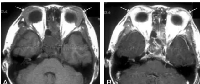
Kimura Disease: CT and MR Imaging Findings
Full text
Figure

Related documents
In order to achieve low carbon and energy conservation for buildings, in this paper, the energy-saving control system of new buildings was studied, combined with multi
Microscopic studies: Results of comparative study regarding leaf, stem, root of the four plants with their transverse microscopic sections have been given in table
Compounds XVg, XVh and Xvi were not active against gram positive bacteria, where as compounds XVo and XVp were not active against gram negative bacteria.Among the
Conclusion : This study showed Mirtazapine versus Citalopram was more effective with faster effect onset on MDD with anxiety symptoms.. Ali
Our ELISA data demonstrated that the expres- sion level of SOCS-3 was remarkably increased in the plasma samples derived from the patients with AMI compared to those from
While there is no sub- stantial change of retinal structure after expo- sure to hypergravity (+10 Gz/3 min), the expres- sion levels of the Müller cell marker GFAP and the
zidovudine based HAART regimen and proposed that the HAART naïve clients have a five. times tendency of developing anaemia than those on zidovudine based
Short vertical lines indicated complementary nucleotides while dark spot indicated uncomplimentary nucle- otides; (B) Relative luciferase activity in MC3T3-E1 cells
