ISSN Online: 2160-889X ISSN Print: 2160-8881
DOI: 10.4236/opj.2018.81001 Jan. 9, 2018 1 Optics and Photonics Journal
Granulocyte Colony—Stimulating Factor
Multiplies Normal Blood ROS Generation
at Less than 1 µg/l
Thomas Stief
Institute of Laboratory Medicine and Pathobiochemistry, University Hospital of Giessen & Marburg, Giessen & Marburg, Germany
Abstract
Background: The neutrophils (PMN) are our main blood cells to combat
fun-gi, bacteria, and fibrin. For normal function, an activated PMN generates a certain concentration of reactive oxygen species (ROS). If the generated blood ROS concentration is too low, then fungi, bacteria or fibrin might threaten the life of the patient, and it could be of great medical interest to stimulate PMN by physiologic drugs. Granulocyte-Colony Stimulating Factor (G-CSF) is a cell hormone that increases the cell number of PMN and that stimulates the individual PMN. The blood ROS generation assay (BRGA) is an innovative physiologic test to monitor the ROS generation of PMN in blood. Here the ROS generating action of G-CSF on normal PMN is quantified. Material andMethods: 40 µl 0 - 10.3 ng/ml (final conc.) G-CSF (in 5% human albumin) in
black Brand® 781608 high quality polystyrene F-microwells was incubated in triplicate with 125 µl Hanks’ balanced salt solution (HBSS; modified without phenol red) and 10 µl normal citrated blood. Immediately (BRGA) or after 60 min (BRGA-60-) 10 µl 5 mM luminol sodium salt in 0.9% NaCl and 10 µl 0 or 36 µg/ml zymosan A in 0.9% NaCl was added. The photons were counted within 0 - 318 min (37˚C) in a photons-multiplying microtiter plate lumino-meter. At about 0.5 t-maxn (0.5 fold the time to normal maximum) theap-prox. SC200 of G-CSF was determined. Results and Discussion: The apap-prox. SC200 of G-CSF on normal blood ROS generation was 0.2 µg/l (=20 IU/ml). In clinical situations where an increased blood ROS generation is pharmaco-logically required, few micrograms of G-CSF could be a sufficient dosage for an adult patient. The BRGA helps to find out the correct stimulating G-CSF dosage for each individual. An enhanced PMN function could favor a better clinical outcome in situations of wanted increase of the innate immunology or in cellular fibrinolysis. G-CSF plasma concentrations of 0.1 - 1 µg/l might fa-vor singlet oxygen generation without immunosuppression or cell fragment- How to cite this paper: Stief, T. (2018)
Granulocyte Colony—Stimulating Factor Multiplies Normal Blood ROS Generation at Less than 1 µg/l. Optics and Photonics Journal, 8, 1-10.
https://doi.org/10.4236/opj.2018.81001
Received: October 24, 2017 Accepted: January 6, 2018 Published: January 9, 2018
Copyright © 2018 by author and Scientific Research Publishing Inc. This work is licensed under the Creative Commons Attribution International License (CC BY 4.0).
http://creativecommons.org/licenses/by/4.0/
DOI:10.4236/opj.2018.81001 2 Optics and Photonics Journal induced thrombin generation.
Keywords
Singlet Oxygen (1ΔO2), Reactive Oxygen Species (ROS), Excited Carbonyl
(R-C = O*), Photon (hν), Phagocytes, Neutrophils (PMN), BRGA, G-CSF
1. Introduction
The main cells of innate immunology are the phagocytes (neutrophils = PMN, monocytes = MØ, dendritic cells = DC). Drugs that enhance the PMN function are of great clinical relevance in many diseases where PMN are needed against the disease [1] [2]. The present study aimed to analyze the drug granulocyte- colony stimulating factor (G-CSF) in the blood ROS generation assay (BRGA)
[3][4], an innovative test for whole blood ROS generation working with lumi-nol-enhanced photons emission primarily by diluted whole blood PMN [5][6] [7], stimulated by typical pathophysiological septic concentrations of the fungal compound zymosan A (ZyA; 1 - 2 µg/ml).
2. Material and Methods
40 µl 0 - 10.3 ng/ml (0 - 974 IU/ml) G-CSF (final conc.) (2nd International WHO Standard, human rDNA derived, protein expressed in E. coli; NIBSC, Potters Bar, UK; article nr. 09/136; 1000 ng G-CSF (containing less than 10 ng LPS [8]), 10 mg arginine, 10 mg phenylalanine, 5 mg trehalose, 2 mg human al-bumin, 0.01% Tween 20® dissolved in 500 µl H2O followed by 500 µl 5% human
albumin (CSL Behring, Marburg, Germany) in black high quality flat bottomed polystyrene microwells (Brand, Wertheim, Germany; article nr. 781608), diluted with 5% human albumin, were incubated in triplicate with 125 µl Hanks’ ba-lanced salt solution (HBSS; modified without phenol red; SAFC Bios-ciences-Sigma, Deisenhofen, Germany; article nr. 55037C-1000 ML) and 10 µl freshest normal blood anticoagulated with 11 mM sodium citrate (within 30 min after withdrawal). Immediately (BRGA) or after 60 min (BRGA-60-) 10 µl 5 mM luminol sodium salt (Sigma, Deisenhofen, Germany) in 0.9% NaCl and 10 µl 0 or 36 µg/ml zymosan A (Sigma) in 0.9% NaCl were added. The photons were counted within 0 - 318 min (37˚C) in a photons-multiplying microtiter plate lumi-nometer (LUmo; anthos, Krefeld, Germany) with an integration time of 0.5 s per well. The intra-assay coefficients of variation were less than 10%. At about 0.5 t-maxn
(0.5 fold the time to normal maximum) the approx. SC200 of G-CSF was determined. HBSS consisted of 185.4 mg/l CaCl2·2H2O, 200 mg/l MgSO4·7H2O, 400 mg/l
KCl, 60 mg/l KH2PO4, 350 mg/l NaHCO3, 8000 mg/l NaCl, 90 mg/l Na2HPO4,
1000 mg/l glucose, pH 7.0 - 7.4. Expressed in molarity, the concentrations of the HBSS components are: 1.3 mM Ca2+, 0.8 mM Mg2+, 5.8 mM K+, 143 mM Na+,
144 mM Cl−, 1.6 mM 2 4
SO −, 0.4 mM 2 4
H PO−, 0.6 mM 2
4
DOI: 10.4236/opj.2018.81001 3 Optics and Photonics Journal 5.6 mM glucose.
3. Results
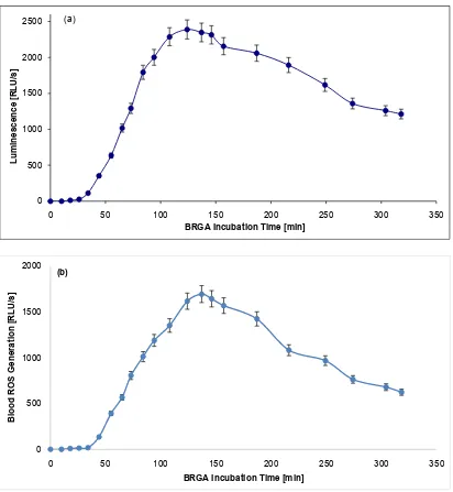
[image:3.595.94.506.188.635.2]In albumin samples, the BRGA maximum of 2389 RLU/s was reached after 124 min. In NaCl samples, the maximum of 1694 RLU/s was reached after 137 min. At 318 min, the blood ROS generation was 51% or 37% of the maximum, re-spectively (Figure 1).
Figure 1. Blood ROS generation in presence of albumin or 0.9% NaCl in BRGA. 40 µl 5% human albumin (Figure 1(a)) or 0.9% NaCl for control (Figure 1(b)) in black Brand® 781608 high quality polystyrene F-microwells were incubated in triplicate with 125 µl Hanks’ balanced salt solution (HBSS; modified without phenol red) and 10 µl normal citrated blood. 10 µl 5 mM luminol so-dium salt in 0.9% NaCl and 10 µl 36 µg/ml zymosan A in 0.9% NaCl were added. The photons were counted within 0 - 318 min (37˚C) in a photons-multiplying microtiter plate luminometer (LUmo). In albumin samples the maximum of 2389 RLU/s was reached after 124 min, in NaCl samples the maximum of 1694 RLU/s was reached after 137 min. At 318 min the blood ROS gen-eration was 51% or 37% of the maximum, respectively. The experiment was repeated twice, the standard deviations were <10%.
0 500 1000 1500 2000 2500
0 50 100 150 200 250 300 350
Lu
mi
nescen
ce [
R
LU
/s]
BRGA Incubation Time [min]
(a)
0 500 1000 1500 2000
0 50 100 150 200 250 300 350
B
lo
od
R
O
S
G
en
er
at
io
n
[R
LU
/s
]
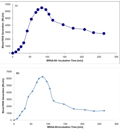
DOI:10.4236/opj.2018.81001 4 Optics and Photonics Journal In albumin samples, the BRGA-60-maximum of 6502 RLU/s was reached after 84 min. In NaCl samples, the maximum of 6254 RLU/s was reached after 84 min, too. At 264 min, the blood ROS generation was 43% or 22% of the maxi-mum, respectively (Figure 2). This means that a protein-poor environment faci-litates the down-regulation of the ROS generation.
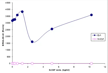
[image:4.595.108.491.201.617.2]In the BRGA, the approx. SC200 was 0.2 ng/ml G-CSF (=20 IU/ml) (Figure 3). In the BRGA-60-, there appeared an approx. IC50 of 2 ng/ml G-CSF. Higher conc. of G-CSF again stimulated the ROS generation (Figure 4). This means that
Figure 2. Blood ROS generation in presence of albumin or 0.9% NaCl in BRGA-60-. 40 µl 5% human albumin (Figure 2(a)) or 0.9% NaCl for control (Figure 2(b)) in black Brand® 781608 high quality polystyrene F-microwells were incubated in triplicate with 125 µl Hanks’ balanced salt solution (HBSS; modified without phenol red) and 10 µl normal citrated blood. After 60 min 10 µl 5 mM luminol sodium salt in 0.9% NaCl and 10 µl 36 µg/ml zymosan A in 0.9% NaCl were added. The photons were counted within 0 - 264 min (37˚C) in a photons-multiplying microtiter plate luminometer (LUmo). In albumin samples the maximum of 6502 RLU/s was reached after 84 min, in NaCl samples the maximum of 6254 RLU/s was reached after 84 min, too. At 264 min the blood ROS generation was 43% or 22% of the maximum, respectively. The experiment was repeated twice, the standard deviations were < 10%.
0 1000 2000 3000 4000 5000 6000 7000
0 50 100 150 200 250 300
B lo od R O S G en er at io n [R LU /s ]
BRGA-60- Incubation Time [min] (a) 0 1000 2000 3000 4000 5000 6000 7000
0 50 100 150 200 250 300
B lo od R O S G en er at io n [R LU /s ]
DOI: 10.4236/opj.2018.81001 5 Optics and Photonics Journal
Figure 3. Approx. SC200 of G-CSF in BRGA. 40 µl 0 - 10.3 ng/ml (final conc.) G-CSF (in 5% human albumin) in black Brand® 781608 high quality polystyrene F-microwells were incubated in triplicate with 125 µl Hanks’ balanced salt solution (HBSS; mod-ified without phenol red) and 10 µl normal citrated blood. 10 µl 5 mM luminol sodium salt in 0.9% NaCl and 10 µl 0 or 36 µg/ml zymosan A in 0.9% NaCl were added. The photons were counted at 44 min (37˚C); approx. SC200 = 0, 2 ng/ml = 20 IU/ml.
Figure 4. Approx. IC50 of G-CSF in BRGA-60-. 40 µl 0 - 10.3 ng/ml (final conc.) G-CSF (in 5% human albumin) in black Brand® 781608 high quality polystyrene F-microwells were incubated for 60 min (37˚C) in triplicate with 125 µl Hanks’ balanced salt solu-tion (HBSS; modified without phenol red) and 10 µl normal citrated blood. 10 µl 5 mM luminol sodium salt in 0.9% NaCl and 10 µl 0 or 36 µg/ml zymosan A in 0.9% NaCl were added. The photons were counted at 42 min (37˚C). There appeared an approx. IC50 of 2 ng/ml G-CSF. Higher conc. of G-CSF again stimulated the ROS generation.
0 500 1000 1500 2000 2500 3000
0 1 2 3 4 5 6 7 8 9 10 11
BRG
A
-44 [
R
LU
/s]
G-CSF conc. [ng/ml]
ZyA no ZyA
0 500 1000 1500 2000 2500 3000 3500 4000 4500
0 2 4 6 8 10 12
BRG
A
-60
-42
[R
LU
/s]
G-CSF conc. [ng/ml]
[image:5.595.111.488.398.652.2]DOI:10.4236/opj.2018.81001 6 Optics and Photonics Journal
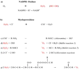
Figure 5. Biogenesis of ROS/photons by activated neutrophils.
within the first incubation time of one hour (37˚C) in the BRGA-60-, G-CSF seems to be inactivated to some extent.
4. Discussion
By contrast, in the BRGA, very low concentrations of G-CSF stimulate blood ROS generation. This could be of pharmacologic interest: in clinical situations where an increased blood ROS generation is pharmacologically required, few micrograms of G-CSF could be a sufficient dosage for an adult patient. The BRGA helps to find out the correct stimulating G-CSF dosage for each individual. An enhanced PMN function could favor a better clinical outcome in situations of wanted increase of the innate immunology or in cellular fibrinolysis [9]-[17].
DOI: 10.4236/opj.2018.81001 7 Optics and Photonics Journal monocytes/dendritic cells the generation of immune suppressive interleukin-10, on endothelial cells the release of von Willebrand factor and F8, on hepatocytes the release of fibrinogen [21]. There could be an enhanced generation of throm-bin/systemically circulating micro-thrombi [14] [22]. Thus, respective blood hemostasis, a G-CSF dosage of about 300 µg seems to be “too much of a good thing”. The present work indicates that a G-CSF plasma concentration around 1 ng/ml (injection of about 3 µg G-CSF,
i.e. 100 fold less than currently used)
might favour the physiologic singlet oxygen generation (Figure 5) against pa-thogens without pathologic thrombin generation or immune suppressive side ef-fects [23]-[47]. The BRGA is a powerful tool to compare new analogues of G-CSF (e.g. the E. coli product filgrastim or the CHO product lenograstim). Dose-finding studies are highly indicated to establish the range of beneficial G-CSF concentrations for each individual patient.References
[1] Stief, T.W. (2008) Neutrophil Granulocytes in Hemostasis. Hemostasis Laboratory, 1, 269-289.
[2] Seguchi, H. and Kobayashi, T. (2002) Study of NADPH Oxidase-Activated Sites in Human Neutrophils. Journal of Electron Microscopy, 51, 87-91.
https://doi.org/10.1093/jmicro/51.2.87
[3] Stief, T. (2013) The Routine Blood ROS Generation Assay (BRGA) Triggered by Typical Septic Concentrations of Zymosan A. Hemostasis Laboratory, 6, 89-98. [4] Stief, T. (2013) Pathophysiologic Routine Blood Tests for the Generation of Reactive
Oxygen Species. Hemostasis Laboratory, 6, 141-153.
[5] Stief, T.W. (2013) Reactive Oxygen Species Generation in Diluted Whole Blood An-ticoagulated by Citrate, EDTA, or Heparin. Hemostasis Laboratory, 6, 155-174. [6] Nathan, C.F. (1987) Neutrophil Activation on Biological Surfaces. Massive
Secre-tion of Hydrogen Peroxide in Response to Products of Macrophages and Lympho-cytes. Journal of Clinical Investigation, 80, 1550-1600.
https://doi.org/10.1172/JCI113241
[7] Goodridge, H.S., Wolf, A.J. and Underhill, D.M. (2009) Beta-Glucan Recognition by the Innate Immune System. Immunological Reviews, 230, 38-50.
https://doi.org/10.1111/j.1600-065X.2009.00793.x
[8] Stief, T. (2013) Quantification of Active LPS in Unknown Matrices by the Oxidative Limulus Test. Hemostasis Laboratory, 6, 343-350.
[9] Stief, T.W. and Fareed, J. (2000) The Antithrombotic Factor Singlet Oxygen/Light (1O
2/hν). Clinical and Applied Thrombosis /Hemostasis, 6, 22-30.
[10] Stief, T.W., Fu, K., Doss, M.O. and Fareed, J. (1999) The Anti-Thrombotic Factor Singlet Oxygen (1O
2) Induces Selective Thrombolysis in vivo by Massive Phagocyte
Infiltration into the Thrombus. XVII Congress of the International Society on Thrombosis and Haemostasis, 14-21 August 1999, Washington DC.
[11] Stief, T.W. (2000) The Blood Fibrinolysis/Deep-Sea Analogy: A Hypothesis on the Cell Signals Singlet Oxygen/Photons as Natural Antithrombotics. Thrombosis Re-search, 99, 1-20. https://doi.org/10.1016/S0049-3848(00)00213-9
DOI:10.4236/opj.2018.81001 8 Optics and Photonics Journal
[13] Stief, T.W. (2004) Regulation of Hemostasis by Singlet-Oxygen (1∆O
2). Current
Vascular Pharmacology, 2, 357-362. https://doi.org/10.2174/1570161043385420 [14] Stief, T. (2013) Micro-Thrombi Stimulate Blood ROS Generation. Hemostasis
Laboratory, 6, 315-325.
[15] Ringelstein, E.B., Thijs, V., Norrving, B., Chamorro, A., Aichner, F., Grond, M., Saver, J., Laage, R., Schneider, A., Rathgeb, F., Vogt, G., Charissé, G., Fiebach, J.B., Schwab, S., Schäbitz, W.R., Kollmar, R., Fisher, M., Brozman, M., Skoloudik, D., Gruber, F., Serena Leal, J., Veltkamp, R., Köhrmann, M. and Berrouschot, J. (2013) AXIS 2 Investigators. Granulocyte Colony-Stimulating Factor in Patients with Acute Ischemic Stroke: Results of the AX200 for Ischemic Stroke Trial. Stroke, 44, 2681-2687. https://doi.org/10.1161/STROKEAHA.113.001531
[16] Lu, F., Nakamura, T., Toyoshima, T., Liu, Y., Shinomiya, A., Hirooka, K., Okabe, N., Miyamoto, O., Tamiya, T., Keep, R.F. and Itano, T. (2014) Neuroprotection of Granulocyte Colony-Stimulating Factor during the Acute Phase of Transient Fore-brain Ischemia in Gerbils. Brain Research, 1548, 49-55.
https://doi.org/10.1016/j.brainres.2013.12.010
[17] Welte, K. (2014) G-CSF: Filgrastim, Lenograstim and Biosimilars. Expert Opinion on Biological Therapy, 14, 983-993.https://doi.org/10.1517/14712598.2014.905537
[18] Kawakami, M., Tsutsumi, H., Kumakawa, T., Abe, H., Hirai, M., Kurosawa, S., Mo-ri, M. and Fukushima, M. (1990) Levels of Serum Granulocyte-Stimulating Factor in Patients with Infections. Blood, 76, 1962-1964.
[19] De Haas, M., Kerst, J.M., van der Schoot, C.E., Calafat, J., Hack, C.E., Nuijens, J.H., Roos, D., van Oers, R.H. and von dem Borne, A.E. (1994) Granulocyte Colo-ny-Stimulating Factor Administration to Healthy Volunteers: Analysis of the Im-mediate Activating Effects on Circulating Neutrophils. Blood, 84, 3885-3894. [20] Pauksen, K., Elfman, L., Ulfgren, A.K. and Venge, P. (1994) Serum Levels of
Gra-nulocyte-Colony Stimulating Factor (G-CSF) in Bacterial and Viral Infections, and in Atypical Pneumonia. British Journal of Haematology, 88, 256-260.
https://doi.org/10.1111/j.1365-2141.1994.tb05015.x
[21] Anderlini, P. and Champlin, R.E. (2008) Biologic and Molecular Effects of Granu-locyte Colony—Stimulating Factor in Healthy Individuals: Recent Findings and Current Challenges. Blood, 111, 1767-1772.
https://doi.org/10.1182/blood-2007-07-097543
[22] Kang, H.J., Kim, H.S., Zhang, S.Y., Park, K.W., Cho, H.J., Koo, B.K., Kim, Y.J., Soo Lee, D., Sohn, D.W., Han, K.S., Oh, B.H., Lee, M.M. and Park, Y.B. (2004) Effects of Intracoronary Infusion of Peripheral Blood Stem-Cells Mobilised with Granulo-cyte-Colony Stimulating Factor on Left Ventricular Systolic Function and Resteno-sis after Coronary Stenting in Myocardial Infarction: The MAGIC Cell Randomised Clinical Trial. The Lancet, 363, 751-756.
https://doi.org/10.1016/S0140-6736(04)15689-4
[23] Subramaniam, R., Barnes, P.F., Fletcher, K., Boggaram, V., Hillberry, Z., Neuenschwander, P. and Shams, H. (2014) Protecting against Post-Influenza Bac-terial Pneumonia by Increasing Phagocyte Recruitment and ROS Production. The Journal of Infectious Diseases, 209, 1827-1836.https://doi.org/10.1093/infdis/jit830
[24] Sun, K. and Metzger, D.W. (2014) Influenza Infection Suppresses NADPH Oxi-dase-Dependent Phagocytic Bacterial Clearance and Enhances Susceptibility to Secondary Methicillin-Resistant Staphylococcus aureus Infection. The Journal of Immunology, 192, 3301-3307.https://doi.org/10.4049/jimmunol.1303049
DOI: 10.4236/opj.2018.81001 9 Optics and Photonics Journal
on Protective Pulmonary Humoral Immunity during Primary Influenza a Virus In-fection. Journal of Virology, 84, 5007-5014.https://doi.org/10.1128/JVI.02408-09
[26] Kanlop, N., Thommasorn, S., Palee, S., Weerateerangkul, P., Suwansirikul, S., Chat-tipakorn, S. and ChatChat-tipakorn, N. (2011) Granulocyte Colony-Stimulating Factor Stabilizes Cardiac Electrophysiology and Decreases Infarct Size during Cardiac Ischaemic/Reperfusion in Swine. Acta Physiologica, 202, 11-20.
https://doi.org/10.1111/j.1748-1716.2011.02259.x
[27] Stief, T. (2013) Neutrophils’ Photons and Opsins (Preface). In: Photonic Hemosta-sis, Physiology of Light Signals in the Neutrophil, Nova Science Publishers, New York, vii-xvii.
[28] Stief, T. (2013) Blood Neutrophils See UV Light: 340 nm Primes ROS Generation Nearly Half as Strong as 405 nm. Hemostasis Laboratory, 6, 389-403.
[29] Fuhler, G.M., Blom, N.R., Coffer, P.J., Drayer, A.L. and Vellenga, E. (2007) The Reduced GM-CSF Priming of ROS Production in Granulocytes from Patients with Myelodysplasia Is Associated with an Impaired Lipid Raft Formation. Journal of Leukocyte Biology, 81, 449-457.https://doi.org/10.1189/jlb.0506311
[30] Olivetta, E., Pietraforte, D., Schiavoni, I., Minetti, M., Frederico, M. and Sanchez, M. (2005) HIV-1 Nef Regulates the Release of Superoxide Anions from Human Macrophages. Biochemical Journal, 390, 591-602.
https://doi.org/10.1042/BJ20042139
[31] Hoggatt, J. and Pelus, L.M. (2014) New G-CSF Agonists for Neutropenia Therapy.
Expert Opinion on Investigational Drugs, 23, 21-35.
https://doi.org/10.1517/13543784.2013.838558
[32] Bath, P.M., Sprigg, N. and England, T. (2013) Colony Stimulating Factors (Includ-ing Erythropoietin, Granulocyte Colony Stimulat(Includ-ing Factor and Analogues) for Stroke. The Cochrane Database of Systematic Reviews, 6, CD005207.
[33] Moazzami, K., Roohi, A. and Moazzami, B. (2013) Granulocyte Colony Stimulating Factor Therapy for Acute Myocardial Infarction. The Cochrane Database of Syste-matic Reviews, 5, CD008844.https://doi.org/10.1002/14651858.CD008844.pub2
[34] Stief, T. (2006) G-CSF Enhances Cellular Fibrinolysis. Clinical and Applied Thrombosis/Hemostasis, 12, 122.https://doi.org/10.1177/107602960601200123
[35] Gong, Y. and Hoover-Plow, J. (2012) The Plasminogen System in Regulating Stem Cell Mobilization. Journal of Biomedicine and Biotechnology, 2012, Article ID: 437920.https://doi.org/10.1155/2012/437920
[36] Kuritzkes, D.R. (2000) Neutropenia, Neutrophil Dysfunction, and Bacterial Infec-tion in Patients with Human Immunodeficiency Virus Disease: The Role of Granu-locyte Colony—Stimulating Factor. Clinical Infectious Diseases, 30, 256-260.
https://doi.org/10.1086/313642
[37] Hartung, T. (1999) Granulocyte Colony—Stimulating Factor: Its Potential Role in Infectious Disease. AIDS, 13, S3-S9.
[38] Hübel, K., Dale, D.C. and Liles, W.C. (2002) Therapeutic Use of Cytokines to Mod-ulate Phagocyte Function for the Treatment of Infectious Diseases: Current Status of Granulocyte Colony-Stimulating Factor, Granulocyte-Macrophage Colo-ny-Stimulating Factor, Macrophage ColoColo-ny-Stimulating Factor, and Interfe-ron-Gamma. The Journal of Infectious Diseases, 185, 1490-1501.
https://doi.org/10.1086/340221
DOI:10.4236/opj.2018.81001 10 Optics and Photonics Journal
E., Savarino, A. and Palamara, A.T. (2013) A Candidate Anti-HIV Reservoir Com-pound, Auranofin, Exerts a Selective “Anti-Memory” Effect by Exploiting the Base-line Oxidative Status of Lymphocytes. Cell Death & Disease, 4, e944.
https://doi.org/10.1038/cddis.2013.473
[40] Stief, T.W., Slenczka, W., Renz, H. and Klenk, H.D. (2001) Singlet Oxygen (1O 2)
Generating Chloramines at Concentrations That Are Tolerable for Normal Hemo-stasis Function Inactivate the Lipid Enveloped Vesicular Stomatitis Virus in Human Blood. 3rd Symposium on the Biology of Endothelial Cells, Pathophysiology of the Endothelium: Vascular and Infectious Diseases, Giessen, 24-26 May 2001, Abstr. D10.
[41] Stief, T.W. (2008) Hemostasis Tolerable Singlet Oxygen—A Perspective in AIDS Therapy. Hemostasis Laboratory, 1, 21-40.
[42] Stief, T.W. (2003) Singlet Oxygen—Oxidizable Lipids in the HIV Membrane, New Targets for AIDS Therapy? Medical Hypotheses, 60, 575-577.
https://doi.org/10.1016/S0306-9877(03)00046-X
[43] Stief, T.W. (2010) Singlet Oxygen and Thrombin Generation: 0.5-1 mM Chloramine as Anti-Viral Therapy. Hemostasis Laboratory, 3, 311-324.
[44] Li, C., Lu, L., Zhang, J., Huang, S., Xing, Y., Zhao, M., Zhou, D., Li, D. and Meng, A. (2015) Granulocyte-Stimulating Factor Exacerbates Hematopoietic Stem Cell Injury after Irradiation. Cell & Bioscience, 5, 65.
https://doi.org/10.1186/s13578-015-0057-3
[45] Carrão, A.C., Chilian, W.M., Yun, J., Kolz, C., Rocic, P., Lehmann, K., van den Wijngaard, J.P., van Horssen, P., Spaan, J.A., Ohanyan, V., Pung, Y.F. and Busch-mann, I. (2009) Stimulation of Coronary Collateral Growth by Granulocyte Stimu-lating Factor: Role of Reactive Oxygen Species. Arteriosclerosis, Thrombosis, and Vascular Biology, 29, 1817-1822.https://doi.org/10.1161/ATVBAHA.109.186445
[46] Stief, T. and Cimpean, C.M. (2013) Singlet Oxygen—Oxidized Human Albumin Stimulates Blood ROS Generation. Hemostasis Laboratory, 6, 423-435.