Re
search Paper
Seed Surface Micro Morphological Features of the Holoparasitic
Angiosperm Aeginetia spp. (Orobanchaceae) in South India
C R VIJAY1, M C THRIVENI2,* and G R SHIVAMURTHY2
1Maharani’s Science College for Women, JLB Road, Mysuru 570 009, India
2Plant Parasite Interaction Laboratory, Department of Studies in Botany, Manasagangotri, University
of Mysore, Mysuru 570 006, India
(Received on 26 May 2016; Revised on 29 June 2016; Accepted on 15 July 2016)
The seed surface features of three Indian species of the genus Aeginetia (Orobanchaceae) was studied with the help of light microscopy and Scanning Electron Microscopy to understand the comparative morphology of the seed coat surface, and its utilization as a source of taxonomic evidence at the species level. The seeds of the three taxa show marked differences in their size, shape and surface ornamentation. Although Aeginetia pedunculata and A. sessilis show resemblances in their morphological and floral characters but differ in their seed coat sculpture. In A. indica the cells of the seed coat are alveolar and look like empty bunch of baskets with ribbed to scalariform thickenings on their lateral walls. In A. pedunculata outer tangential walls are retained but for a central ovoid depression or pore. In A. sessilis, however, the cells of the seed coat surface have a shallow depression. The micropylar end of the seed is smooth surfaced without any ornamentation. The comparative morphology of the seed coat surface in these three species of Aeginetia is discussed and its use as a source of taxonomic evidence is presented.
Keywords: Aeginetia spp.; Orobanchaceae; Root Parasite; SEM Studies; Seed Coat Morphology
*Author for Correspondence: E-mail: thrivenimc@gmail.com
Introduction
Scanning Electron Microscopic studies on the seed coat morphology of Angiosperms have contributed significantly to the systematics of flowering plants. The SEM studies on seed coat surface of parasitic flowering plants are relatively scanty. Chuang and Heckard (1972) described four types of seed coat ornamentation in Cordylanthus. Musselman and Mann (1976) studied the seed surface characteristics of 23 species of Scrophulariaceae and two species of Orobanchaceae. Olsen and Olsen (1980) while studying the seed coat morphology of Boschniakia
hookeri concluded that the seed surface structure
probably helps in the maintenance of early phase of host root/parasite connection. Based on seed coat morphology, Chuang and Heckard (1983) distinguished different species and infra generic groups in Orthocarpus. The Scanning Electron Microscopic study of seed coat morphology in parasitic plants like
Agalinis by Canne (1979), surface features of Striga
seeds by Musselman and Parker (1981), seed coat ornamentation in Striga hermonthica by Jones and Safa (1982) and on Orobanche by Plaza et al. (2004) are said to be of taxonomic significance. Heywood (1969) highlighted the importance of seed characters in detailed taxonomic studies.
The genus Aeginetia L. (Orobanchaceae) comprises 10 Indomalayan species (Airy Shaw 1973). Three species viz., A. indica L., A. pedunculata Wall. and A. sessilis Shiva & Raja are reported from India (Vijay 2007). All are leafless annual herbaceous holoparasitic Angiosperms. In recent years A. sessilis was reported as a new taxon from Kerekatte, Kuduremukha ranges of Western Ghats (Shivamurthy & Rajanna 1994). Morphologically A. indica is clearly distinct from the other two taxa while there are some morphological resemblances between A. pedunculata and A. sessilis. There was no information on the seed Proc Indian Natn Sci Acad 83 No. 1 March 2017 pp. 197-201
coat morphology of these three species at the Scanning Electron Microscope level and hence the present study.
Materials and Methods
Seeds of the three species of Aeginetia were collected from the grass lands and forests near Kerekatte of Kuduremukha ranges of Western Ghats during September-October 2005-2007. Ripe fruits were separated from the plants and shade dried. Seeds were separated manually. For Scanning Electron Microscopic observation, mature seeds were dried to a critical point in a biorad CPD 750 apparatus. Seeds were then mounted on specimen stubs with a double adhesive tape and coated with gold in an Edwards S150B sputter coater. Examination and photomicrography of the seeds were done by using a LEO 435VP Scanning Electron Microscope at 20kV.
Results
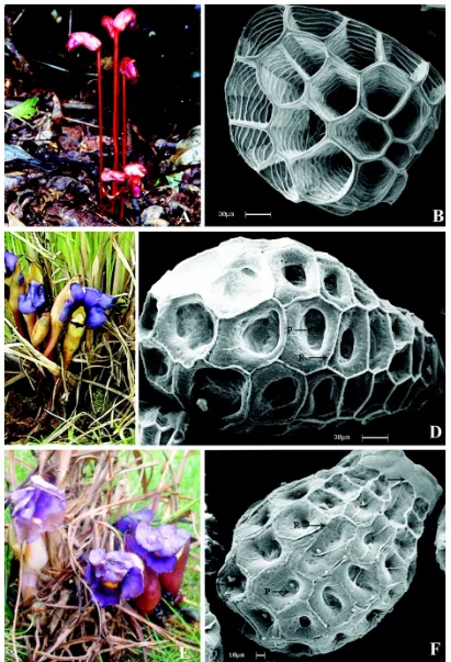
The seeds of A. indica are ovoid (Fig. 1B), yellowish and measure 232- 279µm in length and 155- 187µm in diameter. The integument transforms into the seed coat and their cells are polygonal in outline. The radial and inner tangential walls of these cells gradually undergo thickening due to the deposition of additional cell wall materials to produce rib like anastomosing bands (Fig. 1B). During maturity the outer peripheral (surface) walls and the protoplasmic contents of these cells gradually disintegrate completely and a ‘deep well’ like depression can be seen. The rim of each of these cells is highly thickened due to additional cell wall materials. Under the electron microscope the mature seed coat surface appears reticulate and the whole seed looks like a bunch of polygonal baskets held firmly by the ridges of the polygonal cell walls (Fig. 1B).
In A. pedunculata (Fig. 1C) the seeds are brown and somewhat top shaped with the micropylar end more pointed. The seed size ranges from 274-356µm in length and 198 232µm in diameter. Seed coat surface cells at the micropylar end are smaller and become somewhat narrower at the lower end (Fig. 1D). The surface of the seed coat shows reticulate pattern with the raised ridges. Unlike in A.
indica, the outer tangential walls of each of the cells
of the seed coat are found to be somewhat hard, as a result of which they remain intact in a mature seed except at the centre where it disintegrates to create an ovoid deep perforation. The outer rim of each of the cells is raised and forms distinct hexagonal to pentagonal ridge. All along the rim of the tangential walls minute punctae are noticed (Fig. 1D).
[image:2.612.67.548.627.740.2]The seeds of A. sessilis (Fig. 1F) are obovate, brown to black and measure from 247-298µm in length and 174-206µm in diameter. The cells of the seed coat are polygonal, highly thickened and the seed coat show reticulate pattern on the outer surface. The boundaries of the reticulum are slightly raised. Some of the outer tangential walls on the surface of the seed coat cells are intact where as in others they show a round to oval shaped shallow depression (Fig. 1F). Punctae are scattered on the ridge of the cell walls. At the micropylar end of the seed, there is a smooth surfaced collar without any ornamentation (Table 1).
Discussion
In Aeginetia the seed coat develops from the cells of the integument. Due to the continued deposition of additional cell wall materials, the cells of the seed coat acquire various patterns. In A. indica, the outer surface walls of the seed coat surface collapse
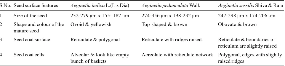
Table 1: Differences in the seed micromorphological features of Aeginetia spp.
S.No. Seed surface features Aeginetia indica L.(L x Dia) Aeginetia pedunculata Wall. Aeginetia sessilis Shiva & Raja
1 Size of the seed 232-279 µm x 155- 187 µm 274-356 µm x 198-232 µm 247-298 µm x 174-206 µm
2 Shape and colour of the Ovoid & yellowish Top shaped & brown Obovate & brown mature seed
3 Seed coat surface Reticulate & polygonal Reticulate with ridges raised Reticulate & boundaries of reticulum are slightly raised
4 Seed coat cells Alveolar & look like empty Aereolate with reticulate network Polygonal, edges with slightly
completely and disappear at maturity of seeds. On the other hand in A. pedunculata the outer surface walls of seed coat are retained but for a central ovoid deep depression or pore, while in A. sessilis the cells of the seed coat surface are provided with a shallow depression.
The seeds of the three taxa of the present study show marked differences in their size, shape and sculpture of the seed coat and this could be used to distinguish one from the other. The study also showed that the seeds of A. pedunculata are larger than A.
sessilis. In A. pedunculata and A. sessilis, the plants
look morphologically similar except for lack of peduncle and just one (rarely up to 3) flower/s in the latter. In A. sessilis the cells of the seed coat on the surface especially the polygonal edges of the reticulum have slightly raised ridges. The depression in the centre of the outer tangential walls of the cells is shallow. In A. indica the cells of the seed coat are alveolar and look like empty bunch of baskets with ribbed to scalariform thickenings on their lateral walls. Such a seed coat has also been observed in
Boschniakia hookeri (Olsen & Olsen 1980) and
called it as alveolar. This alveolar seed coat helps in dispersal by water and also in anchorage of the host roots.
Based on the SEM studies of the seed coat of
Cordylanthus, Chuang and Heckard (1972)
described four types of ornamentations namely irregularly crested, deeply reticulate, shallowly reticulate and irregularly striate. However in A.
pedunculata the surface of the seed coat was found
to be aereolate with reticulate network. Study carried out by Musselman and Parker (1981) in nine species of Striga revealed that all species have aereolate
surfaces with prominent ridges. They concluded that surface features of seeds are of some taxonomic value in certain species complexes. However, Jones and Safa (1982) contradicted this with their studies on Striga hermonthica and stated that ornamentation on seeds from one species were constant but varied within and between populations probably due to out breeding. Rauh et al. (1975) opined that the pitted or chambered seed surfaces are common features in small seeds, which are dispersed by wind, typical of many Scrophulariaceae, Orobanchaceae and a number of other families and this finding which is also noticed in Aeginetia species of the present study.
Micromorphological studies on the seeds of A.
indica by Anuradha and Kumbhojkar (1996) also
showed that seed coat ornamentation is of systematic importance. Since the seeds are relatively stable in external morphology, the Scanning Electron Microscopic study on seed coat morphology provides effective and simple data to distinguish taxa which are morphologically alike.
The present study helped in proving conclusively that A. pedunculata and A. sessilis, even though show many morphological resemblances, are distinctly different taxa based on their their micro morphological features and they also differ from A. indica.
Acknowledgements
The first author is thankful to UGC for the Faculty Improvement Programme. The second author is grateful to the Council of Scientific and Industrial Research (CSIR), New Delhi for Research Associate Fellowship. The authors are grateful to University of Mysore, Mysuru for facilities.
References
Airy Shaw H K (1973) A Dictionary of Flowering plants and Ferns. 8thEdition, Cambridge University Press, Great
Britain
Anuradha S U and Kumbhojkar M S (1996) Micromorphology of Aeginetia indica L. seed in J Bomb Nat Hist Soc 93 318-320
Canne J M (1979) A light and scanning electron microscope study of seed morphology in Agalinis (Scrophulariaceae) and its taxonomic significance in Syst Bot 4 281-296
Chuang T I and Heckard L R (1972) Seed-coat morphology in Cordylanthus (Scrophulariaceae) and its taxonomic significance in Amer J Bot 59 258-265
Chuang T I and Heckard L R (1983) Systematic significance of seed-surface features in Orthocarpus (Scrophulariaceae-subtribe Castillejinae) Amer J Bot 70 877-890
Heywood V H (1969) Scanning electron microscopy in the study of plant materials in Micron 1 1-14
in Ann Bot 50 629-634
Musselman L J and Mann J R (1976) A survey of surface characteristics of seeds of Scrophulariaceae and Orobanchaceae using scanning electron microscopy in Phytomorph 26 370-378
Musselman L J and Parker C (1981) Surface features of Striga seed (Scrophulariaceae) in Adansonia 20 431-437
Olsen S and Olsen I D (1980) Observation on the biology of Boschniakia hookeri (Orobanchaceae) in Bot Tidssk 75 159-172
Plaza L, Fernandez I, Juan R, Pastor J and Pujadas J (2004) Micro morphological studies on seed of Orobanche species
from the Iberian Peninsula and the Balearic Islands and their systematic significance in Ann Bot 4 167-178
Rauh W, Barthlot W and Ehler N (1975) Morphologie und Funktion der Testa staubformiger Flugsamen in Bot Jahrb Syst 96 353-374
Shivamurthy G R and Rajanna L (1994) A new species of Aeginetia L. (Orobanchaceae) from Western Ghats in Rheedea 4 133-135