
Original Article
Transient Down-Regulation of Nucleoside Transporter 3 Gene
Expression as a Drug Target in
Leishmania
major
Using
Antisense RNA Technology
Farideh TOHIDI 1,2, *Bahram KAZEMI 3,4, Mojgan BANDEHPOUR 3,5, Iraj SHARIFI 1,6,
Mohammad Reza RABIEI 7, Ebrahim SAEDI DEZAKI 8, *Zahra BABAEI 1,6
1. Department of Medical Parasitology and Mycology, School of Medicine, Kerman University of Medical Sciences, Kerman, Iran 2. Infectious Diseases Research Center, Golestan University of Medical Sciences, Gorgan, Iran
3. Cellular and Molecular Biology Research Center, Shahid Beheshti University of Medical Sciences, Tehran, Iran
4. Department of Biotechnology, School of Medicine, Shahid Beheshti University of Medical Sciences, Tehran, Iran
5. Department of Biotechnology, School of Advanced Technologies in Medicine, Shahid Beheshti University of Medical Sciences, Tehran, Iran
6. Leishmaniasis Research Center, Kerman University of Medical Sciences, Kerman, Iran
7. Department of Statistics, School of Mathematical Sciences, Shahrood University of Technology, Shahrood, Iran 8. Department of Medical Parasitology and Mycology, School of Medicine, Shahrekord University of Medical Science, Shahrekord, Iran
Received 16 Feb 2018
Accepted 15 May 2018 Abstract Background: This study was aimed to silencing the Nucleoside transporter 3 (NT3) permease nucleobases involved in the salvage pathway of Leishmania in order to disrupt purine nucleotide uptake in the parasite and consequently, destruction of the parasite.
Methods: Overall, 502 bp fragment of the NT3 gene sequence was designed to pro-duce an antisense transcript upon entry of the vector into the parasite. The NT3 con-struct was transfected into L. major promastigotes and NT3 gene expression was stud-ied in vivo and in vitro conditions.
Results: Relative expression NT3 gene in transgenic Leishmania was decreased in tenth day. The percentages and the number of amastigotes infected macrophages with trans-genic L. major were less than infected macrophages with wild-type strain. Our results in two groups of BALB/c female mice (wild-type strain and mutant, n=4 each group) were showed that size and number of ulcers in BALB/c mice infected with transgenic
Leishmania promastigotes were less than the BALB/c mice infected with wild-type
para-sites.
Conclusion: The results indicate the use of antisense RNA reduces of NT3 gene ex-pression in L. major. More studies are required to obtain a new approach for treating
Leishmania infection.
Keywords:
Nucleoside transporter 3 gene;
Leishmania major; Antisense RNA; Drug target; Gene expression
*Correspondence
Email:
Iranian Society of Parasitology http://isp.tums.ac.ir
Iran J Parasitol
Open access Journal at http://ijpa.tums.ac.ir Tehran University of Medical
Introduction
eishmaniasis is a disease with a broad range of clinical manifestations caused by several species of the ge-nus Leishmania, in the family Trypanosomatidae (1, 2). It is one of the most important parasit-ic diseases in 98 countries and manifests in cutaneous, visceral and cutaneous-mucosal forms (3). The life cycle of these parasites has two stages: amastigote that resides inside the reticuloendothelial cells of the vertebrate host and a slender, promastigote that replicates in medium and in the gut of the sandfly as a bio-logical vector (1). Like all protozoa,
Leishma-nia spp. are unable to synthesize cyclic purines de novo and must import purines from both
their vertebrate and insect hosts (4, 5). In all organisms, purines are responsible for many vital functions, like the synthesis of DNA and RNA as well as precursors of coenzyme A, ATP, GTP, NADH, carbohydrates, lipids and the second messenger cAMP (6, 7). All para-site purine transporters are members of the Equilibrative Nucleoside Transporter SLC29 family in the human genome database (8, 9) or the 2.A57 family in the transporter classifi-cation database (10). Nucleoside transporters are present in mitochondria, cell membranes and lysosomes. Leishmania absorbs purines via purine permease through food and salvage. The first step of salvage is the displacement of purine nucleosides and nucleobases in the plasma membrane of the parasite. These hy-drophilic nucleosides and nucleobases are un-able to be released in the parasitic plasma membrane lipid layers, and proteins or trans-porters are needed to move them. The purine transporters are pharmacologically critical, as well as being important in human pathogenesis because they act as mediators in purine analogue uptake. Non-purine drugs such as allopurinol, which is an
anti-Leishmania drug, are toxic to parasites when
they enter into these organisms via purine transporters (4, 6, 7, 11, 12).
The Leishmania genome has four identified sites for purine transfer, known as nucleoside transporters 1–4 (NT1, NT2, NT3 and NT4) (6). Leishmania major expresses two purine nu-cleobase transporters, nucleoside transporters 3 and 4 (NT3 and NT4). Functional analysis of cloned transporter genes has revealed that purine nucleobases, including adenine, gua-nine, hypoxanthine and xanthine, are the spe-cific targets of NT3 (6, 13, 14). NT3 per-mease expresses a large quantity of mRNA during the growth of parasites and has a high affinity for purine nucleobases. Although NT3 permease nucleobases are not essential for the parasite, NT3 gene deletion may re-duce parasite viability and lead to the death of the parasite. These events underline the es-sential role of nucleobase transporters of
Leishmania in purine uptake in both vertebrate
and invertebrate hosts (14).
For this reason, this research was carried out to knocking-down the NT3 permease nu-cleobases that are involved in the salvage pathway of Leishmania in order to disrupt pu-rine nucleotide uptake in the parasite, result-ing in the death of the parasite.
Materials and Methods
NT3 plasmid construct
The 502 bp sequence (5′ end) of NT3 gene (Accession Number: XM_ 001681813) was considered as antisense. This antisense was synthesized by Generay Technology, Shanghai Co, China. Gene was subcloned into pcDNA 3.1 Neomycin vector by Hind III and EcoRI restriction enzymes. E. coli strain TOP 10 was selected and recombinant plasmid was trans-formed into it. Subsequently, plasmid was ex-tracted and confirmed by Hind III and EcoRI restriction analysis. Presence of NT3 gene was confirmed by PCR method with following primers:
F: 5΄- GGCGTACCTCTACTTCAGCC-3΄, R: 5΄- TTGATGGCACGGTACTCACC-3΄.
Culture of Leishmania major
L. major promastigotes (MRHO/IR/75/ER)
were grown in complete DMEM culture me-dium containing 10% inactivated foetal bovine serum and 100U/ml penicillin and 100 μg/ml streptomycin and incubated at 25 °C. After 24–48 h, the cells were used for electro-poration (15).
Transfection
L. major promastigotes at stationary phase
with concentration of 1 × 10⁶/ml were washed in PBS buffer and resuspended in electroporation buffer (20mM HEPES, pH 7.2, 137mM NaCl, 5 mM KCl, 0.7 mM Na2HPO4, and 6mM Glucose). Overall, 350 μl promastigotes were transferred to a 0.2 cm electroporation cuvette (Eppendorf, Germa-ny) containing 2 ng/μl plasmid DNA harbouring the antisense NT3 gene. Electro-poration was performed at 1000 V with a ca-pacitance of 160 μs. Transfected cells were incubated on ice for 10 min, then transferred to 2 ml complete DMEM medium without antibiotics, and finally incubated at 25 °C for 20-24 h. On the second day, after centrifuga-tion of cell cultures and removal of the super-natant, the precipitate was dissolved in the suspension obtained and transferred into 1cc complete DMEM medium containing 50 μg/ml G418 antibiotics (Sigma, USA) and in-cubated at 25 ºC. This culture medium was checked every day for cell number, morpholo-gy and motility under a microscope (15-17). Wild-type L. major as positive control was electroporated.
Quantitative real time-RT PCR
Total RNA was extracted from the mutant and wild-type promastigotes on 3, 7, 10, 15 and 20 d after transfection by total RNA Puri-fication kit (Jena Bioscience, Germany). The cDNA synthesized was performed using Ac-cuPower RT PreMix (Bioneer, Korea). The
rRNA 45 gene of L. major was used as a housekeeping gene (18). The following PCR cycling conditions: denaturation 55 °C for 30 sec; amplification for 12 cycles of 20 °C (30 sec), 42 °C (4 min) and 95 °C (5 min). Real-time PCR was performed with 1 μl of cDNA, 2 μl of 5 pmol primers and 5 μl 2 × Greenstar qPCR Master Mix (Bioneer, Korea) and 2 μl of deionized water in a final volume of 10 μl. The real-time PCR program in the Rotor gene 6000 (Qiagen, USA) was as follows: 94 °C for 2 min and 94 °C for 20 sec; 40 cycles of 53 °C for 20 sec and 72 °C for 30 sec. Relative quan-tity (RQ) values were calculated using the 2 – ΔΔCt method.
Western blot analysis
Pellets of mutant and wild-type Leishmania promastigotes (after transfection) were col-lected by centrifugation on day 3, 7, 10, 15 and 20; proteins were extracted into lysis buffer and separated by SDS-PAGE in 10% acryla-mide gel (19). The protein bands were trans-ferred to a nitrocellulose membrane. NT3-specific sheep antibody was used as a primary antibody with titer of 1:1000 (Genscript, USA). Rabbit anti-sheep IgG antibody conjugated with alkaline phosphatase (as a 1:10000 dilu-tion) was used for the detection of NT3 pro-tein.
Macrophage infection assay
percent-age of infected macrophpercent-ages and the mean number of amastigotes per infected macro-phages in one hundred macromacro-phages were cal-culated. The number of amastigotes in phages and the percentage of infected macro-phages were counted.
Infectivity in BALB/c mice
Two groups (n = 4) of BALB/c female mice (4–6 wk of age), 20-25 gr and were used. Mice of one group were infected with wild-type and another group were infected with mutant promastigotes. Mice for macroscopic exami-nation with respect to ulcer(s) incubation pe-riod; count and diameter form each group was studied. Wild-type and transgenic pro-mastigotes a concentration of 1.5 × 10⁸ (21) were injected subcutaneously into the base of the tails of mice in a 0.1 ml volume (22). The mice were examined macroscopically for 4 wk after infection.
Ethics Statements
The Ethics Committee on Laboratory Ani-mals at Kerman University of Medical Scienc-es (KMU) approved the study protocols of this project (Code of Number ethics: 92/78, 2013). This research was performed in com-pliance with the recommendations and guide-lines for the use and surveillance of laboratory animals of International Institute.
Neverthe-less, all efforts were made to minimize animal suffering.
Statistical analysis
Data analysis was performed using SPSS version 22.0 (SPSS Inc., Chicago, IL, USA) and ANOVA was used for statistically signifi-cant differences (P<0.05).
Results
NT3 fragment antisense was cloned in pcDNA 3.1 Neomycin+ vector. By using NT3-specific primers, PCR was performed and the 500-bp cloned fragment was isolated.
NT3 gene expression
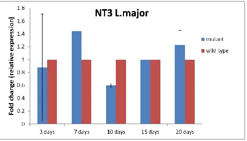
Real time-RT PCR results analysis on days 3, 7, 10, 15 and 20 after transfection revealed that NT3 gene expression in transgenic
Leish-mania was reduced on the tenth day, but it had
upward trend up to the twentieth day. There was a significant difference in increased ex-pression of this gene on day 7 and reduction of its expression on tenth day in mutant para-site (P<0.05) (Fig. 1).
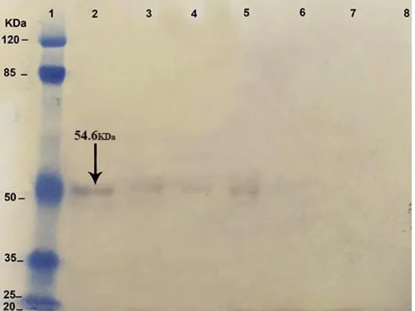
NT3 protein identity by Western blotting NT3 gene in transgenic Leishmania was pressed on 20, 15, and 10 d (Fig 2) and its ex-pression was too low (Fig. 3).
Fig. 2: SDS-PAGE of NT3 gene expression in transgenic Leishmania on 3, 7, 10, 15 and 20 days after transfec-tion. Wild-type as positive control (lane1), mutant strain in 20th (lane2), 15th (lane3), 10th (lane4), 7th (lane5), 3th (lane6)
days after electroporation
Fig. 3: Western blot analysis of NT3 gene expression in transgenic Leishmania on 3, 7, 10, 15 and 20 d after
transfection. This result confirmed NT3 protein expression in the previous test. Total protein extracted from 1×10⁶ cells of wild-type promastigotes and transgenic promastigotes were electrophoresed and subjected to western blot analysis with anti NT3 Ab. NT3 protein is 54.6 KDa. Protein Marker (lane1), wild-type as posi-tive control (lane2), mutant strain in 20th (lane3), 15th (lane4), 10th (lane5), 7th (lane6), 3th (lane7) d after elec-troporation and NT3 construct as negative control (lane8). The result of this experiment shows that NT3 gene
Macrophages infection
The percentages of infected macrophages in contact with the mutant L. major and wild-type strain were evaluated after 24, 48 and 72 h. The infected macrophages with the mutant parasites at the three-time points were lower than that of the wild-type strain. The statistical analysis showed a significant difference (P<0.05) between percentages of the infected macrophages in the mutant L. major and the wild-type strains (Fig. 4).
The number of amastigotes infected macro-phages with transgenic L. major was lower than that of the wild-type strain. The statistical analysis showed a significant difference (P<0.05) between the number of amastigotes infected macrophages in the mutant L. major and the wild-type strains (Fig. 5). In the light microscopy analysis, no difference was found in the shape and size (by eyepiece micrometer) of amastigotes in the mutant parasite and wild-type strain.
Fig. 4: Percent of infective macrophage in contact with the mutant type with NT3 Leishmania major and wild
type. The percentages of infected macrophages in the mutant strains was 15 ± 7.07% (n=2), 27.5± 3.5% (n=2) and 32.5 ± 3.5% (n=2) at 24, 48 and 72 h, respectively. In contrast, the percentage of infected macro-phages in the wild-type was 62.5± 3.5% (n=2), 75 ± 7.07% (n=2, and 85 ± 7.07% (n=2) at 24, 48 and 72 h,
respectively
Fig. 5: The number of amastigotes in macrophages infected with transgenic Leishmania major. The number of amastigotes in macrophages infected with transgenic L. major was 17.5±3.5 (n=2), 25±7.07 (n=2), and 32.5±3.5 (n=2) in 24, 48 and 72 h, respectively; on the other hand, the number of amastigotes in the wild-type
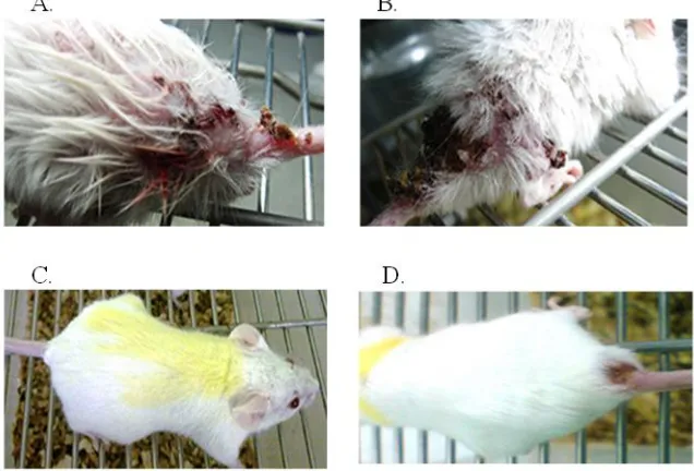
The lesions in BALB/c mice, created within 30 d in the mice infected with the wild type (Fig. 6 A) but in the mice infected with mutant parasites any lesions were not created within 30 d (Fig. 6 B). It was created within 70 d in the mice infected with mutant parasites (Fig. 6
C,D within 70 d in the mice infected with the wild type). More than one ulcer with a diame-ter greadiame-ter than 5 mm were found in the mice receiving the wild type, while one ulcer with a diameter of less than 5 mm was observed in the mice infected with the mutant strain.
Fig. 6: Compare BALB/c mice infected lesions in wild type and transgenic parasites. The lesions were created
within 30 d in the mice injected with wild type (B) and were not created within 30 d in the mice injected with mutant parasites (A). It was created during 70 d in the mice infected with mutant parasites (C, D lesions70 d in the mice infected with wild type). Many lesions were seen with a diameter of more than 5 mm in the mice infected with wild type. While, one lesion with a diameter of less than 5 mm was observed in the mice infected
with the NT3 mutant strain
Discussion
In L. major, NT3 gene is the essential trans-porter for purine nucleobases and it has many roles in the biology of L. major (14). In this study, antisense RNA was used to down-regulating NT3 gene expression. Antisense RNA usually creates a barrier to the transmis-sion of genetic information from RNA to pro-teins (23). In this research, expression of this gene was evaluated after transfection with mu-tant Leishmania in vitro and in vivo.
Antisense has been effective on the expres-sion of this gene since the tenth day. Although on the seventh day, there was a significant in-crease in the expression of the gene (P<0.05),
on the tenth day, the antisense had a signifi-cant decreasing on the expression of the gene (P<0.05). Since the proliferation of pro-mastigotes is performed on every 7 h, the anti-sense effect is temporary so, parasites lose plasmid and normally express this gene. Con-sequently, the gene expression from the tenth day shows an increasing trend.
of L. major promastigotes. There is the tion of the infected macrophages and reduc-tion in the number of amastigotes inside the infected macrophages with mutant Leishmania. On the other hand, in the infected macro-phages with mutant Leishmania, there is re-duced the ability of proliferation of parasites or virulence of parasites. Macrophages stimu-late the cellular response and eventually elimi-nate amastigotes inside the macrophages (24, 25). In this study, size and number of ulcers in BALB/c mice were investigated in vivo. Size and number of ulcers in BALB/c mice infect-ed with transgenic Leishmania promastigotes were less than the BALB/c mice infected with wild-type parasites. However, the incubation period of ulcers in BALB/c mice infected with transgenic Leishmania promastigotes was longer than the BALB/c mice infected with wild-type parasites. In this case, our results are agreement with the research of Mottram (26). Our results showed the reduction of the ex-pression the gene and the virulence of para-sites may be caused by antisense RNA.
Conclusion
We were able to temporarily downregulate gene expression of NT3. Further studies are required to provide new targets for interven-tional therapy in Leishmania infection.
Acknowledgements
This article was extracted from the Ph.D. thesis by Farideh Tohidi. The authors would like to thank the Iran National Science, Tech-nology Fund Deputy of Presidency (Code: 92000589) and Deputy of Research and Tech-nology, Kerman University of Medical Scienc-es, Kerman, Iran for financial supports. We also appreciate the contribution of the Cellular and Molecular Biology Research Center of Shahid Beheshti University of Medical Scienc-es, Tehran, Iran.
Conflict of interest
The authors have declared no conflict of in-terest.
References
1. Alvar J, Ve´lezI D, Bern C, Herrero M, Desjeux PH, Cano J, Jannin J, Boer M. The WHO Leishmaniasis control team, Leishmani-asis worldwide and global estimates of its inci-dence. PLOS ONE. 2012; 7 (5), e35671. 2. Herwaldt BL. Leishmaniasis. Lancet. 1999; 354:
1191–1199.
3. Murray HW, Berman JD, Davies CR, Saravia NG. Advances in Leishmaniasis. Lancet. 2005; 366(9496):1561-1577.
4. Carter NS, Rager N, Ullman B. Purine and pyrimidine transport and metabolism. In: Mo-lecular and Medical Parasitology. Marr J.J, Niel-sen T, Komuniecki R. (eds). London, Academ-ic Press. 2003. p. 197–223.
5. Berens R L, Krug E C, Marr J J. In: Purine and Pyrimidine Metabolism. Marr J J and Muller M (eds), Academic, New York, USA. 1995. p. 89– 117.
6. Landfear SM, Ullman B, Carter N, Sanchez MA. Nucleoside and Nucleobase Transporters in parasitic protozoa. Eukaryot Cell. 2004: 245– 254.
7. Martinez S, Marr JJ. Allopurinol in the treat-ment of American cutaneous leishmaniasis. N Engl J Med.1992.326: 741–744.
8. Hediger MA. Bioparadigms. 2004. http://www.bioparadigms.org
9. Stein A, Vaseduvan G, Carter NS, Ullman B, Landfear SM, Kavanaugh MP. Equilibrative nucleoside transporter family members from Leishmania donovani are electrogenic proton symporters. J Biol Chem. 2003. 278(37):35127-34.
10. TCDB. Transporters classification database. 2007.
https://academic.oup.com/nar/article/44/D1 /D372/2502612
12. Nelson DJ, Bugge CJ, Elion GB, Berens RL, Marr JJ. Metabolism of pyrazolo (3, 4-d) Py-rimidines in leishmania braziliensis and leishmania donovani. Allopurinol, Oxipurinol and 4-aminopyrazolo (3, 4-d) pyrimidine. J Biol Chem. 1979.254:3959-3964.
13. Iovannisci DM, Kaur K, Young L, Ullman B. Genetic analysis of nucleoside transport in Leishmania donovani. Mol Cell Biol. 1984. 4: 1013–1019.
14. Ortiz D, Sanchez MA, Pierce S et al. Molecular genetic analysis of purine nucleobase transport in Leishmania major. Mol Microbio. 2007. 64(5):1228–1243.
15. Beverley SM, Clayton CE. Transfection of Leishmania and Trypanosoma brucei by electro-poration. Methods Mol Biol. 1993. 21:333-48. 16. Potter H, Heller R. Transfection by
electro-poration. Curr Protoc Mol Biol. 2003. U–9.3. 17. Liang X, Potter J, Kumar S et al. Rapid and
highly efficient mammalian cell engineering via
Cas9 protein transfection. J Biotechnol. 2015. 208: 44-53.
18. Ouakad M, Bahi-Jaber N, Chenik M, Dellagi K, Louzir H. Selection of endogenous reference genes for gene expression analysis in Leishmania major developmental stages. Parasitol Res. 2007. 101(2): 473-7.
19. Sambrook J, Fritsch E, Maniatis T. Molecular Cloning: A Laboratory Manual. Cold Spring
Harbor, Cold Spring Harbor Laboratory 1, New York, N.Y., USA. 1989.
20. Liu W, Boitz JM, Galazka J, Arendt CS, Carter NS, Ullman B. Functional characterization of nucleoside transporter gene repiacements in Leishmania donovani. Mol Biochem Parasitol. 2006. 150(2): 300-307.
21. Zhang WW, Matlashewski G. Loss of viru-lence in Leishmania donovani deficient in an amastigote-specific protein, A2. Proc Natl Acad Sci USA. 1997. 94: 8807–8811.
22. Nicolas L, Prina E, Lang T, Milon G. Real-Time PCR for detection and quantitation of Leishmania in mouse tissues. J Clin Microbiol. 2002. 40(5), 1666–1669.
23. Dias N, Stein CA. Antisense oligonucleotides: Basic concepts and mechanisms. Mol Cancer Ther. 2002. 1: 347-355.
24. Mallinson DJ, Coombs GH. Interaction of Leishmania metacyclics with macrophages. Int J Parasitol. 1989. 19: 647–656.
25. Van Assche T, Deschacht M, da Luz RA, Maes L, Cos P. Leishmania-macrophage interactions: insights into the redox biology. Free Radic Biol Med. 2011.51:337–351.
26. Mottram JC, Souza AE, Hutchison JE, Carter R, Frame MJ, Coombs GH. Evidence from dis-ruption of the lmcpb gene array of Leishmania mexicana that cysteine proteinases are virulence factors. Proc Natl Acad Sci USA. 1996. 93(12): 6008-13.

![The EC common policy in science and technology, and the US/EC R&D cooperation. Statement by Gunter Schuster at the EC/US High-Level Consulations. [Washington DC], 25 May 1978](data:image/gif;base64,R0lGODlhAQABAIAAAP///wAAACH5BAEAAAAALAAAAAABAAEAAAICRAEAOw==)