R E S E A R C H
Open Access
Clinical implications of high NQO1 expression in
breast cancers
Yang Yang
1†, Yan Zhang
2†, Qunying Wu
3, Xuelian Cui
1, Zhenhua Lin
1, Shuangping Liu
1*and Liyan Chen
1,4*Abstract
Background:NAD (P) H: quinone oxidoreductase 1 (NQO1) is a xenobiotic metabolizing enzyme that detoxifies chemical stressors and antioxidants, providing cytoprotection in normal tissues. However, high-level expression of NQO1 has been correlated with numerous human malignancies, suggesting a role in carcinogenesis and tumor progression. This study aimed to explore the clinicopathological significance of NQO1 and as a prognostic determinant in breast cancer.
Methods:A total of 176 breast cancer patients with strict follow-up, 45 ductal carcinomain situ(DCIS), 22 hyperplasia and 52 adjacent non-tumor breast tissues were selected for immunohistochemical staining of NQO1 protein. Immunofluorescence staining was also performed to detect the subcellular localization of NQO1 protein in MCF-7 breast cancer cells. Eight fresh breast cancers paired with adjacent non-tumor tissues were quantified using real time RT-PCR (qRT-PCR) and western blot. The correlations between NQO1 overexpression and the clinical features of breast cancer were evaluated using chi-square test and Fisher’s exact tests. The survival rate
was calculated using the Kaplan–Meier method, and the relationship between prognostic factors and patient
survival was also analyzed by the Cox proportional hazards models.
Results:NQO1 protein showed a mainly cytoplasmic staining pattern in breast cancer. The strongly positive rate of NQO1 protein was 61.9% (109/176) in breast cancer, and was significantly higher than in DCIS (31.1%, 14/45), hyperplasia tissues (13.6%, 3/22) and adjacent non-tumor tissues (13.5%, 7/52). High-level expression of NQO1 protein was correlated with late clinical stage, poor differentiation, lymph node metastasis, Her2 expression and disease-free and 10-year overall survival rates in breast cancer. Moreover, multivariate analysis suggested that NQO1 emerged as a significant independent prognostic factor along with clinical stage and Her2 expression status in patients with breast cancer.
Conclusions:High-level expression of NQO1 appears to be associated with breast cancer progression, and may be a potential biomarker for poor prognostic evaluation of breast cancers.
Keywords:Breast cancer, NQO1, Immunohistochemistry, Prognosis
Background
Breast cancer is one of the most common malignancies in women worldwide and the second leading cause of cancer death among women [1,2]. Studies over the past several decades have found that the expression profiles of estrogen receptor (ER), progesterone receptor (PR)
and human epidermal growth factor receptor 2 (Her2)/ neu are closely related with breast cancer, and have been used for predicting the outcome and response to breast cancer therapy [3,4]. Although numerous advances in prevention, surgical resection, and adjuvant chemoradio-therapy have led to a decline in the overall mortality due to breast cancer, the survival rates for patients with metastatic disease have not significantly improved [5,6]. Consequently, the discovery of novel biomarkers in-volved in the diagnosis and progression of breast cancer is of great value.
* Correspondence:liushp@ybu.edu.cn;lychen@ybu.edu.cn
†Equal contributors
1Department of Pathology & Cancer Research Center, Yanbian University Medical College, Yanji 133002, China
4Department of Biochemistry, Yanbian University Medical College, Yanji 133002, China
Full list of author information is available at the end of the article
NAD (P) H: quinone oxidoreductase 1 (NQO1), also known as DT-diaphorase, menadione reductase, or quin-one reductase 1, is a cytoplasmic flavoenzyme encoded by a gene located on chromosome 16q22. NQO1 uses NADH or NADPH as substrates to directly reduce qui-nones to hydroquiqui-nones [7,8]. Functions of NQO1 in-clude xenobiotic detoxification, superoxide scavenging and the maintenance of endogenous antioxidant vita-mins [9]. It is conceivable that NQO1 plays an important role in protecting normal cells against oxidative injury and carcinogenesis. Paradoxically, despite the cellular functions of this “cell protector”, the antioxidant role of NQO1 was suggested by evidence that the disruption of the NQO1 gene or genetic polymorphism increased the risk of chemical-induced toxicity and cancers [10,11]. NQO1 has been found to be expressed at high levels in many human tumors, including breast cancer, melan-oma, lung cancer, cholangiocarcinoma and pancreatic cancer [12-15]. In addition, the high level of NQO1 ex-pression in solid tumors in combination with the ability to reduce many quinine-containing antitumor drugs has dawn attention to NQO1 as a potential molecular target in cancer treatment [16,17]. However, the clinical signifi-cance of NQO1 expression status in breast signifi-cancer re-mains unclear.
In this study, we demonstrated the clinicopathological significance of NQO1 through prognostic evaluation of NQO1 overexpression in breast cancers. The results re-vealed that NQO1 protein is frequently upregulated in breast cancers compared with hyperplasia and adjacent non-tumor breast tissues. These findings indicate that NQO1 may be a good independent predictor of progno-sis for patients with breast cancer.
Materials and methods
Ethics statement
This research complied with the Helsinki Declaration and was approved by the Human Ethics Committee and the Research Ethics Committee of Yanbian University Medical College. Patients were informed that the resected speci-mens were stored by the hospital and potentially used for scientific research, and that their privacy would be main-tained. Follow-up survival data were collected retrospect-ively through medical record analyses.
Clinical samples
Eight fresh breast cancers paired with adjacent non-tumor tissues were snapfrozen in liquid nitrogen and
stored at −80°C until use. The histopathology of each
specimen was reviewed on the hematoxylin and eosin-stained tissue section to confirm diagnosis and tumor content at least 70% of tumor cells in the tissue sample. The study of 176 paraffin embedded breast cancer
sam-ples, as well as 45 ductal carcinoma in situ (DCIS)
samples, 22 hyperplasis and 52 adjacent non-tumor tis-sues were also conducted. These samples were selected randomly from patients who underwent surgery between 2002 and 2009, with strict follow-up for survival status. Clinicopathological classification and staging were deter-mined according to the American Joint Committee on Cancer (AJCC) criteria. Clinical information of the sam-ples is summarized in Table 1.
Immunohistochemical (IHC) analysis
IHC analysis was performed using the DAKO LSAB kit (DAKO A/S, Copenhagen, Denmark). Briefly, to elimin-ate endogenous peroxidase activity, 4 μm thick tissue sections were deparaffinized, rehydrated and incubated with
3% H2O2 in methanol for 15 min at room temperature
(RT). The antigen was retrieved at 95°C for 20 min by pla-cing the slides in 0.01 M sodium citrate buffer (pH 6.0). The slides were then incubated with the NQO1 mo-noclonal antibody (1:200, A180: sc-32793, Santa Cruz Biotechnology, Santa Cruz, CA, USA) at 4°C overnight. After incubation with the biotinylated secondary anti-body at RT for 30 min, the slides were incubated with a streptavidin-peroxidase complex at RT for 30 min. IHC
staining was developed using 3,3′-diaminobenzidine,
and Mayer’s hematoxylin was used for counterstaining. We used tonsil sections as the positive control and mouse IgG as an isotope control. In addition, tissue sec-tions were processed omitting the primary antibody as the negative control.
Two pathologists (Lin Z & Liu S) who did not possess knowledge of the clinical data examined and scored all tissue specimens. In case of discrepancies, a final score was established by reassessment by both pathologists on a double-headed microscope. Briefly, the IHC staining
for NQO1 was semi-quantitatively scored as ‘–’
(nega-tive) (no or less than 5% positive cells),‘+’(5–25% posi-tive cells),‘++’ (26–50% positive cells) and ‘+++’ (more than 50% positive cells). The cytoplasmic expression pat-tern was considered as positive staining. Tissue sections scored as‘++’and‘+++’were considered as strong posi-tives (high level expression) of NQO1 protein.
Immunofluorescence (IF) staining analysis
1:1000, Invitrogen, Carlsbad, CA, USA) for 1 h. After washing with PBS, cells were counterstained with 49-6-diamidino-2-phenylindole (DAPI) (C1006, Beyotime, Shanghai, China) and the coverslips were mounted with Antifade Mounting Medium (P0126, Beyotime) [18]. Finally, the IF signals were visualized under a Leica SP5II CLSM microscope (Heidelberg, Germany) with filters for the corresponding fluorescent stains.
Western blotting
Fresh tissue samples were ground to powder in liquid ni-trogen and lysed with SDS-PAGE sample buffer. Equal
protein samples (20 μg) were separated on 10.5% SDS
polyacrylamide gels and transferred to PVDF membranes
(Immobilon P, Millipore, Bedford, MA, USA). Membranes were blocked with 5% fat-free milk in phosphate-buffered saline with Tween-20 for 1 h at RT. Membranes were incubated with the NQO1 antibody (1:1000) overnight at 4°C, and then with horseradish peroxidase-conjugated goat anti-mouse IgG (CWBIO, China, CW0096A). NQO1 expression was detected using ECL Prime western blotting detection reagent (Amersham) according to the manu-facturer’s instructions. Anti-β-actin mouse monoclonal antibody (CW0096A CWBIO, China) was used as a load-ing control [19].
Quantitative real-time PCR (qRT-PCR)
As described previously [20], total RNA samples from eight of primary tumor materials were extracted using Trizol reagent (Invitrogen, Carlsbad, CA, USA) accord-ing to the manufacturer’s instructions. The extracted
RNA was pretreated with RNase-free DNase, and 2 μg
RNA from each sample was used for cDNA synthesis primed with random hexamers. For the PCR amplification of NQO1 cDNA, an initial amplification step using NQO1 specific primers was performed with denaturation at 95°C for 15 min, followed by 38 denaturation cycles at 95°C for 30 s, primer annealing at 60°C for 30 s, and a primer ex-tension phase at 72°C for 30 s. Upon the completion of the cycling steps, a final extension step at 72°C for 7 min was conducted before the reaction mixture was stored at 4°C. Real-time PCR was then employed to determine the fold increase of NQO1 mRNA in each of the primary breast tumors relative to the paired adjacent non-tumor tissue taken from the same patient. Double-stranded DNA specific expression was tested by the comparative Ct
method using 2-ΔΔCt. Primers were as follows: NQO1
5′-GGC AGA AGA GCA CTG ATC GTA-3′, and 5′
-TGA TGG GAT -TGA AGT TCA TGG C-3′; GAPDH 5′
-CAT CAC -CAT CTT CCA GGA GCG-3″, and 5′-TGA
CCT TGC CCA CAG CCT TG-3′. Expression data were
normalized to the geometric mean of GAPDH to control the variability in expression levels. All experiments were performed in triplicate.
Statistical analysis
Statistical analyses were performed using SPSS 17.0 soft-ware. Correlation between NQO1 expression and clini-copathological characteristics was evaluated using theχ2 test and Fisher’s exact tests. Disease-free survival (DFS) and 10-year overall survival (OS) after tumor removal were calculated using the Kaplan-Meier method, and dif-ferences in survival curves were analyzed using the Log-rank tests. Multivariate analysis was performed using the Cox proportional hazards regression model on all sig-nificant characteristics measured for univariate analysis. P< 0.05 was considered statistically significant.
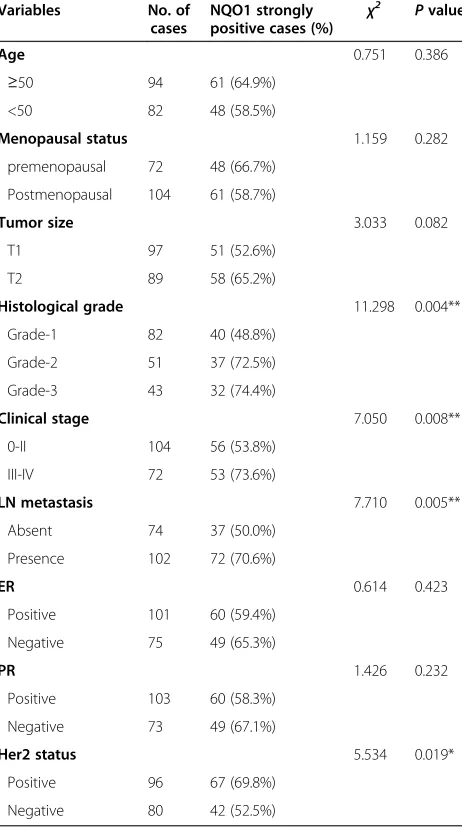
Table 1 Correlation between NQO1 protein expression and the clinicopathological parameters of breast cancer
Variables No. of cases
NQO1 strongly positive cases (%) χ
2 Pvalue
Age 0.751 0.386
≥50 94 61 (64.9%)
<50 82 48 (58.5%)
Menopausal status 1.159 0.282 premenopausal 72 48 (66.7%)
Postmenopausal 104 61 (58.7%)
Tumor size 3.033 0.082
T1 97 51 (52.6%)
T2 89 58 (65.2%)
Histological grade 11.298 0.004**
Grade-1 82 40 (48.8%)
Grade-2 51 37 (72.5%)
Grade-3 43 32 (74.4%)
Clinical stage 7.050 0.008**
0-II 104 56 (53.8%)
III-IV 72 53 (73.6%)
LN metastasis 7.710 0.005**
Absent 74 37 (50.0%)
Presence 102 72 (70.6%)
ER 0.614 0.423
Positive 101 60 (59.4%)
Negative 75 49 (65.3%)
PR 1.426 0.232
Positive 103 60 (58.3%)
Negative 73 49 (67.1%)
Her2 status 5.534 0.019*
Positive 96 67 (69.8%)
Negative 80 42 (52.5%)
Results
NQO1 mRNA and protein expression in breast cancers
NQO1 mRNA levels were examined in eight pairs of breast cancers and adjacent non-tumor breast tissues using qRT-PCR. The results revealed that the relative mRNA ex-pression level of NQO1 was significantly upregulated in cancers compared with adjacent non-tumor tissues (Figure 1A). Western blot data also demonstrated that NQO1 protein was highly expressed in breast cancer tissues compared with adjacent non-tumor tissues (Figure 1B).
To determine the subcellular localization of NQO1 protein, IF staining for NQO1 protein was performed in MCF-7 breast cancer cells. The staining results clearly showed that NQO1 protein is mainly located in the cytoplasm in MCF-7 breast cancer cells (Figure 2).
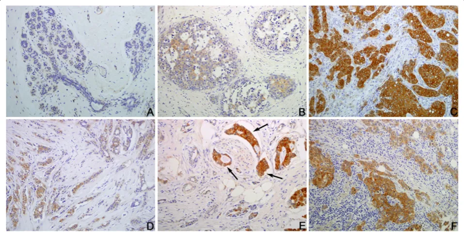
IHC staining also showed that NQO1 protein is mainly located in the cytoplasm of breast cancer cells (Figure 3). The positive rate of NQO1 protein expression was 84.7% (149/176) in breast cancers, which was sig-nificantly higher than that in hyperplasia (36.7%, 8/22) and adjacent non-tumor tissues (30.8%, 16/52) (P< 0.001). Similarly, the strongly positive rate of NQO1 expression was 61.9% (109/176) in breast cancers, which was also sig-nificantly higher than that in hyperplasia (13.6%, 3/22) and adjacent non-tumor tissues (13.5%, 7/52) (P< 0.001). More importantly, the positive rate of NQO1 protein in DCIS was also significantly higher (51.1%, 23/45) than hyperpla-sia (36.7%, 8/22) and adjacent non-tumor tissues (30.8%, 16/52) (Table 2). These data suggest that overexpression of NQO1 may be an early event that can be detected dur-ing the early stage of breast cancer.
Clinicopathological significance of NQO1 protein overexpression in breast cancers
To evaluate the role of NQO1 protein in breast cancer progression, the correlation between NQO1 expression and clinical features of patients was analyzed. As sum-marized in Table 1, there were no significant correlations between the expression level of NQO1 protein and pa-tient age, menopausal status, tumor size, ER levels or PR levels in patients with breast cancer. However, the strongly positive rate of NQO1 protein was significantly higher in Grade 2 and Grade 3 breast cancers than in
Grade 1 cases (P= 0.004), and it was also higher in
breast cancers with lymph node metastasis than in cases without metastasis (P= 0.005). In addition, overexpres-sion of NQO1 showed a correlation with the clinical stage of breast cancer, which was higher in advanced stage (stage III–IV) breast cancers than in early stage (stage I–II) cases (P= 0.008). Furthermore, the strongly positive rate of NQO1 protein was higher in cancer cases with high Her2 expression compared to those with low Her2 expression.
Figure 1Overexpression of NQO1 mRNA and protein in breast cancer tissues.Expression of NQO1 mRNA and protein in breast cancers tissues (T) and adjacent non-tumor tissues (ANT) were examined by qPCR(A)and western blotting(B). Data in(A)represent fold change of relative NQO1 mRNA expression normalized to GAPDH levels. Error bars represent the standard deviation of the mean (SD) calculated from three parallel experiments. *P< 0.05.
Association between NQO1 expression and prognosis of breast cancer patients
Univariate analysis demonstrated that histological grade (P= 0.004), clinical stage (P= 0.008), LN metastasis (P=
0.005), Her2 expression levels (P= 0.019), and NQO1
expression status were significantly associated with DFS and 10-year OS in patients with breast cancer (Table 3). These data suggest that NQO1 could be a valuable prog-nostic factor in breast cancer. Further multivariate ana-lysis using the Cox proportional hazards model revealed that NQO1 overexpression emerged as a significant inde-pendent prognostic factor for survival along with clinical stage and Her2 expression in breast cancer (P= 0.040).
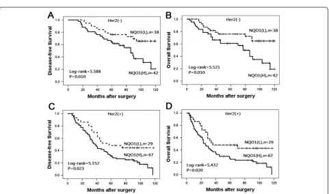
To further substantiate the importance of high NQO1 expression in breast cancer progression, we analyzed DFS and 10-year OS of 176 breast cancer cases using the Kaplan–Meier method and found that patients with high NQO1 expression had lower DFS and 10-year OS than those with low NQO1 expression (bothP< 0.0001) (Figure 4). In addition, the expression of NQO1 was strongly associated with DFS and 10-year OS rates of patients with both early-stage tumors (P = 0.024) and late-stage tumors (P= 0.015) (Figure 5). Similarly, for pa-tients with either Her2 low or high expression, high NQO1 expression showed significantly worse DFS and
10-year OS than those with low NQO1 expression (P=
0.010 andP= 0.023, respectively) (Figure 6).
Discussion
NQO1 was first identified by Ernster and Navazio in the late 1950s [21]. After decades of research, considerable data has demonstrated that NQO1 can protect against natural and exogenous quinones. NQO1 expression in most human tissue types also suggests that it may func-tion primarily in an antioxidant capacity in these cells. Notably, NQO1 is also upregulated in various mamma-lian cancers. Lyn-Cook et al. demonstrated that NQO1 expression is higher in pancreatic adenocarcinomas compared to non-tumor tissues [22].
Wakai et al. demonstrated strong IHC staining of NQO1 in intrahepatic cholangiocarcinoma (ICC), whereas
Figure 3Immunohistochemical staining for NQO1 protein expression. (A)NQO1 staining is negative in non-tumor tissue.(B)Weakly positive NQO1 protein signals in breast hyperplasia.(C)Strongly positive NQO1 protein signal in breast cancer cases with metastasis.(D)Weakly positive NQO1 protein signal in invasive ductal breast cancers without metastasis.(E)Strongly positive NQO1 protein in the cancer cells metastatic to blood vessels (arrows).(F)Strongly positive NQO1 protein signal in the metastatic cancer loci in lymph node. Original magnification, A: ×100; B–F: ×200.
Table 2 NQO1 expression in breast cancers
Diagnosis No. of cases
Positive cases Positive cases rates
Strongly positive rates - + ++ +++
Breast cancers 176 27 40 62 47 84.7%** 61.9%**
DCIS 45 22 9 10 4 51.1%* 31.1%*
Hyperplasia 22 14 5 3 0 36.7% 13.6%
Adjacent non-tumor
52 36 9 7 0 30.8% 13.5%
DCIS: ductal carcinomain situ.
Positive rate: percentage of positive cases with +, ++, and +++ staining score. Strongly positive rate: (high-level expression) percentage of positive cases with ++ and +++ staining score.
the non-tumor bile ducts and liver parenchyma were weakly stained. Cheng et al. showed that NQO1 expression is significantly increased in primary melanomas compared with dysplastic nevi and this may occur in the initiation stage of melanoma development [23]. A recent focus of current research has been the identification of polymor-phisms in NQO1, which have been demonstrated as an in-creased risk of some tumors. Ouerhani et al. reported that the NQO1C609T genotype was overrepresented in acute lymphoblastic leukemia patients and was associated with an aggravating effect compared to the reference group with
NQO1 C609C genotype [24]. Jamieson et al. reported the NQO1 SNP (rs1800566) was associated with a poorer out-come and a lower likelihood of having a treatment delay [25]. These findings indicated that NQO1 may have roles in carcinogenesis and tumor progression. However, the clinico-pathological significance of NQO1 protein expression in breast cancer is less clear.
In this study, we performed IHC staining of NQO1 pro-tein in breast cancer tissue. In agreement with previous studies [13,15], we found that staining of NQO1 is mainly localized in the cytoplasm, and these observations were Table 3 Univariate and multivariate survival analyses (Cox regression model) of various factors in 176 breast cancer patients
Factors B SE Wald HR (95%CI) Pvalue
Univariate
Age 0.2650.265 0.153 2.991 1.303 (0.095–1.758) 0.084
Menopausal status 0.219 0.154 2.037 1.245 (0.921–1.683) 0.154
Tumor size 0.283 0.154 3.389 1.328 (0.982–1.795) 0.066
Histological grade 0.218 0.099 4.843 1.244 (1.024–1.510) 0.028
Clinical stage 1.017 0.169 36.097 2.766 (1.985–3.855) 0.000
LN metastasis 0.382 0.158 5.858 1.465 (1.075–1.996) 0.016
ER 0.190 0.153 1.525 1.209 (0.895–1.633) 0.217
PR 0.114 0.154 0.548 1.121 (0.829–1.515) 0.459
Her2 0.550 0.155 12.600 1.733 (1.279–2.437) 0.000
NQO1 0.447 0.157 8.055 1.563 (1.148–2.128) 0.005
Multivariate
Histological grade 0.207 0.109 3.629 1.230 (0.994–1.521) 0.057
Clinical stage 0.906 0.175 26.929 2.475 (1.758–3.485) 0.000
LN metastasis 0.222 0.168 1.756 1.249 (0.889–1.736) 0.185
Her2 0.394 0.161 5.990 1.484 (1.082–2.035) 0.014
NQO1 0.372 0.181 4.216 1.450 (1.017–2.067) 0.040
B: Coefficient; SE: standard error; Wald: Waldstatistic; HR: hazard ratio.
Figure 5Kaplan–Meier survival curves of in early and late stage patients. (A)and(B)show comparison of DFS and 10-year OS, respectively, in NQO1 (L) and (H) patients of early stage.(C)and(D)show comparison of DFS and 10-year OS, respectively in NQO1 (L) and (H) patients of late stage.
consistent with our IF staining results in MCF-7 breast cancer cells. Compared with adjacent non-tumor tissues, NQO1 protein was found to be significantly up-regulated in breast cancer using IHC. Western blot and qRT-PCR results also demonstrated that the mRNA and protein levels of NQO1 in four cases of fresh breast cancer sam-ples were elevated compared with the adjacent non-tumor tissues. Furthermore, our IHC results showed that the positive rate of NQO1 protein in DCIS was also signifi-cantly higher than either hyperplasia or adjacent normal tissues, indicating that NQO1 upregulation may occur in the initiation stage of breast cancer progression. These findings suggest that NQO1 protein level might be used as an early diagnostic indicator of this disease.
Despite the strong association between NQO1 expres-sion and cancer, there have been few reports of NQO1 protein expression-based outcomes in tumor patients. Mikami K et al. reported that the expression and enzyme activity of NQO1 is not only upregulated in colon cancer cell lines and colorectal tumors, but also significantly greater in tumors with nodal metastases than those without metastases [26], while Gan et al. reported higher expression of NQO1 protein in lower-grade and superfi-cial bladder tumors compared with high-grade and inva-sive tumors [27]. In the present study, we found that the NQO1 expression level was markedly associated with histological grade (P = 0.004), clinical stage (P= 0.008),
LN metastasis (P= 0.005) and Her2 expression levels
(P= 0.019), suggesting that NQO1 upregulation
pro-motes the invasion and/or metastasis of breast cancer cells. These finding indicate that NQO1 might be useful as a poor prognostic biomarker of breast cancer. More-over, Buranrat et al. demonstrated a significant associ-ation between high level of NQO1 expression and short overall survival time of cholangiocarcinoma patients, which raises the exciting possibility of using NQO1 as a tumor marker [14]. Additionally, an association was found by Awadallah et al. between high level of NQO1 expression and short overall survival of pancreatic can-cer patients [15]. In our previous study, we found that high level of NQO1 protein significantly associated with shortened survival of patients with gastric adenocarcin-oma [28]. However, the alternative hypothesis seems to be true with low NQO1 expression evaluated by IHC in intrahepatic cholangiocarcinoma (ICC) cases predicting poor prognosis [29]. The conflicting conclusions may be due to the different study populations, which also highlights the need to evaluate the biomarkers under relevant circumstances. In the present study, univariate survival analysis revealed that tumor histological grade, clinical stage, LN metastasis, Her2 expression level and NQO1 expression status are all significantly related with DFS and10-year OS rates of patients with breast cancer (P< 0.05). Further multivariate survival analysis
showed that NQO1 expression was one of the independent prognostic factors, along with tumor clinical stage and Her2 status. Moreover, finding tumor-selective therapies for breast cancer is of utmost importance. Our study with NQO1 protein expression in breast cancers also indicated that as an exploitable cancer target, NQO1 might improve patient management and outcome by personalized therapy. A comprehensive analysis of the molecular mechanism of NQO1 involved in the tumorigenesis and progression of breast cancer is essential.
Conclusions
In summary, NQO1 plays a key role in the progression of breast cancer, and high level of NQO1 protein is strongly associated with advanced stage, lymph node metastasis, Her2 overexpression and shortened survival of patients with breast cancer. The high proportion and prognostic value of NQO1 expression suggests that NQO1 may be a significant biomarker and a potential therapeutic target for patients with breast cancer.
Competing interests
The authors declare that they have no competing interests.
Authors’contributions
YY and YZ contributed equally to this work. All authors read and approved the final manuscript.
Acknowledgments
This study was supported by grants from the National Natural Science Funds of China (No. 31301065) and The Projects of Research & Innovation of Jilin Youth Leader and Team (No. 20130521017JH).
Author details
1Department of Pathology & Cancer Research Center, Yanbian University Medical College, Yanji 133002, China.2Department of Breast Surgery, The Second Hospital of Jilin University, Changchun 130041, China.3Department of Pathology, Yanbian Women and Children’s Hospital, Yanji 133002, China. 4Department of Biochemistry, Yanbian University Medical College, Yanji 133002, China.
Received: 4 January 2014 Accepted: 27 January 2014 Published: 5 February 2014
References
1. Stopeck AT, Brown-Glaberman U, Wong HY, Park BH, Barnato SE, Gradishar WJ, Hudis CA, Rugo HS:The role of targeted therapy and biomarkers in breast cancer treatment.Clin Exp Metastasis2012,29(7):807–819. 2. Kamel M, Shouman S, El-Merzebany M, Kilic G, Veenstra T, Saeed M, Wagih M,
Diaz-Arrastia C, Patel D, Salama S:Effect of tumour necrosis factor-alpha on estrogen metabolic pathways in breast cancer cells.J Cancer2012,
3:310–321.
3. Thorat D, Sahu A, Behera R, Lohite K, Deshmukh S, Mane A, Karnik S, Doke S, Kundu GC:Association of osteopontin and cyclooxygenase-2 expression with breast cancer subtypes and their use as potential biomarkers.
Oncol Lett2013,6(6):1559–1564.
4. Camerini A, Donati S, Viacava P, Siclari O, Puccetti C, Tartarelli G, Valsuani C, De Luca F, Martini L, Cavazzana A, Amoroso D:Evaluation of HER2 and p53 expression in predicting response to docetaxel-based first-line chemotherapy inadvanced breast cancer.J Exp Clin Cancer Res2011,
30:38.
5. Charpentier M, Martin S:Interplay of stem cell characteristics, EMT, and microtentacles in circulating breast tumor cells.Cancers2013,
6. Cuppone F, Bria E, Vaccaro V, Puglisi F, Fabi A, Sperduti I, Carlini P, Milella M, Nisticò C, Russillo M, Papaldo P, Ferretti G, Aapro M, Giannarelli D, Cognetti F:Magnitude of risks and benefits ofthe addition of bevacizumab to chemotherapy for advanced breast cancerpatients: meta-regression analysis of randomized trials.J Exp Clin Cancer Res2011,30:54. 7. Zhu CL, Huang Q, Liu CH, Lin XS, Xie F, Shao F:NAD (P) H: quinone
oxidoreductase 1 (NQO1) C609T gene polymorphism association with digestive tract cancer:a meta-analysis.Asian Pac J Cancer Prev2013,
14(4):2349–2354.
8. Ross D, Kepa JK, Winski SL, Beall HD, Anwar A, Siegel D:NAD (P) H:quinine oxidoreductase 1 (NQO1): chemoprotection, bioactivation, gene regulation and genetic polymorphisms.Chem Biol Interact2000,129:77–97. 9. Siegel D, Gustafson DL, Dehn DL, Han JY, Boonchoong P, Berliner LJ, Ross
D:NAD (P) H:quinone oxidoreductase 1: role as a superoxide scavenger.
Mol Pharmacol2004,65:1238–1247.
10. Su XL, Yan MR, Yang L:Qimuge-Suyila: NQO1 C609T polymorphism correlated to colon cancer risk in farmers from western region of Inner Mongolia.Chin J Cancer Res2012,24(4):317–322.
11. Radjendirane V, Joseph P, Lee YH, Kimura S, Klein-Szanto AJ, Gonzalez FJ, Jaiswal AK:Disruption of the DT diaphorase (NQO1) gene in mice leads to increased menadione toxicity.J Biol Chem1998,273:7382–7389. 12. Garate M, Wani AA, Li G:The NAD (P) H:Quinone Oxidoreductase 1 induces cell cycle progression and proliferation of melanoma cells.
Free Radic Biol Med2010,48:1601–1609.
13. Siegel D, Franklin WA, Ross D:Immunohistochemical detection of NAD (P) H:quinone oxidoreductase in human lung and lung tumors.Clin Cancer Res1998,4(9):2065–2070.
14. Buranrat B, Chau-In S, Prawan A, Puapairoj A, Zeekpudsa P, Kukongviriyapan V:NQO1 Expression correlates with cholangiocarcinoma prognosis.
Asian Pac J Cancer Prev2012,13(Suppl):131–136.
15. Awadallah NS, Dehn D, Shah RJ, Russell Nash S, Chen YK, Ross D, Bentz JS, Shroyer KR:NQO1 expression in pancreatic cancer and its potential use as a biomarker.Appl Immunohistochem Mol Morphol2008,16:24–31. 16. Siegel D, Yan C, Ross D:NAD (P) H: quinone oxidoreductase 1 (NQO1) in
the sensitivity and resistance to antitumor quinones.Biochem Pharmacol
2012,83(8):1033–1040.
17. Bey EA, Reinicke KE, Srougi MC, Varnes M, Anderson VE, Pink JJ, Li LS, Patel M, Cao L, Moore Z, Rommel A, Boatman M, Lewis C, Euhus DM, Bornmann WG, Buchsbaum DJ, Spitz DR, Gao J, Boothman DA:Catalase abrogates β-lapachone-induced PARP1 hyperactivation–directed programmed necrosis in NQO1-positive breast cancers.Mol Cancer Ther2013,
12(10):2110–2120.
18. Jin J, Jin T, Quan M, Piao Y, Lin Z:Ezrin overexpression predicts the poor prognosis of gastric adenocarcinoma.Diagn Pathol2012,7:135. 19. Liu S, Li L, Zhang Y, Zhao Y, You X, Lin Z, Zhang X, Ye L:The oncoprotein
HBXIP uses two pathways to up-regulate S100A4 in promotion of growth and migration of breast cancer cells.J Biol Chem2012,
287(36):30228–30239.
20. Kong J, Li Y, Liu S, Jin H, Shang Y, Quan C, Li Y, Lin Z:High expression of ezrin predicts poor prognosis in uterine cervical cancer.BMC Cancer
2013,13(1):520.
21. Ernster L, Navazio F:Soluble diaphorase in animal tissues.Acta Chem Scand1958,12:595.
22. Lyn-Cook BD, Yan-Sanders Y, Moore S, Taylor S, Word B, Hammons GJ:
Increased levels of NAD (P) H: quinone oxidoreductase 1 (NQO1) in pancreatic tissues from smokers and pancreatic adenocarcinomas: a potential biomarker of early damage in the pancreas.Cell Biol Toxicol
2006,22:73–80.
23. Cheng Y, Li J, Martinka M, Li G:The expression of NAD (P) H:quinone oxidoreductase 1 is increased along with NF-kappaB p105/p50 in human cutaneous melanomas.Oncol Rep2010,23(4):973–979.
24. Ouerhani S, Cherif N, Bahri I, Safra I, Menif S, Abbes S:Genetic polymorphisms of NQO1, CYP1A1 and TPMT and susceptibility to acute lymphoblastic leukemia in a Tunisian population.Mol Biol Rep2013,40(2):1307–1314. 25. Jamieson D, Cresti N, Bray J, Sludden J, Griffin MJ, Hawsawi NM, Famie E,
Mould EV, Verrill MW, May FE, Boddy AV:Two minor NQO1 and NQO2 alleles predict poor response of breast cancer patients to adjuvant doxorubicin and cyclophosphamide.Pharmacogenet Genomics2011,
21(12):808–819.
26. Mikami K, Naito M, Ishiguro T, Yano H, Tomida A, Yamada T, Tanaka N, Shirakusa T, Tsuruo T:Immunological quantitation of DT-diaphorase in
carcinoma cell lines and clinical colon cancers: advanced tumors express greater levels of DT-diaphorase.Jpn J Cancer Res1998,89:910–915. 27. Gan Y, Mo Y, Kalns JE, Lu J, Danenberg K, Danenberg P, Wientjes MG, Au JL:
Expression of DT-diaphorase and cytochrome P450 reductase correlates with mitomycin C activity in human bladder tumors.Clin Cancer Res2001,
7:1313–1319.
28. Lin L, Qin Y, Jin T, Liu S, Zhang S, Shen X, Lin Z:Significance of NQO1 overexpression for prognostic evaluation of gastric adenocarcinoma.
Exp Mol Pathol2013. DOI: 10.1016 /j.yexmp. 2013.12.008.
29. Wakai T, Shirai Y, Sakata J, Matsuda Y, Korita PV, Takamura M, Ajioka Y, Hatakeyama K:Prognostic significance of NQO1 expression in intrahepatic cholangiocarcinoma.Int J Clin Exp Pathol2011,4(4):363–370.
doi:10.1186/1756-9966-33-14
Cite this article as:Yanget al.:Clinical implications of high NQO1 expression in breast cancers.Journal of Experimental & Clinical Cancer Research201433:14.
Submit your next manuscript to BioMed Central and take full advantage of:
• Convenient online submission
• Thorough peer review
• No space constraints or color figure charges
• Immediate publication on acceptance
• Inclusion in PubMed, CAS, Scopus and Google Scholar
• Research which is freely available for redistribution