R E S E A R C H
Open Access
Construction of a recombinant eukaryotic
expression vector containing PHD3 gene and its
expression in HepG2 cells
Qi-Lian Liang
1*†, Zhou-Yu Li
2†, Yuan Zhou
1, Qiu-Long Liu
1, Wen-Ting Ou
1and Zhi-Gang Huang
3Abstract
Prolyl hydroxylase domain 3 (PHD3) is a hypoxia inducible factor-α(HIFα) regulator; it degrades HIFαin the presence of oxygen. Recently, there have been an increasing number of studies about the role of PHD3 in proliferation and apoptosis of cancer cells. However, most of the evidence for the role of PHD3 is observational, and little is known of the molecular mechanism. In our current study, we constructed a recombinant eukaryotic expression vector containing the PHD3 gene and detected its biological activity in human hepatoma cell line (HepG2 cells). We successfully constructed a recombinant pcDNA 3.1(+)-PHD3 plasmid; the results showed that PHD3 overexpression could inhibit the proliferation of HepG2 cells and induce apoptosis by activating caspase-3 activity. Our study has provided preliminary materials and data for further investigation of the effect of PHD3 on HepG2 cells.
Keywords:Prolyl hydroxylase domain 3 (PHD3), Hepatocellular cancer (HCC), Hypoxia inducible factor (HIF), Caspase-3
Introduction
There are three prolyl hydroxylase domain proteins (PHDs), PHD1, PHD2 and PHD3, that are the key re-gulators of degradation of hypoxia inducible factor (HIF) in mammals. They are known as HIF-prolyl hydroxylase (HPHs) in Drosophila and egg-laying nine (EGLN or EGL-9) inC. elegans[1,2]. PHD1 and PHD2 mRNAs are highly expressed in placenta, and PHD3 mRNA is highly expressed in both placenta and heart [3]. In the presence of oxygen, two of the proline residues of HIFαare hydro-xylated by PHDs, which allows specific recognition and binding of von Hippel-Lindau tumor suppressor protein (pVHL) and then leads to the subsequent ubiquitination and proteosomal degradation of HIFα [4]. In addition, PHDs play a novel role in tumor progression and develop-ment [5], especially PHD3. Recently, an increasing num-ber of studies have indicated that PHD3 is involved in the development and prognosis of cancer [6-10] and also
appears to induce apoptosis in cancer cells [11-13]. How-ever, most of these studies are observational, and know-ledge of PHD3’s molecular mechanism is still limited. In our current study, we constructed a eukaryotic expression vector containing the PHD3 gene and detected its expres-sion in human hepatoma cell line (HepG2) cells to estab-lish a foundation for future studies.
Materials and methods
Materials
Plasmid pcDNA 3.1(+) was obtained from the Central Laboratory of Affiliated Hospital of Guangdong Medical College (Guangdong, China). E. coli DH5α was gained from the Pathogenic Biology Laboratory of Guangdong Medical College. Human hepatoma cells (HepG2) were obtained from the Laboratory of Hepatobiliary Surgery. Placenta tissue and the written informed consent for this tissue were obtained from the Operating Room of Affiliated Hospital of Guangdong Medical College. RNAiso Plus, High Fidelity Prime Script™ RT-PCR Kit, TaKaRa Agarose Gel DNA Purification Kit Ver.2.0, DL10,000 DNA Marker, DNA A-Tailing Kit, pMD19-T Simple Vector, DNA Ligation Kit Ver.2.0, Hind III, Xho I, * Correspondence:[email protected]
†Equal contributors
1
Department of Oncology, Affiliated Hospital of Guangdong Medical College, Zhanjiang 524001, China
Full list of author information is available at the end of the article
© 2012 liang et al.; licensee BioMed Central Ltd. This is an Open Access article distributed under the terms of the Creative Commons Attribution License (http://creativecommons.org/licenses/by/2.0), which permits unrestricted use, distribution, and reproduction in any medium, provided the original work is properly cited.
TaKaRa MiniBEST Plasmid Purification Kit Ver.2.0 and SYBR® Prime Script® RT-PCR Kit II (Perfect Real Time) were purchased from TAKARA (Japan). Neonatal Bovine Serum was acquired from Hangzhou Sijiqing Biological Engineering Materials Co., Ltd (China). Dulbecco's modi-fied Eagle’s medium(DMEM)was purchased from Hyclone Company (USA). Lipofectamine™ 2000 was purchased from Invitrogen Biotechnology (USA). DMSO was pur-chased from Sigma (USA). 3-(4,5-Dimethyl-2-Thiazolyl)-2,5-Diphenyl Tetrazolium Bromide (MTT) was purchased from Sangon Biotech (Shanghai) Co., Ltd (China). Pri-mary rabbit polyclonal anti-EGLN3 antibody was pur-chased from Jiamay Biotech Company (China). Primary rabbit polyclonal anti-Caspase-3 antibody was purchased from Zhongshan Goldenbridge Biotechnology CO., LTD (China). Primary rabbit polyclonal anti-tubulin antibody, a BCA protein assay kit and BeyoECL Plus were purchased from Beyotime Institute of Biotechnology (China).
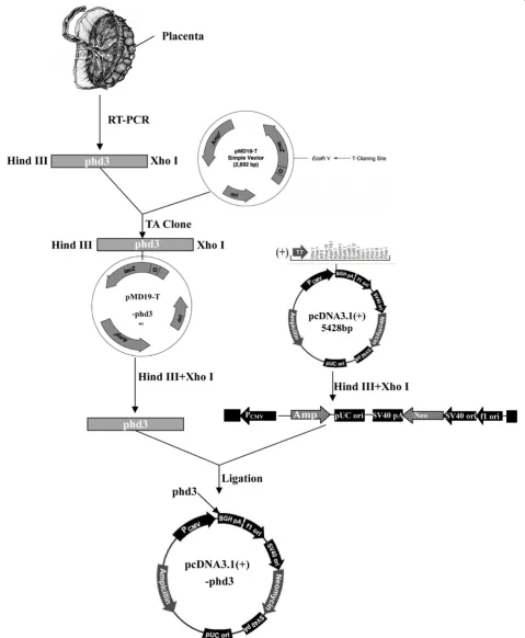
Vector construction
Total RNA extraction and PHD3 cDNA synthesis
Total RNA from placental tissue was extracted with RNAiso Plus according to the manufacturer’s instructions. First, 1μg of total RNA was used to synthesize full-length PHD3 CDS with High Fidelity Prime Script™RT-PCR Kit. A pair of specific primers, containing Hind III and Xho I restriction enzyme cutting sites, were designed: forward 50-CCCAAGCTTGATGCCCCTGGGACACATCAT-30 and reverse 50 -CCGCTCGAGTCAGTCTTCAGTGAGGG-CAGA-30.
Purification of PHD3 cDNA and ligation with pMD19-T simple vector
The RT-PCR products were separated with 1.5% agarose gel electrophoresis, and the target fragments were re-trieved and purified by TaKaRa Agarose Gel DNA Purifi-cation Kit v.2.0. The target fragments were polyadenylated using DNA A-Tailing Kit; these fragments were then
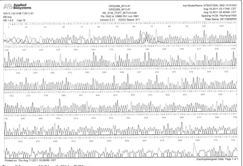
ligated into pMD19-T Simple Vector with DNA Ligation Kit v.2.0 (TA Clone). The recombinant pMD19-T-PHD3 was transformed into E. coli DH5α competent cells for amplification. Recombinant vectors were isolated from transformants by TaKaRa MiniBEST Plasmid Purification Kit v.2.0, and the pMD19-T-PHD3 was sequenced by an ABI 377 DNA sequencer (Applied Biosystems, USA).
Construction of recombinant pcDNA 3.1(+)-PHD3 eukaryotic expression vector
The pMD19-T-PHD3 plasmids were digested by Hind III and Xho I restriction enzymes, and the target frag-ments (full length PHD3 cDNAs) were isolated and pu-rified. The pcDNA 3.1(+) eukaryotic expression vectors were also digested by Hind III and Xho I and then li-gated into PHD3 cDNA with DNA Ligation Kit v.2.0. The recombinant pcDNA 3.1(+)-PHD3 was amplified in
E. coliDH5αcompetent cells, and isolated with TaKaRa MiniBEST Plasmid Purification Kit v.2.0. The correct pcDNA 3.1(+)-PHD3 plasmid sequence was verified by restriction enzyme mapping and DNA sequencing.
A Schematic representation of the construction of the recombinant pcDNA 3.1(+)-PHD3 eukaryotic expression vector is presented in Figure 1.
Expression of the recombinant pcDNA 3.1(+)-PHD3 eukaryotic expression vector in HepG2 cells
Cell transfection
HepG2 cells were cultured in DMEM containing 10% Neonatal Bovine Serum at 37°C in a humidified atmos-phere of 5% CO2. Cells were passaged and plated (12-well plates for mRNA assay, 6-well plates for western blot and 96-well plates for growth curve assay) for 24 hours before transfection at 80%–90% confluence. Cells were divided into four groups: no treatment (Normal), Lipofectamine™ 2000 (LP2000), Lipofectamine™ 2000 + pcDNA 3.1(+) (PC3.1) and Lipofectamine™ 2000 + pcDNA 3.1(+)-PHD3 (PHD3). Transfection was carried out according to
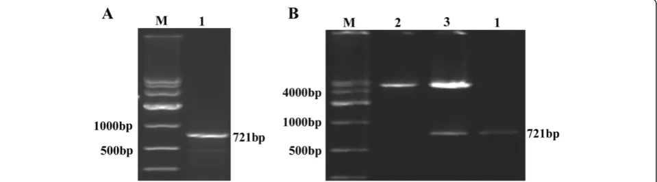
Figure 2Identification of PHD3.(A) Electrophoresis of full-length target gene RT-PCR product; M: DNA Marker DL10,000, 1: PHD3. (B) Hind III and Xho I digestion and electrophoresis of pcDNA 3.1(+)-PHD3 eukaryotic expression vector; M: DNA Marker DL10,000, 1: PHD3, 2: pcDNA 3.1(+) plasmid digested by Hind III and Xho I, 3: pcDNA 3.1(+)-PHD3 plasmid digested by Hind III and Xho I.
lianget al. Journal of Experimental & Clinical Cancer Research2012,31:64 Page 3 of 7
Lipofectamine™2000 instructions. Forty-eight hours after transfection, cells were collected to conduct subsequent assays.
Detection of PHD3 mRNA by quantitative real time RT-PCR Total RNA was isolated from transfected cells by RNAiso Plus, and 500 ng of total RNA was analyzed
with SYBR® Prime Script® RT-PCR Kit II on a LightCy-cler480 (Roche, Switzerland) according to manufacturer’s instructions. The primers were as follows: PHD3 forward 5’- CATCAGCTTCCTCCTGTC-3’, reverse 5’- CCACCA TTGCCTTAGACC-3’and β-actin forward 5’- CTGTGC CCATCTACGAGG-3’, reverse 5’- ATGTCACGCACGAT TTCC-3’. The data were analyzed using Ct method.
Figure 4Expression and biological activity of PHD3. (A) PHD3 mRNA was measured by quantitative real-time RT-PCR.Cells transfected with PHD3 significantly overexpressed PHD3, compared with the control groups (allP=0.00). (BandC) PHD3 protein was analyzed by western blot. Cells transfected with PHD3 significantly overexpressed PHD3, compared with the control groups (allP=0.00). Normal: no treatment, LP2000: Lipofectamine™2000, PC3.1: Lipofectamine™2000+pcDNA 3.1(+), PHD3: Lipofectamine™2000+pcDNA 3.1(+)-PHD3.#P<0.05 indicates statistically
significant differences in comparison to PHD3-transfected cells.
Western blot assay
After transfection, cells were collected and lysed, and the protein concentration was detected by BCA protein assay kit. Supernatants were loaded on a 12%SDS–PAGE gel, and they were then wet transferred onto PVDF membranes. The membranes were incubated with their respective primary antibodies, followed by incubation with HRP-conjugate secondary antibodies. The bands were visualized with BeyoECL Plus and exposed to X-ray film.
Cell proliferation assay
To analyze the effects of PHD3 on proliferation of HepG2 cells, MTT assay was performed. Cells were cul-tured in 96-well plates, and a total cell number was detected every 12 hours. At each time point, twenty μl of MTT (5 mg/ml) was added to each well, and incubated at 37°C for 4 hours. The supernatant was discarded, and 150μl of DMSO was added to each well. The absorbance (OD value) of the cells was measured using a micro plate reader (Thermo, USA) with a 492 nm filter.
Statistical analysis
The data were presented as mean ± SD based on three in-dependent experiments. Statistical comparisons between two groups were made by Student’s t test, and the cell growth curve was analyzed with multivariate analysis of variance (MANOVA). Statistical analyses were performed by using SPSS 13.0 software for windows (SPSS Inc., USA). Statistical significance was defined asP< 0.05.
Results
Evaluation of RT-PCR product and recombinant pcDNA 3.1(+)-PHD3 eukaryotic expression vector
The RT-PCR products were loaded on 1.5% agarose gels, and the band for full-length PHD3 cDNA was located at 721 bp (Figure 2A). After the PHD3 cDNA fragment was inserted into the pcDNA 3.1(+) plasmid (5428 bp), the fragment was confirmed by Hind III and Xho I digestion and electrophoresis (Figure 2B). Additionally, the cDNA was confirmed by DNA sequencing, as shown in Figure 3.
mRNA and protein expressions of PHD3 in HepG2 cells
After transfection, the expression of PHD3 was analyzed by quantitative real-time RT-PCR and western blot. The results showed that the PHD3 transfected group
Figure 6Activation of caspase-3. Cells transfected with the cleaved 17 kD active caspase-3 fragment of PHD3 expressed more protein than the control groups (allP=0.00).Normal: no treatment, LP2000: Lipofectamine™2000, PC3.1: Lipofectamine™2000+pcDNA 3.1(+), PHD3: Lipofectamine™2000+pcDNA 3.1(+)-PHD3.#P<0.05 indicates statistically significant differences in comparison to PHD3-transfected cells.
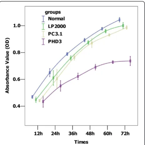
Figure 5HepG2 cell growth curves. Compared with the control groups, PHD overexpression significantly inhibited cell proliferation (allP=0.00).Normal: no treatment, LP2000: Lipofectamine™2000, PC3.1: Lipofectamine™2000+pcDNA 3.1(+), PHD3: Lipofectamine™2000+pcDNA 3.1(+)-PHD3.
lianget al. Journal of Experimental & Clinical Cancer Research2012,31:64 Page 5 of 7
overexpressed more PHD3(all P= 0.00), when compa-red with the control groups (Figure 4A, Figure 4B and Figure 4C).
Effect of PHD3 on proliferation of HepG2 cells
The OD value of each group was obtained by measuring it every 12 h after transfection, for up to 72 h. Cell pro-liferation curves were depicted with mean OD values of each time point. As shown in Figure 5, the pcDNA 3.1 (+)-PHD3 transfected group grew slower than the con-trol groups (allP= 0.00)
Effect of PHD3 on apoptosis of HepG2 cells
To investigate whether PHD3 has an effect on inducing apoptosis in HepG2 cells, caspase-3 assays were per-formed. We found that PHD3 overexpression increased caspase-3 activity (all P= 0.00), and the cleaved 17 kD active caspase-3 fragment was visualized by western blot analysis (Figure 6A and Figure 6B).
Discussion
PHD3 was originally considered an HIFα regulator; it played a vital role in the progression and prognosis of cancer by targeting the degradation of HIFα. Recently, a number of studies have shown that PHD3 was closely related to cancer, independent of its hydroxylase activity. Chen, S et al. [8] found that PHD3 was highly expressed in lung cancer (NSCLC), associating with early-stage and well differentiated tumors. Fox, S. B et al. [14] showed that PHD3 expression was significantly increased after therapy with epirubicin, alone or in combination with tamoxifen, in patients with T2-4 N0-1 breast cancer; however, PHD3 expression was not relevant in treatment response and survival. Su, C et al. [6] also demonstrated that the ex-pression of PHD3 was significantly increased from non-cancerous mucosa to cancer, and its high expression correlated with well differentiated tumors. In contrast, Couvelard, A et al. [10] discovered that high nuclear PHD3 expression related to poor survival in patients with pancreatic endocrine tumors. Gossage, L et al. [9] also found that PHD3 expression in tumor tissue indicated a worse overall disease-free survival in ampullary adenocar-cinomas and pancreatic adenocaradenocar-cinomas. These studies suggested that the role of PHD3 varied from one cancer type to another and that it could be a predictor for treat-ment and prognosis of cancer. With an increased under-standing of PHD3, more attention has been focused on its ability to suppress tumor growth [11-13]; however, little is known about PHD3’s exact mechanism. In pancreatic cells overexpressing PHD3, Su, Y et al. [13] found that apop-tosis increased sharply in the presence of nerve growth factor by the activation of caspase-3. Tennant, D. A et al. [12] demonstrated PHD3-mediated alpha-ketoglutarate-induced apoptosis in three human colorectal cancer cell
lines (HCT116, A431 and A375). In colorectal cancer cells, PHD3 inhibits cell growth by blocking IKKβ/NF-κB signaling [11].
So far, the relationship between PHD3 and hepatocel-lular cancer (HCC) is still unclear. To clarify the effect of PHD3 on HCC, we constructed a recombinant eukar-yotic expression vector containing PHD3 and detected its biological activities in HepG2 cells. The results showed that pcDNA3.1(+)-PHD3 was successfully constructed, and PHD3 could be overexpressed in HepG2 cells after transient transfection. To investigate whether PHD3 can inhibit HepG2 cells, we carried out a cell growth curve assay and found that PHD3 arrested cell proliferation. Moreover, we analyzed caspase-3 activity and clarified whether PHD3 had an effect on apoptosis. We found that the cleaved 17 kD active caspase-3 fragment was signifi-cantly overexpressed in PHD3 group, which is in line with previous studies [13,15].
In conclusion, we constructed a recombinant eukar-yotic expression vector, pcDNA3.1(+)-PHD3, showing that PHD3 overexpression can inhibit proliferation and induce apoptosis in HepG2 cells. Our study has provided pre-liminary data for further research of stably transfecting pcDNA3.1(+)-PHD3 into HepG2 cell and clarifying the mechanism of PHD3-induced apoptosis.
Competing interests
The authors declared that they have no competing interest.
Authors’contributions
Qi-Lian Liang conceived and designed the study, and drafted the manuscript. Zhou-Yu Li carried out molecular genetic studies and drafted the manuscript. Yuan Zhou Qiu-Long Liu1 and Wen-Ting Ou contributed to cell culture, cell transfection and western blot respectively. Zhi-Gang Huang participated in statistical analyses. All authors read and approved the final manuscript.
Acknowledgments
This work was supported by a grant from the Science and Technology Innovation Fund of Guangdong Medical College, China (No. STIF201126) and Excellent Master’s Thesis Fostering Fund of Affiliated Hospital of Guangdong Medical College, China (No.YS1108).
Author details
1Department of Oncology, Affiliated Hospital of Guangdong Medical College, Zhanjiang 524001, China.2Department of Radiotherapy, Affiliated Tumor Hospital of Guangzhou Medical College, Guangzhou 510095, China. 3Department of Epidemiology, School of Public Health, Guangdong Medical College, Dongguan 523808, China.
Received: 1 July 2012 Accepted: 11 August 2012 Published: 17 August 2012
References
1. Bruick RK, McKnight SL:A conserved family of prolyl-4-hydroxylases that modify HIF.Science2001,294:1337–1340.
3. Cioffi CL, Liu XQ, Kosinski PA, Garay M, Bowen BR:Differential regulation of HIF-1 alpha prolyl-4-hydroxylase genes by hypoxia in human
cardiovascular cells.Biochem Biophys Res Commun2003,303:947–953. 4. Fong GH, Takeda K:Role and regulation of prolyl hydroxylase domain
proteins.Cell Death Differ2008,15:635–641.
5. Kiss J, Kirchberg J, Schneider M:Molecular oxygen sensing: implications for visceral surgery.Langenbecks Arch Surg2012,397:603–610. 6. Su C, Huang K, Sun L, Yang D, Zheng H, Gao C, Tong J, Zhang Q:
Overexpression of the HIF hydroxylase PHD3 is a favorable
prognosticator for gastric cancer.Med Oncol2012, [Epub ahead of print]. 7. Peurala E, Koivunen P, Bloigu R, Haapasaari KM, Jukkola-Vuorinen A:
Expressions of individual PHDs associate with good prognostic factors and increased proliferation in breast cancer patients.Breast Cancer Res Treat2012,133:179–188.
8. Chen S, Zhang J, Li X, Luo X, Fang J, Chen H:The expression of prolyl hydroxylase domain enzymes are up-regulated and negatively correlated with Bcl-2 in non-small cell lung cancer.Mol Cell Biochem2011, 358:257–263.
9. Gossage L, Zaitoun A, Fareed KR, Turley H, Aloysius M, Lobo DN, Harris AL, Madhusudan S:Expression of key hypoxia sensing prolyl-hydroxylase PHD1, -2 and−3 in pancreaticobiliary cancer.Histopathology2010, 56:908–920.
10. Couvelard A, Deschamps L, Rebours V, Sauvanet A, Gatter K, Pezzella F, Ruszniewski P, Bedossa P:Overexpression of the oxygen sensors PHD-1, PHD-2, PHD-3, and FIH Is associated with tumor aggressiveness in pancreatic endocrine tumors.Clin Cancer Res2008,14:6634–6639. 11. Xue J, Li X, Jiao S, Wei Y, Wu G, Fang J:Prolyl hydroxylase-3 is
down-regulated in colorectal cancer cells and inhibits IKKbeta independent of hydroxylase activity.Gastroenterology2010,138:606–615.
12. Tennant DA, Gottlieb E:HIF prolyl hydroxylase-3 mediates alpha-ketoglutarate-induced apoptosis and tumor suppression.J Mol Med (Berl)
2010,88:839–849.
13. Su Y, Loos M, Giese N, Hines OJ, Diebold I, Gorlach A, Metzen E, Pastorekova S, Friess H, Buchler P:PHD3 regulates differentiation, tumour growth and angiogenesis in pancreatic cancer.Br J Cancer2010,103:1571–1579. 14. Fox SB, Generali D, Berruti A, Brizzi MP, Campo L, Bonardi S, Bersiga A, Allevi
G, Milani M, Aguggini S, Mele T, Dogliotti L, Bottini A, Harris AL:The prolyl hydroxylase enzymes are positively associated with hypoxia-inducible factor-1alpha and vascular endothelial growth factor in human breast cancer and alter in response to primary systemic treatment with epirubicin and tamoxifen.Breast Cancer Res2011,13:R16.
15. Buchler P, Gukovskaya AS, Mouria M, Buchler MC, Buchler MW, Friess H, Pandol SJ, Reber HA, Hines OJ:Prevention of metastatic pancreatic cancer growth in vivo by induction of apoptosis with genistein, a naturally occurring isoflavonoid.Pancreas2003,26:264–273.
doi:10.1186/1756-9966-31-64
Cite this article as:lianget al.:Construction of a recombinant eukaryotic expression vector containing PHD3 gene and its expression in HepG2 cells.Journal of Experimental & Clinical Cancer Research201231:64.
Submit your next manuscript to BioMed Central and take full advantage of:
• Convenient online submission
• Thorough peer review
• No space constraints or color figure charges
• Immediate publication on acceptance
• Inclusion in PubMed, CAS, Scopus and Google Scholar
• Research which is freely available for redistribution
Submit your manuscript at www.biomedcentral.com/submit
lianget al. Journal of Experimental & Clinical Cancer Research2012,31:64 Page 7 of 7