Brassica oleracea var. botrytis Linn.
Suganya.D1, Hussain Ali Fathima.M2 and Kanimozh.Ki3
1,2
PG & Research Department of Microbiology, Jamal Mohamed College (Autonomous) Tiruchirappalli-620020
3
PG& Research Department of Botany & Microbiology, AVVM.Shri Pushpam College (Autonomous)
ABSTRACT
Objective: To investigate the antibacterial activity and phytochemical analysis of Brassica oleraceae.
Methods: The antibacterial activity was evaluated using agar well diffusion and microdilution methods against the bacterial (E.coli, Proteus, S.aureus, Klebsiella and Pseudomonas) iolates. The extraction of the vegetable was carried using the solvent namely acetone. Phytochemical, FT-IR and HPLC analysis was carried out in the acetone extract.
Results: Acetone extract showed maximum activity against E.coli, Proteus, S.aureus, Klebsiella and Pseudomonas. Phytochemical analysis of Brassica oleraceae extracts showed the presence of secondary metabolites like alkaloids, flavonoids, glycosides, phenolic compounds, saponins, tannins, terpenoids, carbohydrates and anthraquinone. The FT-IR and HPLC revealed different characteristic peak values with various functional compounds in the extracts.
Conclusion: From this study, it can be concluded that Brassica oleraceae exhibits antibacterial activity against certain microorganisms.
Keywords:Brassica oleraceae, Brassicaceae, Antibacterial, Phytochemical analysis, HPLC and FT-IR analysis
I. INTRODUCTION
Cauliflower is one of several vegetables in the species Brassica oleracea, in the
family Brassicaceae. It is an annual plant that reproduces by seed. Typically, only the head (the white curd) is eaten. The cauliflower head is composed of a white inflorescencemeristem. Cauliflower heads
resemble those in broccoli, which differs in having flower buds. Brassica oleracea also includes broccoli,brussels sprouts, cabbage, collard greens, and kale, though they are of different cultivar groups.
Vegetables have been analyzed as potent medicine and man is able to obtain from them a wondrous assortment of industrial chemicals. In recent years population continues to explode and microbial disaster may occur. So vegetables with possible antimicrobial activity should be tested against an appropriate microbial model to confirm its activity and to ascertain the parameter associated with it. Cruciferous vegetables are one of the dominant food crops which have high vitamin C, soluble fibre and contain multiple nutrients and photochemical with potential anticancer properties.
been made to evaluate the antimicrobial activity and phytochemical analysis of Brassica oleracea
extracts against the pathogenic microbes.
The Brassicaceae family comprises many commonly consumed vegetables, condiments, forages and oil containing plants, such as cabbage, broccoli, cauliflower, Brussels sprouts and rape. They are rich in glucosinolates. Glucosinolates (alkyl-N-hydroximine sulphate esters with a β-D thioglucopyranosid group attached to the hydroximine carbon in Z-configuration relative to the sulphate group) have been reported to have detrimental activity against various types of cancers such as breast, lung and colon. These are also reported to have antibacterial and fungistatic activity. Over 120 different glucosinolates have been identified to this date. Glucosinolates may breakdown by the action of the endogenous enzyme myrosinase (thioglucoside glucohydrolase) to form isothiocyanates, nitriles thiocyanates, indoles and oxazolidinethiones. Isothiocyanates and indoles in particular have been implicated to have anticarcinogenic properties. There are clear indications that they block tumour initiation by modulating the activities of Phase I and Phase II biotransformation enzymes and increase the antioxidant effect and suppress tumors by forcing tumor cells to go for apoptosis.
In 2013, global production of cauliflowers (combined for production reports with broccoli) was 22.3 million tonnes, with China and India together accounting for 76% of the total (table).Secondary producers, each having about 0.5 million tonnes annually, were Spain, Mexico and Italy.
100 grams of raw white cauliflower provides 25 calories, is low in fat, carbohydrates, dietary fiber and protein (table). It has a high content (20% or more of the Daily Value, DV) of vitamin C and moderate levels (10-19% DV) of several B vitamins and vitamin K.
Phytochemicals
Cauliflower contains several phytochemicals, common in the cabbage family, that are under
preliminary research for their potential properties,
including isothiocyanates and glucosinolates.Boiling reduces the levels of cauliflower compounds, with losses of 20–30% after five minutes, 40–50% after ten minutes, and 75% after thirty minutes. However, other preparation methods, such as steaming, microwaving, and stir frying, have no significant effect on the compounds.
History
Cauliflower is a highly modified plant with a long history. The oldest record of cauliflower dates back to the 6th century B.C. In the 2nd century, Pliny included what he called cyma among his descriptions of cultivated plants in Natural History: "Ex omnibus brassicae generibus suavissima est cyma, ("Of all the varieties of cabbage the most pleasant-tasted is cyma"). Pliny's descriptions likely refer to the flowering heads of an earlier cultivated variety of Brassica oleracea, but come close to
describing modern cauliflower. In the 12th century, three varieties were described in Spain as introductions from Syria, where it had doubtless been grown for more than a thousand years. It is found in the writings of the Arab botanists Ibn al-'Awwam and Ibn al-Baitar, in the 12th and 13th centuries when its origins were said to be Cyprus.
François Pierre La Varenne employed chouxfleurs in Le cuisinier françois. They were
introduced to France from Genoa in the 16th century, and are featured in Olivier de Serres' Théâtre de l'agriculture (1600), as cauli-fiori "as the Italians call it, which are still rather rare in France; they hold
an honorable place in the garden because of their delicacy", but they did not commonly appear on grand tables until the time of Louis XIV. It was introduced in India in 1822 from England by the British.
II. MATERIALS AND METHODS Vegetable Collection
before being stored at room temperature until further used. The date, place and information of vegetable collection were recorded.
Extraction and sample preparation
Broccoli vegetables were purchased from the local market and the florets were removed from the head, dried, pulverized and extracted with solvents of increasing polarity such as acetone at room temperature for 48 hours.
The extracts were filtered using Whatman No.1 fliter paper and concentrated to dryness under reduced pressure in a rotary evaporator and stored in sterile vials at 4ºC until used.
Test organisms
Bacillus species, Escherichia coli, Staphylococcus aureus, Proteus species and Pseudomonas
species.
ANTIMICROBIAL ASSAY
Well diffusion method
The agar well diffusion method was employed for the determination of antimicrobial activity of the extracts. To brief, wells were made in Muller Hinton agar plates (Himedia, Mumbai, India) using cork borer (5mm diameter) and the inoculum containing 50 µl of bacteria and fungi were swabbed on the above plates with a sterile swabs separately. 20 µl of the Brassica oleracea extracts, control
(DMSO) and standard antibiotics (4mg of Chloramphenical and nystatin) (Himedia, Mumbai, India) was filled in wells with the help of micropipette separately. The plates were then incubated at 37º C for 24 hours for bacteria and at room temperature (25 -30º C) for five days for fungal strains. The samples were tested in duplicates and the diameter for the zone of inhibition was measured as millimeter (mm).
Microdilution method
The minimum inhibitory concentration (MIC) was determined by micro dilution method using the serially diluted Brassica oleraceae extracts. The acetone extract was diluted to different concentrations in sterile Muller Hinton broth. The microorganism suspension of 50µl was added to the broth dilutions and was incubated at 37ºC for 24 hours. The MIC values were taken as the lowest concentration of the extract in the well of the microtitre plate that showed no turbidity after incubation. The turbidity of the wells in the microtitre plate was interpreted as visible growth of microorganisms.
Phytochemical analysis
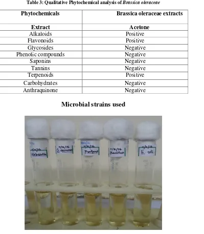
Phytochemical analysis of the solvent extracts of Broccoli was performed by the following standard procedures (Trease and Evans, 2002; Harborne,1998 and Sofowara,1993). In brief, 0.5 ml of extract was added with a drop or two of Mayer’s reagent by the side of the test tube and the formation of white or creamy precipitate indictaes the presence of alkaloids.
Adding 1ml of extract with ammonia and conc. Sulphuric acid and disaapearance of yellow colour on standing indicates flavonoids.
Formation of brown ring at the interface by the addition of 2ml of glacial acetic acid followed by few drops of ferric chloride solution and 1ml of conc.Sulphuric acid to the extracts revealed the presence of glycosides.
Adding few drops of neutral ferric chloride to the extract and the development of dark green colour indicates the presence of the phenolic compounds
Existence of froth formation during warming and vigorous shaking indicates saponins.
Appearance of brownish green or blue black colouration after adding 0.1% ferric chloride to the cooled extract indicates tannis.
Boil the extract with the little amount of Benedict’s solution; if glucose is present the colour change from blue to opaque green, then to yellow and finally to red indicates the presence of carbohydrates.
Boil the test material with 1ml of dil.sulphuric acid in test tube for 5 minutes, centrifuge and filter while hot, filtrate, cool and shake with an equal volume of dichloromethane which separates the lower dichloromethane layer and shake with half of its volume with dil.ammonia , the red colour is produced in the ammonical layer indicates anthraquinone.
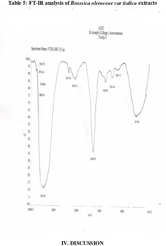
FT-IR Analysis
FT-IR analysis (Fourier Transform Infrared) is perhaps the most powerful tool for identifying the types of chemical bonds (functional groups). The wavelength of light absorbed is characteristic of the chemical bond which can be seen in the annotated spectrum. By interpreting the infrared absorption spectrum, the chemical bonds in a molecule can be determined.
For the FT-IR study, dried powder of acetone extarct (10mg) of Brassica oleraceae was taken in a mortar and pestle and ground with 2.5mg of dried potassium bromide (KBr). The powder so obtained was filled in a 2mm internal diameter micro-cup and loaded onto FT-IR set at 26ºC ± 1ºC. The samples were scanned using infrared in the range of 3500-500 cm-1 using Fourier Transform Infrared Spectrometer (Shimadzu, IR Affinity 1, and Japan). The spectral data obtained were compared with the reference chart to identify the functional groups present in the sample.
HPLC Analysis
The High-performance liquid chromatography coupled with diode array detector (HPLC-DAD) analysis of polyphenolic compounds of vegetable extracts was measured according to the existing method in the laboratory, (Jaiswal et al.,2011a).
In brief, the HPLC system consisted of a reversed-phase HPLC column on an Alliance HPLC (e2695 separations modules; waters, Milford, MA, USA) equipped with an autosampler and controller with dual pump, a 2998 photodiode array detector and the Empower software.
An Atlantis C18 column (250x4.6mm, 5µm particle size) from waters was used for the polyphenolic separation at 25ºC. Mobile phase used was similar to as reported in earlier studies (Jaiswal et al., 2011a).
The chromatograms ware monitored at 280nm [hydroxy benzoic acid (HBA)], 320nm [hydroxy cinnamic acid (HCA)], 360nm (flavonea and flavonols) and 520nm (anthocyanins); complete spectral data were recorded in the range of 220-600nm.
The HPLC-DAD recorded UV-vis spectrum of each peak of the chromatogram which allowed explicit attribution of each chromatographic peak to distinct class of PPs as each class exhibits a characteristic UV-vis spectrum. Different groups of PPs were identified by comparing their UV-vis spectra with spectra of reference compounds and reported values. Polyphenolic profiles at 280nm for the studied Brassica vegetables are presented here.
III. RESULTS
Antibacterial activity of Brassica oleraceae
The results of agar well diffusion method and the microdilution method of Brassica oleraceae
using the solvent acetone as depicted in Table 1.
Table 1: Antibacterial activity of the acetonic extracts of Brassica oleraceae using agar well diffusion method Zone of inhibition in diameter (mm)
Microorganisms Acetone
Escherichia coli 27
Pseudomonas 26
Bacillus 20
Klebsiella 22
Table 2: Minimum Inhibitory Concentration (MIC) of Brassica oleraceae against the tested microorganisms
Microorganisms Acetone extract (mg/ml)
40 20 10 5
Escherichia coli 0.54 0.40 0.26 0.20
Pseudomonas 0.27 0.21 0.17 0.19
Bacillus 0.32 0.27 0.21 0.15
Staphylococcus aureus 0.26 0.28 0.20 0.19
Klebsiella 0.17 0.26 0.21 0.18
The minimum inhibitory concentration (MIC) of the extracts to inhibit the microorganisms was determined by using the microdilution method.
Table 3: Qualitative Phytochemical analysis of Brassica oleraceae
Phytochemicals Brassica oleraceae extracts
Extract Acetone
Alkaloids Positive
Flavonoids Positive
Glycosides Negative
Phenolic compounds Negative
Saponins Negative
Tannins Negative
Terpenoids Positive
Carbohydrates Negative
Anthraquinone Negative
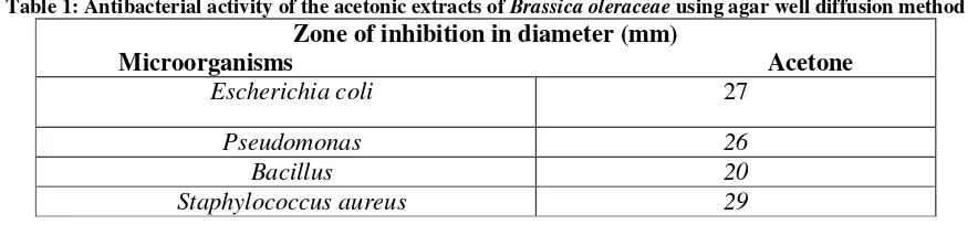
Plate 1: Well diffusion of Brassica oleraceae extracts by using the bacterial strains
A B
C
D
E
Plate2: Microdilution of Brassica oleraceae extracts by using the bacterial strains
Plate 3: Phytochemical Analysis of Brassica oleraceae.L
Alkaloids [Mayer’s Test] Flavonoids Glycosides
Terpenoids Carbohydrates Anthraquinone
Table 4: High Performance Liquid Chromatography (HPLC) of Brassica oleraceae extracts
Table 5: FT-IR analysis of Brassica oleraceae var italica extracts
IV. DISCUSSION
In recent years the use of plants as source of drugs has been increased for the treatment of infectious diseases. Hence, there is a need to move towards the traditional medicine which can serve as novel therapeutics. Numerous studies have highlighted the potential importance of vegetables as a source of medicine which has been inherited as an important component of the health care system in India. Vegetable extracts are given singly or as concoctions for the treatment of microbial diseases. In the present study notable activity was observed against all the tested micro organisms. In an overview of the bioactivity data obtained from the current study, it can be highlighted that the tested extracts have potential to inhibit bacteria and fungi. Pseudomonas aeruginosa exhibited more inhibitory
synthesis is an alternative target for antifungal agents because it is unique to the pathogen. Because of its uniqueness, fungal cell wall offers a unique target site for developing new antibiotic. The phytochemical obtained from the vegetables have a potential role in health care industries and also serve as a lead chemicals for new drug development with diverse range of antimicrobial properties. Bioactive substances from this plant can therefore be employed in the formulation of antimicrobial drugs for the treatment of various bacterial and fungal infections. Phytochemical agents act as antimicrobial agent by inhibiting the extracellular enzyme acting on the substrates required for microbial growth or by inhibiting oxidative phosphorylation of microbial metabolism. The phytochemical contents of the leafy vegetables serve as supplements for food and also have the potential to improve the health status of its users as a result of the presence of various compounds vital for good health. Hence, the recent research showed that the complex mixture of phytochemicals in vegetables provides a better protective effect on health than single phytochemicals. Therefore, the complex components of cauliflower extract needs to be scrutinized in depth, in order to find out the best mixture of effective components that had role in the currently shown antibacterial and antifungal activity. The presence of characteristic functional groups may be responsible for the medicinal properties of Brassica oleracea which contain high therapeutic content. Determination of respective antimicrobial potential and toxicological evaluation of these extracts with the view to formulate novel chemotherapeutic agents to be used future is worth mentioning.
To conclude, the present bioprospecting study justifies the medicinal uses of the vegetables and also reveals the potentialities to isolate promising natural compound for the management of the bacterial infectious disease.
IV. ACKNOWLEDGEMENT
The authors wish to place their record of thanks to the authorities of Jamal Mohamed College (Autonomous), Trichy, for providing a good infrastructure facility.
BIBLIOGRAPHY
[1] Park K. 2003 Textbook of preventive and social medicine. 19th ed. Banarsidan Bhanol Publishers, Jabalpur;. [2] Fliri AF, Loging WT, Thadeio PF, Volkmann RA. Analysis of drug – induced effect patterns to link structure and
side effects of medicines. Nature Chemical Biology 2005; 1: 389 – 397.
[3] Niraimathi Kl, Karunanithi M, Brindha P. (2012) Phytochemical and in-vitro screening of aerial parts of Cleome
viscosa Linn.extracts (Capparidaceae). International Journal of Pharmacy and Pharmaceutical Sciences; 4(2): 27-30.
[4] Ambrose CB, Tang L. Cruciferous vegetable intake and cancer prevention: role of nutrigenetics. Cancer Prevention Research (Phila Pa) 2009; 2(4): 298-300.
[5] Stoke EJ1975. Clinical Bacteriology. 4th ed. Edward Arnold publishers.
[6] Singh G, Kumar P and Jindal A. (2012) Antibacterial potential of sterols of some medicinal plants. International
Journal of Pharmacy and Pharmaceutical Sciences; 4(3): 159 -162.
[7] Trease GS, Evans HC. 1978. Text book of Pharmacognosy. 9th ed. Baitar Zindall and Co. London.
[8] Naumann D, Helm D, Labischinski H, Giesbrecht P. 1991b The characterization of microorganisms by Fourier‐transform infrared spectroscopy (FT‐IR). In: Nelson WH, editor. In Modern Techniques for Rapid
Microbiological Analysis. New York: VCH Publishers;. pp. 43–96.
[9] Sherman JM, Hodge HM. 1936 The bactericidal properties of certain plant juices. Journal of Bacteriology; 31: 96 – 98.
[10] Shrisha DL, Raveesha KA, Nagabhushan S. 2011. Bioprospecting of selected medicinal plants for antibacterial activity against some pathogenic bacteria. Journal of medicinal plants research; 5(17): 4087 – 4093.
[11] Vidula RD, Pragna DD. 2000 An agar planted assay to detect cell wall active antifungal agent. Current Science; 78 (5): 615 – 617.
[12] Umamaheswari N, Rhama S, Kannahi M, Madhavan S. 2010.Antifungal activities of Clitoria ternatea leaf extract.
Advances in Plant Sciences; 23 (II): 377 – 378.
[13] Chu YF, Sun J, Wu X, Liu RH. 2002 Antioxidant and antiproliferative activities of common vegetables. Journal of
Agricultural Food Chemistry; 50: 6910 – 6914.
[15] Apak, R., K. Güçlü, M. Özyürek, S.E. Karademir and E. Erça. 2006. The cupric ion reducing antioxidant capacity and polyphenolic content of some herbal teas. Int. J. Food Sci. Nut. 57: 292-304.
[16] Arabshahi-Delouee S. and A. Urooj. 2007. Antioxidant properties of various solvent extracts of mulberry (Morus indica L.) leaves. Food Chem. 102: 1233-1240.
[17] Baardseth, P. 1989. Effect of selected antioxidants on the stability of dehydrated mashed potatoes. Food Addit. Contam. 6: 201-207.
[18] Bast, A., G.R.M.M. Haenen and C.J.A. Doelman. 1991. Oxidants and antioxidants: state of the art. Am. J. Med. 91: 2-13.
[19] Beauchamp, C. and I. Fridovich. 1971. Superoxide dismutase: improved assays and an assay applicable to acrylamide gels. Anal. Biochem. 44: 276-287.
[20] Blois, M.S. 1958. Antioxidant determinations by the use of a stable free radical. Nature 26, 1199-1200.
[21] Brand-Williams, W., M.E. Cuvelier and C. Berset. 1995. Use of a free radical method to evaluate antioxidant activity. Lebensm. Wiss. Techonol. 28: 25-30.
[22] Büyükokuro¤lu, M.E., ‹. Gülçin, M. Oktay and Ö.‹. Küfrevio¤lu. 2001.
[23] In vitro antioxidant properties of dantrolene sodium. Pharmacol.Res. 44: 491-495. [24] Chung, Y.C., C.T. Chang, W.W. Chao, C.F. Lin and S.T. Chou. 2002.
[25] Antioxidative activity and safety of the 50% ethanolic extract from red bean fermented by Bacillus subtilis IMR-NK1. J. Agric. Food Chem. 50: 2454-2458.
[26] Cook, N.C. and S. Samman. 1996. Flavonoids: chemistry, metabolism, cardioprotective effects and dietary sources.
J. Nut. Biochem. 7:66-76.
[27] Cos, P., L.Y. Ying, M. Calomme, J.H. Hu, K. Cimanga, B. Van Poel, L. Pieters, A.J. Vlietinck and D.V. Berghe. 1998. Structure activity relationships and classification of flavonoids as inhibitors of xanthine oxidase and superoxide scavengers. J. Nat. Prod. 61:71-76.
[28] Demo, A., P. Kefalas and D. Boskou. 1998. Nutrient antioxidants in some herbs and Mediterranean plant leaves. Food Res. Int. 32:351–354.
[29] Dinis, T.C.P., V.M.C. Madeira and L.M. Almeida. 1994. Action of phenolic derivates (acetoaminophen, salycilate, and 5-aminosalycilate) as inhibitors of membrane lipid peroxidation and as peroxyl radical scavengers. Arch. Biochem. Biophys. 315: 161-169.
[30] Duh, P.D., Y.Y. Tu and G.C. Yen. 1999. Antioxidant activity of water extract of harng jyur (Chrysanthemum morifolium Ramat). Lebensm. Wiss. Technol. 32: 269-277.
[31] Elmastafl, M., ‹. Gülçin, Ö. Beydemir, Ö.‹. Küfrevio¤lu and H.Y. Aboul-Enein. 2006. A study on the in vitro antioxidant activity of juniper (Juniperus communis L.) seeds extracts. Anal. Lett. 39: 47-65.
[32] Elmastas, M., ‹. Gülçin, Ö. Iflıldak, Ö.‹. Küfrevio¤lu, K. ‹bao¤lu and H.Y.Aboul-Enein. 2006b. Antioxidant capacity of bay (Laurus nobilisL.) leave e extracts. J. Iran. Chem. Soc. 3: 258-266.
[33] Elmastafl, M., ‹. Gülçin, L. Öztürk and ‹. Gökçe. 2005. Investigation of antioxidant properties of spearmint (Mentha spicata L.). Asian J.Chem. 17: 137-148.
[34] Finefrock, A.E., A.I. Bush and P.M. Doraiswamy. 2003. Current statusof metals as therapeutic targets in Alzheimer’s disease. J. Am.Geriat. Soc. 51: 1143-1148.
[35] Fogliano, V., V. Verde, G. Randazzo and A. Ritieni. 1999. Method for measuring antioxidant activity and its application to monitoring the antioxidant capacity of wines. J. Agric. Food Chem. 47: 1035-1040.
[36] Grice, H.C. 1986. Safety evaluation of butylated hydroxytoluene (BHT) in the liver, lung and gastrointestinal tract. Food Chem. Toxicol.24: 1127-1130.
[37] Gülçin, ‹. 2005. The antioxidant and radical scavenging activities of black pepper (Piper nigrum) seeds. Int. J. Food
Sci. Nut. 56: 491-499.
[38] Gülçin, ‹. and A. Dafltan. 2007. Synthesis of dimeric phenol derivatives and determination of in vitro antioxidant and radical scavenging activities. J. Enzym. Inhib. Med. Chem. 22: 685-695.