Aleksander Łukasiewicz
1, 2, A–d, Tomasz drewa
2, A, d–fSynthetic Implants in Hernia Surgery
1 department of Vascular Surgery, Regional Specialist Hospital in Grudziądz, Poland
2 Tissue Engineering department, Nicolaus Copernicus University, Ludwik Rydygier Collegium Medicum in Bydgoszcz, Poland
A – research concept and design; B – collection and/or assembly of data; C – data analysis and interpretation;
D – writing the article; E – critical revision of the article; F – final approval of article; G – other
Abstract
Abdominal wall hernia correction is one of the most common surgical procedures. 85,000 hernia operations are performed in Poland each year. Modern techniques of abdominal wall reconstruction utilize surgical implants for fascial defect closure. In the 70s and the 80s of the last century, these techniques gained widespread acceptance among surgeons. Significant improvement of results in terms of recurrences was observed. Treatment of large abdominal wall defects became possible. Three types of surgical implants were developed early: polipropylene (PP), poliethylene (PE) and politetrafluoroethylene (PTfE). Unfortunately, negative effects of implanted mate-rial soon became apparent. Excessive native tissues inflammatory response to the implanted matemate-rial, leading to multiple complications was observed. Recurrences due to fibrosis, chronic regional pain, stiffness of the operation site, intestinal adhesions and fistulas, infertility and infections were reported. In some cases the use of standard synthetic implant was contraindicated. Analyzing drawbacks of the standard hernia implants, the medical industry developed new materials to improve treatment results. The most popular, currently utilized synthetic materials, are presented in this review in the context of clinical results (Adv Clin Exp Med 2014, 23, 1, 135–142).
Key words: abdominal hernia, surgery, bioprosthesis.
Adv Clin Exp Med 2014, 23, 1, 135–142 ISSN 1899–5276
REVIEWS
© Copyright by Wroclaw Medical University
The primary aim of abdominal hernia recon-struction is the closure of a fascia defect to avoid potentially deadly complications of hernia incar-ceration and to relieve symptoms accompanying the protrusion of abdominal viscera through the defective abdominal wall. According to the utilized surgical technique, closure under tension or ten-sion-free repair can be distinguished [1].
Closure under tension requires direct approx-imation of fascia margins using a surgical suture. The suture line tension and intrinsic weakness of native tissues in hernia patients result in increased hernia recurrence rates, especially in large (over 5 cm diameter) fascia defects. Considering infe-rior treatment results, indications for this tech-nique are currently confined to small abdominal and inguinal hernias and patients of a significant risk of operative site infection (e.g. strangulated hernia).
The tension-free repair of abdominal wall her-nia requires a surgical implant to fill the fascia
defect without tension. The idea of utilizing the surgical graft comes from the knowledge that her-nia occurrence is only a local manifestation of gen-eral disorder that affects all the connective tissue in hernia patients. Abdominal wall fascias are defec-tive and weakened comparing those in healthy in-dividuals and therefore require additional support to assure favorable outcome. The awareness of ex-ternal fascia support necessity in hernia treatment was already evident in late 80s of the 19th century; however, the development of surgical techniques was hampered by the lack of adequate biomateri-als until the second half of the 20th century. It was then that the first synthetic implants were utilized clinically in hernia reconstruction [2]. Currently, tension-free repair dominates hernia treatment in developed countries including Poland [3].
– Sufficient mechanical strength – Chemical stability
– Lack of carcinogenic properties – Easy sterilization
– Ability to limit foreign body reaction – fabrication in required size and shape – Infection resistance
– Biomechanical properties resembling native tissues.
despite many years of laboratory and clini-cal research, the ideal implant production is still the “Holy Grail” of medical industry. Current-ly, 3 major types of implants are utilized in her-nia surgery:
1. Standard non-absorbable synthetic implants. 2. Composite synthetic implants.
3. Bioimplants, mainly composed of derived from human or animal tissues collagen matrices.
The first 2 types, thanks to favorable properties and low costs, gained widespread acceptance in the surgical community.
Standard Synthetic Implants
Standard synthetic implants are currently the most popular prostheses utilized in hernia surgery. They were developed early, in the 50s and 60s of the last century and vast experience in treatment was gained since then. Nowadays, 3 types of mate-rials are utilized widely: polipropylene (PP), poly-ester (PET) and polytetrafluoroethylene (PTfE).
Polypropylene Implants
PP meshes, due to extensive surgical experi-ence, unique properties and low costs are the most commonly utilized implants in hernia surgery [6]. Polypropylene is derived from polymerization of propylene, a derivative of propane. It is a hydro-phobic, resistant to enzymatic degradation poly-mer that is usually manufactured in a form of monofilament mesh. It induces an intensive fi-brous reaction of the surrounding tissues that re-sults in the formation of a strong and resistant to hernia relapse scar. On the other hand, this pro-cess leads to significant (even 30% in some stud-ies) mesh area reduction and sometimes hernia re-currence. PP is susceptible to oxidative stress and subsequent filament damage, a phenomenon occa-sionally observed on microscopy leads to reduced prosthesis durability [7].
Currently utilized PP meshes are divided into 2 categories: heavy-weight and light-weight; how-ever, a strict distinction is problematic [8]. At first, the breakdown criterion was the weight. Implants
over 80 g per m2 were referred to as heavy-weight, the rest was described as light-weight. It was soon recognized, however, that the implant properties depend not only on weight alone but also on its structure, compliance, elasticity and pore size. Ac-cording to these criteria, the term heavy-weight was ascribed to stiff, non-elastic implants with small, below 1 mm pore size. Tensile strength of such implants significantly exceeds physiologic re-quirements (approximately 16 N/cm) [9]. Heavy weight implants induce excessive tissue reaction and fibrosis. This leads to increased stiffness of ab-dominal wall, frequent foreign body sensation and chronic pain. Light-weight meshes have a larger, over 1 mm, pore diameter. They are readily in-corporated and induce less inflammatory reac-tion than standard heavy-weight grafts. The ten-sile strength is similar to physiologic and elasticity is significantly greater than in standard implants (20–35% elongation under physiologic stress). The stiffness of the abdominal wall is less pronounced when light-weight grafts are utilized in hernia re-construction. foreign body sensation and chron-ic pain are reduced. Patients report less consump-tion of analgesics at the early postoperative period, quicker ambulation and return to work [10].
However, light-weight prostheses are not with-out drawbacks. There are reports that reduced ten-sile strength has a negative impact on the hernia recurrence and migration rate [11]. Studies were published that link the use of light-weight prosthe-ses with reduced male fertility and increased peri-toneal adhesions after operation [12].
Polipropylene is relatively resistant to infec-tion. Monofilament mesh structure and large fila-ment interstices allow for easy penetration of anti-biotic and host immune cells as well as reduce the adhesion of bacteria to the mesh structure. debri-dement with systemic antibiotic is a popular treat-ment modality of a limited mesh infection. Par-tial excision of macroscopically infected portion of the implant was also a suggested effective measure in such situations [13]. Currently, diverse types of polypropylene implants prepared for different clinical scenarios are available for surgeons.
Polyester Implants
readily adapts to the natural curvature of the ab-dominal wall. PET implants induce significant fi-brotic reaction of surrounding tissues that facili-tates permanent implant incorporation. Compared to PP meshes, PET prostheses possess superior me-chanical features especially pliancy that makes the implantation technique easier. They are less prone to postoperative retraction, a process that is signif-icant in PP implants. If used intraperitoneally, they induce extensive adhesions with viscera and, there-fore, such placement is contraindicated [15]. Some authors reported increased susceptibility of PET implants to infection and increased rate of intes-tinal bowel fistula while comparing them with PP meshes [16]. Standard PET prostheses are maly used in open surgical techniques that do not in-volve abdominal cavity e.g. Lichtenstein inguinal hernia correction [17].
Polytetrafluoroethylene
Implants
Polytetrafluoroethylene (PTfE) is a synthet-ic polymer produced in the process of polymer-ization of tetrafluoroethylene. PTfE is hydropho-bic: neither water nor water-containing substances wet PTfE, as fluorocarbons demonstrate mitigat-ed London dispersion forces due to the high elec-tronegativity of fluorine. This property determines the resistance of PTfE implants to the formation of adhesions with abdominal viscera [18]. It is non-reactive, partly because of the strength of car-bon–fluorine bonds. Stability of PTfE implants is similar to that offered by PP meshes. fibers in the PTfE prostheses are closely packed (pores below 75 nm). Therefore, they are considered micropore implants that are rather encapsulated than pene-trated by fibroblasts after implantation. during the healing processes, connective tissue forms a fi-brous envelope around the whole implant that sub-sequently contracts decreasing the implant surface causing folding of the prosthesis [19]. This may lead to the implant malfunction and an increased recurrence rate that reaches 20% after ventral her-nia repairs [20]. In the treatment of abdominal wall defects, PTfE prostheses have been utilized for many years. Thanks to their specific anti-ad-hesive properties, they are used when contact of the implant with viscera is anticipated and when intraperitoneal techniques of hernia correction are utilized. A serious drawback of PTfE prostheses is their relative susceptibility to infection. Some series reported even over 10% of implant-related infections [21].
Modern Synthetic Implants
in Abdominal Wall
Reconstructions
Application of the above-mentioned standard surgical synthetic implant in potentially contami-nated operative field carries a high risk of implant infection, abscess or purulent fistula formation, sometimes, subsequent serious complications and even death. furthermore, PP and PET implants, if placed in peritoneal cavity, are prone to viscer-al adhesion formation, which subsequently may lead to ileus or enteric fistula. The quest for a safe and effective hernia implant has yielded a produc-tion of numerous new biomaterials with modified properties to achieve better results.
These new biomaterials can be generally as-signed to one of 2 classes:
A. Absorbable surgical implants.
B. Composite implants that consist of at least 2 components:
– stable and nonabsorbable (most often PP), that is permanent scaffold ensuring integ-rity of the implant and adequate tensile strength.
– anti-adhesive (either absorbable or not) that protects the implant from adhesion formation and in some implants serves as an additional temporary scaffold that reduces the amount of nonabsorbable component while maintaining the struc-tural strength facilitating implantation.
Absorbable Synthetic
Implants
the “open abdomen” treatment and has many ad-vantages. It reduces the risk of complications in the peritoneal cavity and frequency of intestinal fistula as well as abates wound edges retraction that com-plicates the definite closure of abdominal wound. The application of an absorbable implant, defer-ring the final procedure, allows the definite treat-ment to be performed for a patient in better gener-al and locgener-al conditions and with a decreased risk of significant complications [24]. PGA prostheses are often utilized in conjunction with negative pres-sure wound therapy systems, since they are water permeable. This strategy further improves treat-ment results in complicated cases. Yet another dis-tinctive feature of absorbable meshes, contrary to permanent ones, is that they do not require remov-al in case of infection.
Composite Implants
Composite implants are utilized in various clinical scenarios. The most popular indications for application are:
– when intraperitoneal on-lay mesh procedure (IPOM) is performed to correct abdominal wall hernia. This technique facilitates the surgical pro-cedure since no extensive preperitoneal dissection is required to secure the implant in place. It also re-duces the duration of the procedure and decreases the risk of complications
– it is impossible to interpose the implant and abdominal viscera with a layer of native tissues, like in extensive abdominal wall defects
– when it is paramount to reduce the amount of permanent synthetic material in the wound to reduce probability of chronic pain as in case of in-guinal herniorrhaphy.
Composite implants can be generally classified into one of 2 groups:
– the first, partially absorbable, where a non-absorbable scaffold (most often PP) is coated with a layer of absorbable polymer. Such a coating in-creases the tensile strength of the implant at the moment of implantation, allowing a reduced amount of permanent material in the wound. fur-ther, it protects the implant material when it comes in contact with viscera in the case of intraabdom-inal placement. The nonabsorbable component is then encased with connective tissue fibers by the time the coating is completely absorbed.
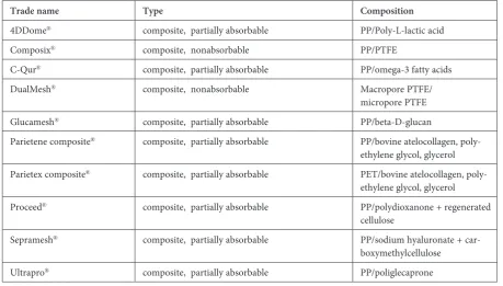
– the second, entirely nonabsorbable, with di-verse inner and outer surface. The outer (fascial) surface is composed of material that has proper-ties (either intrinsic or structural) inducing con-nective tissue cells migration and enhancing fibro-sis. The inner (visceral) surface is smooth, resistant to adhesions formation and most often composed of PTfE. Such implants are used in the IPOM pro-cedures (Table 1).
Table 1. Composite implants utilized in humans
Trade name Type Composition
4ddome® composite, partially absorbable PP/Poly-L-lactic acid
Composix® composite, nonabsorbable PP/PTfE
C-Qur® composite, partially absorbable PP/omega-3 fatty acids
dualMesh® composite, nonabsorbable Macropore PTfE/
micropore PTfE Glucamesh® composite, partially absorbable PP/beta-d-glucan
Parietene composite® composite, partially absorbable PP/bovine atelocollagen,
poly-ethylene glycol, glycerol Parietex composite® composite, partially absorbable PET/bovine atelocollagen,
poly-ethylene glycol, glycerol
Proceed® composite, partially absorbable PP/polydioxanone + regenerated
cellulose
Sepramesh® composite, partially absorbable PP/sodium hyaluronate +
Partially Absorbable
Composite Implants
It is important to realize that partially absorb-able implants vary among themselves in many ways: chemical composition, 3 dimensional structure, weight of permanent material, absorption time of absorbable component. Numerous animal studies were performed to compare these materials. Also, much clinical research comparing standard pros-theses was done to confirm favorable outcomes (Ta-ble 2). Clinical experience indicates that composite implants are a valuable alternative to standard sur-gical prostheses utilized in standard sursur-gical treat-ment. They are essential in the latest minimally in-vasive surgical techniques, such as single incision laparoscopic surgery (SILS) or natural orifice trans-luminal endoscopic surgery (NOTES). Early and mid-term clinical results indicate that the reduction of the amount of permanent component in the com-posite implants benefits the treatment. It abates the frequency and intensity of postoperative pain and allows quicker mobilization and faster convales-cence. Actual benefits of reduced discomfort extend to even 6 months of postoperative period [27, 31].
Undoubtedly, a significant advantage of the partially absorbable implants, when compared with standard polypropylene surgical meshes, is the re-duced influence on abdominal wall motility and elasticity. Experimental studies revealed that ab-dominal wall elongates approximately 11–32% un-der physiologic tension (approximately 16 N/cm). If high pressure is applied, the difference in dimen-sions may reach even 100% [38]. In such circum-stances, implantation of standard, rigid prosthesis induces significant tension at the fixation points. This leads to increased inflammation and fibrosis and – as a result – rigidity of abdominal wall, for-eign body sensation, chronic pain and possibility of recurrence. Modern composite implants in most cases pose elasticity comparable to that of the ab-dominal wall. This allows the incorporation of the prostheses without excessive tension and fibrosis, which probably determines improved early post-operative course. Certainly, it is the structure and amount of the permanent component that affects long-term operation outcomes but a significant re-duction of the non-absorbable scaffold would be difficult without absorbable component reinforc-ing it at the time of surgery [39].
Non-absorbable
Composite Implants
Basic structural component of non-absorb-able implants is PTfE. Its continuous layer on the visceral side of the prosthesis determines its
anti-adhesive properties and allows safe intraperitoneal placement. The outer layer varies in the composi-tion. In some grafts it also consists of PTfE but in such a case the outer surface of the graft is rough and wrinkled, with larger pores to allow some infil-tration of the native connective tissue to reinforce defect correction (e.g. dualmesh™). In others, the outer layer is made of polypropylene, a polymer inducing fibrosis and incorporation of the pros-thesis (e.g. Composix™) (Table 1). Clinical results of these grafts application were compared in the clinical trial by Bondi et al. [40]. despite signifi-cant differences in handling and rigidity in favor of dualmesh™, results were similar with both im-plants. According to these authors, the final out-come of the abdominal wall defect correction de-pends less on the type of implant but rather on the meticulous surgical technique especially adherence to current recommendations of 4 cm excess of the prosthesis to secure wall defect.
Modern synthetic composite implants allow further development of surgical minimally inva-sive techniques. A surgeon is no longer obligated to perform extensive surgical dissection of the ab-dominal wall to assure proper prosthesis coverage by native tissues, since the risk of visceral adhesions and complications is significantly diminished. It shortens the duration of operation, decreases post-operative pain, allows rapid postpost-operative ambu-lation and early return to work. This way despite increased costs of the composite grafts, the overall economic impact is favorable.
Summary
Table 2. Clinical application results of the use of composite implants in abdominal wall reconstructions. AW d – abdominal wall defect, NA – data not available, PET – polyester, PP – polipropylene, RCT – randomized controlled trial Author
Publication year
Study type Material type Defect type Patients / Procedures number
Reccurence (%)
Infection (%)
Seroma
(%)
Observation time
Comment
Amma- turo
[25]
2010
Prospective observational
PET/bovine atelo -collagen Small ventral hernia 100 6 (6) 4 (4) 8(8) 12 months Clean operative field, Large fascia defects Johanet [26] 2006
Prospective cohort
PET/bovine atelo -collagen Small umbili -cal hernia 259 4 (2) 2 (1) 36 (18) 12 months 19 patients lost at follow-up Liu [27] 2011
Prospective observational PP/polydioxanone + regenerated
cel -lulose Large ventral hernia 36 0 2 (5.6%) 6 (16.7) 28 months Clean-contaminated operative field in 22,2% Langen- bach [28] 2006 d ouble blind RCT 1-PP monofilament 2-PP multifilament 3-PP/polyglactin
Inguinal hernia
90 (30 sub -jects in 3 groups) NA NA NA 12 weeks Pain and daily activity restric tion more frequent in group than groups 2 and 3
Torcivia [29]
2011
Randomized study
1-PP standard 2- pp/beta-d -glucan
Inguinal hernia
47 (24/23) NA NA NA 30 days More pain in group 1 on postop day Moreno- Egea [30] 2010
Prospective observational
PET/bovine atelo -collagen Ventral her -nia 200 11(6.25) 0 6 (3) 60 months 1 case of ileus unrelated to mesh Śmie- tański [31] 2009
Prospective cohort PP/ poliglecaprone Inguinal hernia
242 4 (1.6) NA NA 36 months Lack of data on other compli cations Iannitti [32] 2008
Prospective cohort
PP/PT fE Ventral her -nia 455 6 (1.3) 6 (1.3) 22 (4.8) 29 months 3 cases of ileus (1 operated)
Bernard [33]
2007
Prospective cohort
PP/PT fE Large ventral hernia 61 3(4.9) 2 (3.2) 1 (1.6) 35 months 3 cases of ileus (1 operated) Olmi [34] 2006
Prospective cohort
PET/bovine
atelo
-collagen
Ventral hernia
and AW d 178 4 (2.5) 1 (0,6) 7 (4.4) 29 months 10% of cases- primary abdom inal wall defects
Berrevoet [35]
2009
Prospective cohort PP/polydioxanone + regenerated
cel -lulose Ventral her -nia 114 4 (3.5) 0 12 (10.5) 27 months 1 case of ileus related to the mesh Balique [36] 2005
Prospective cohort
PET/bovine atelo -collagen Ventral her -nia 80 5 (6.7) 2 (0.3) 13 (16.3) 48 months 2 cases of ileus unrelated the mesh
Briennon [37]
2011
Prospective cohort
References
[1] Kingsworth A, LeBlanc K: Hernias: inguinal and incisional. Lancet 2003, 362, 1561–1571.
[2] Usher FC, Fries JG, Ochsner JL, Tuttle LL Jr: Marlex mesh, a new plastic mesh for replacing tissue defects. Clinical studies. Arch Surg 1959, 78, 138–145.
[3] Narodowy Fundusz Zdrowia – strona internetowa: NFZ statystyka JGP. http://prog.nfz.gov.pl/jgp/KatalogJGP. aspx. access on 21.05.2012.
[4] Cumberland VH: A preliminary report on the use of a prefabricated nylon weave in the repair of ventral hernia. Med J Aust 1952, 1, 143–144.
[5] Hamer-Hodges DW, Scott NB: Surgeon’s workshop. Replacement of an abdominal wall defect using expanded PTfE sheet (Gore-tex). J R Surg Edinb 1985, 30, 65–67.
[6] Shankaran V, Weber DJ, Reed RL 2nd, Luchette FA: A review of available prosthetics for ventral hernia repair. Ann Surg 2011, 253, 16–26.
[7] Mary C, Marois Y, King MW, Laroche G, Douville Y, Martin L, Guidoin R: Comparison of the in vivo behavior of polyvinylidene fluoride and polypropylene sutures used in vascular surgery. ASAIO J 1998, 44, 199–206.
[8] Bringman S, Conze J, Cuccurullo D, Deprest J, Junge K, Klosterhalfen B, Parra-Davila E, Ramshaw E, Schumpelick V: Hernia repair: the search for ideal meshes. Hernia 2010, 14, 81–87.
[9] Klinge U, Klosterhalfen B, Birkenhauer V, Junge K, Conze J, Schumpelick V: Impact of polymer pore size on the interface scar formation in a rat model. J Surg Res 2002, 103, 208–214.
[10] Smietański M, Bury K, Smietańska IA, Owczuk R, Paradowski T; Polish Hernia Study Group: five-year results of a randomised controlled multi-centre study comparing heavy-weight knitted versus low-weight, non-woven polypropylene implants in Lichtenstein hernioplasty. Hernia 2011, 15, 495–501.
[11] Chui LB, Ng WT, Sze YS, Yuen KS, Wong YT, Kong CK: Prospective, randomized, controlled trial comparing lightweight versus heavyweight mesh in chronic pain incidence after TEP repair of bilateral inguinal hernia. Surg Endosc 2010, 24, 2735–2738.
[12] Peeters E, Spiessens C, Oyen R, De Wever L, Vanderschueren D, Penninckx F, Miserez M: Laparoscopic ingui-nal hernia repair in men with lightweight meshes may significantly impair sperm motility: a randomized controlled trial. Ann Surg 2010, 252, 240–246.
[13] Medberry CJ, Tottey S, Jiang H, Johnson SA, Badylak SF: Resistance to infection of five different materials in a rat body wall model. J Surg Res 2012, 173, 38–44.
[14] Cozad MJ, Grant DA, Bachman SL, Grant DN, Ramshaw BJ, Grant SA: Materials characterization of explanted polypropylene, polyethylene terephthalate, and expanded polytetrafluoroethylene composites: spectral and ther-mal analysis. J Biomed Mater Res B Appl Biomater 2010, 94, 455–462.
[15] Burger JW, Halm JA, Wijsmuller AR, ten Raa S, Jeekel J: Evaluation of new prosthetic meshes for ventral hernia repair. Surg Endosc 2006, 20, 1320–1325.
[16] Leber GE, Garb JL, Alexander AI, Reed WP: Long-term complications associated with prosthetic repair of inci-sional hernias. Ann Surg 1998, 133, 378–382.
[17] Poghosyan T, Veyrie N, Corigliano N, Helmy N, Servajean S, Bouillot JL: Retromuscular mesh repair of midline incisional hernia with polyester standard mesh: monocentric experience of 261 consecutive patients with a 5-year follow-up. World J Surg 2012, 36, 782–790.
[18] Voskerician G, Rodriguez A, Gingras PH: Macroporous condensed poly(tetra fluoro-ethylene). II. In vivo effect on adhesion formation and tissue integration. J Biomed Mater Res A 2007, 82, 426–435.
[19] Orenstein SB, Saberski ER, Kreutzer DL, Novitsky YW: Comparative analysis of histopathologic effects of syn-thetic meshes based on material, weight, and pore size in mice. J Surg Res 2012, 176, 423–429.
[20] Trupka AW, Schweiberer L, Hallfeldt K, Waldner H: Management of large abdominal wall hernias with foreign implant materials (Gore-Tex patch). Zentralbl Chir 1997, 122, 879–884.
[21] McGinty JJ, Hogle NJ, McCarthy H, Fowler DL: A comparative study of adhesion formation and abdominal wall ingrowth after laparoscopic ventral hernia repair in a porcine model using multiple types of mesh. Surg Endosc 2005, 19, 786–790.
[22] Sriussadaporn S, Sriussadaporn S, Pak-art R, Krittayakirana K, Prichayuhd S: Planned ventral hernia with absorbable mesh: a life-saving method in relaparotomy for septic abdomen. J Med Assoc Thai 2010, 93, 449–456.
[23] Prichayudh S, Sriussadaporn S, Samorn P, Pak-Art R, Sriussadaporn S, Kritayakirana K, Capin A: Management of open abdomen with an absorbable mesh closure. Surg Today 2011, 41, 72–78.
[24] Sikkink CJ, Vries de Reilingh TS, Malyar AW, Jansen JA, Bleichrodt RP, van Goor H: Adhesion formation and reherniation differ between meshes used for abdominal wall reconstruction. Hernia 2006, 10, 218–222.
[25] Ammaturo C, Bassi UA, Bassi G: Outcomes of the open mesh repair of large incisional hernias using an intrap-eritoneal composite mesh: our experience with 100 cases. Updates Surg 2010, 62, 55–61.
[26] Johanet H, Dabrowski A, Hauters P; Club Coelio: Repair of large abdominal wall defects using the Proceed™ surgical mesh with open intra-peritonium onlay method. Hernia 2006, 10, 414–418.
[27] Liu F, Li J: Laparoscopic cure of small ventral hernias with composite mesh. Saudi Med J 2011, 32, 504–509.
[28] Langenbach MR, Schmidt J, Zirngibl H: Comparison of biomaterials: three meshes and TAPP for inguinal hernia. Surg Endosc 2006, 20, 1511–1517.
[30] Moreno-Egea A, Bustos JA, Girela E, Aguayo-Albasini JL: Long-term results of laparoscopic repair of incisional hernias using an intraperitoneal composite mesh. Surg Endosc 2010, 24, 359–365.
[31] Smietański M, Bigda J, Zaborowski K, Worek M, Sledziński Z: Three-year follow-up of modified Lichtenstein inguinal hernioplasty using lightweight poliglecaprone/polypropylene mesh. Hernia 2009, 13, 239–242.
[32] Iannitti DA, Hope WW, Norton HJ, Lincourt AE, Millikan K, Fenoglio ME, Moskowitz M: Technique and outcomes of abdominal incisional hernia repair using a synthetic composite mesh: a report of 455 cases. J Am Coll Surg 2008, 206, 83–88.
[33] Bernard C, Polliand C, Mutelica L, Champault G: Repair of giant incisional abdominal wall hernias using open intraperitoneal mesh. Hernia 2007, 11, 315–320.
[34] Olmi S, Erba L, Magnone S, Bertolini A, Croce E: Prospective clinical study of laparoscopic treatment of incision-al and ventrincision-al hernia using a composite mesh: indications, complications and results. Hernia 2006, 10, 243–247.
[35] Berrevoet F, Fierens K, De Gols J, Navez B, Van Bastelaere W, Meir E, Ceulemans R: Multicentric observational cohort study evaluating a composite mesh with incorporated oxidized regenerated cellulose in laparoscopic ventral hernia repair. Hernia 2009, 13, 23–27.
[36] Balique JG, Benchetrit S, Bouillot JL, Flament JB, Gouillat C, Jarsaillon P, Lepère M, Mantion G, Arnaud JP, Magne E, Brunetti F: Intraperitoneal treatment of incisional and umbilical hernias using an innovative composite mesh: four-year results of a prospective multicenter clinical trial. Hernia 2005, 9, 68–74.
[37] Briennon X, Lermite E, Meunier K, Desbois E, Hamy A, Arnaud JP: Surgical treatment of large incisional her-nias by intraperitoneal insertion of Parietex® composite mesh with an associated aponeurotic graft (280 cases). J Visc Surg 2011, 148, 54–58.
[38] Smietański M, Bury K, Tomaszewska A, Lubowiecka I, Szymczak C: Biomechanics of the front abdominal wall as a potential factor leading to recurrence with laparoscopic ventral hernia repair. Surg Endosc 2012, 26, 1461– –1467.
[39] Langenbach MR, Schmidt J, Ubrig B, Zirngibl H: Sixty-month follow-up after endoscopic inguinal hernia repair with three types of mesh: a prospective randomized trial. Surg Endosc 2008, 22, 1790–1797.
[40] Biondi A, Tropea A, Monaco N, Musmeci G, Basile G, Basile F: Laparoscopic treatment of abdominal wall her-nias: prosthesis material comparison Minerva Chir 2011, 66, 547–552.
Address for correspondence:
Aleksander Łukasiewiczdepartment of Vascular Surgery Regional Specialist Hospital L. Rydygiera 15–17
86-300 Grudziądz
Tel.: 56 641 40 82, 56 641 40 89, 505 83 63 85 E-mail: [email protected]
Conflict of interest: None declared