Hady Razak Hady
A–D, F, Maria Sołdatow
B–D, Jerzy Łukaszewicz
B, C,
Magdalena Łuba
B, D, Jacek Pierko
B, Piotr Myśliwiec
B, C, Jerzy R. Ładny
B, C, E,
Jacek Dadan
A, E, FSurgical Treatment of Malignant and Benign Colorectal
Neoplasms Based On Authors’ Clinical Data
Postępowanie chirurgiczne w nowotworach złośliwych i łagodnych
jelita grubego w materiale własnym
1st Department of General and Endocrinological Surgery, Medical University of Bialystok, Poland
A – research concept and design; B – collection and/or assembly of data; C – data analysis and interpretation;
D – writing the article; E – critical revision of the article; F – final approval of article; G – other
Abstract
Background. Colorectal cancer remains a huge diagnostic and therapeutic issue in Poland and worldwide. World epidemiological data indicates a constant increase in morbidity in recent decades.
Objectives. The aim of this research was to present surgical procedures in malignant and benign colorectal neo-plasms based on authors’ clinical data.
Material and Methods. Between 2001 and 2010, in the 1st Department of General and Endocrinological Surgery in Bialystok, 754 patients with malignant colorectal cancer were hospitalized. Precancerous conditions which includ-ed polyps and non-specific bowel inflammations were observinclud-ed in 491 and 52 patients, respectively.
Results. The most frequent location of a malignant colorectal tumor was the rectum – 271 (35%) cases and sig-moid colon – 235 (31%) cases. In 8 cases (1%), a multifocal location of colorectal neoplasm was observed. Similar locations were observed in the case of polyps. They were observed the most frequently in the sigmoid colon – 144 (29.3%) cases and rectum – 122 (24.8%) cases. In the cases of colorectal cancer located in the rectum (271), the most frequently applied procedure was abdomino-perineal amputation – 102 (37.6%) patients (T1-3 N1-2 M0). In sigmoid colon cancer (235 cases), sigmoid colon resection was performed most frequently – in 175 patients (74.5%) (T1-3 N0-2 M0-1). Right hemicolectomy was performed in 120 (T1-4 N0-2 M0-1) patients and left hemicolectomy in 52 (T1-4 N02 M0-1) patients. In 482 cases, endoscopic resection of polyps was performed and in 9 patients resec-tion through laparotomy. The majority of operaresec-tions were performed according to plan, however, many of them were performed in emergency.
Conclusions. Colorectal cancers, irrespectively to their location, develop secretly without any symptoms in the early stages which is the reason why patients contact a doctor in the late stadium of the disease. It is also the cause for a majority of the procedures performed in emergency. The best prognosis and long-term results are obtained with treatment combined with radio- and chemotherapy (Adv Clin Exp Med 2013, 22, 2, 219–227).
Key words: colorectal cancer, surgical treatment.
Streszczenie
Wprowadzenie. Nowotwory jelita grubego pozostają istotnym problemem diagnostycznym i leczniczym zarówno w Polsce, jak i na świecie. Ogólnoświatowe dane wskazują na stały wzrost zachorowalności w ostatnich dekadach.
Cel pracy. Przedstawienie postępowania chirurgicznego w nowotworach złośliwych i łagodnych jelita grubego w materiale własnym.
Materiał i metody. W latach 2001–2010 w I Klinice Chirurgii Ogólnej i Endokrynologicznej w Białymstoku leczo-no 754 chorych z leczo-nowotworem złośliwym jelita grubego. Stany przedrakowe, do których zalicza się polipy i nie-swoiste zapalenia jelita grubego rozpoznano odpowiednio u 491 i 52 chorych.
Wyniki. Najczęstszym umiejscowieniem nowotworu złośliwego jelita grubego była odbytnica – 271 (35%) oraz esica 235 (31%). U 8 (1%) chorych stwierdzono wieloogniskowe umiejscowienie nowotworu jelita grubego. Podobne umiejscowienie dotyczyło polipów jelita grubego. Najczęściej stwierdzano je w esicy u 144 (29,3%) i w odbytnicy u 122 (24,8%) chorych. W przypadku raka jelita grubego umiejscowionego w odbytnicy najczęstszą wykonywaną
Adv Clin Exp Med 2013, 22, 2, 219–227 ISSN 1899–5276
ORIGINAl PAPERS
Colorectal cancer remains a huge diagnostic and therapeutic issue in Poland and worldwide. World epidemiological data indicates a constant increase in morbidity in recent decades. In 2000, in our country, 5837 cases of colorectal cancer in men and 5291 cases in women were registered [1]. In men, the standardized index of morbidity in 100,000 was 12.4 for colon cancer and 9.6 for rectal cancer and in women it was respectively 8.7 and 5.3 [2, 3]. In 2003, in the USA, 150,000 new cases of colorectal cancer were registered – 72,800 in women and 74,700 in men. Respectively, 28,300 women and 28,800 men died [4, 5].
In 2000, in Poland, 4373 men and 4144 wom-en died from colorectal cancer, and the number of new cases is constantly increasing. The stan-dardized index of mortality in men in 100,000 was 11.1 for colon cancer and 4.7 for rectal cancer and for women it was respectively 7.0 and 2.7. The expected rate of mortality from colorectal cancer in the USA will increase from 510,000 in 2000 to 1 million in 2050 [4, 5]. The possibility of survival for 5 years after colon cancer depends on gender and it is 30.8–32.4% and for rectal cancer it is 24–33.2%. (In Western Europe those rates reach 50%.)
Independent of the location, colorectal cancer develops silently without early symptoms which is the reason why patients only visit a doctor in the advanced stage of the disease. Many factors are connected with the incidence of colorectal cancer, among which the most important are age, inflammatory bowel diseases, colorectal polyposis, genetic predisposition to develop colorectal can-cer, family history of hereditary colorectal cancer as well as environmental and dietetic factors: lack of physical activity, obesity, smoking and alcohol consumption [6]. The risk distinctly increases with age. In patients between 45 and 49 years old, the rate of registered cases is twice as high as between 40 and 44 years old and 3 times higher than in patients between 35 and 39 years old. The highest risk is in the 8th decade of life [1]. In order to es-timate the severity, there is a necessity to perform an abdominal and pelvis ultrasonography and/or
CT, thorax RTG and assay of carcinoembryonic antigen (CEA). In some cases, transrectal USG and pelvis MRI are recommended. Abnormalities in laboratory tests result in the majority of patients indicating microcytic anemia and a positive result of a fecal occult blood test. In the case of tumors which are placed low but are movable in per rec-tum examination, a preoperative estimation of lo-cal development is recommended due to its influ-ence on treatment selection. Transrectal USG and pelvis MRI make it possible to evaluate the extent of tumor invasion in rectal tissue and determine indications for preoperative radiotherapy. Specific groups of patients are those suffering from inflam-matory bowel diseases and with genetic predis-position to develop colorectal cancer. Colorectal cancer is the most serious complication of chronic inflammatory bowel diseases, which may appear 7–10 years after recognition of the disease [7–10]. Surgical treatment depends mainly on the sta-dium of the tumor and patient’s clinical state as well as on the occurrence of respiratory and circu-latory system diseases.
The aim of this research was to present surgi-cal procedures in malignant and benign colorectal tumors based on the present clinical data.
Material and Methods
Between 2001 and 2010, in the 1st Depart-ment of General and Endocrinological Surgery in Bialystok, 754 patients with malignant colorectal cancer were hospitalized. The age of patients was between 38 and 92 years old (average age 56.5). This group consisted of 305 (40.5%) women and 449 (59.5%) men. Precancerous conditions which included polyps and non-specific bowel inflam-mations were observed in 543 patients, 17–78 years old (average age 48). Polyps occurred in 491 patients – 213 (43.4%) in women and 278 (56.6%) in men.
The most frequent locations of colorectal can-cer were the rectum – 271 (35%) and sigmoid co-lon – 235 (31%). Other locations, in descending Clin Exp Med 2013, 22, 2, 219–227).
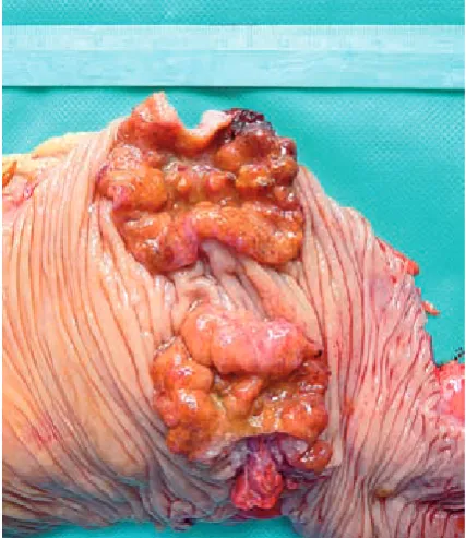
order, were the cecum (Figure 1) and ascending colon – 127 (17%), descending colon – 56 (7.5%), transverse colon – 26 (3.5%), splenic flexure – 19 (2.5%) and hepatic flexure – 12 (1.5%). In 8 (1%) cases, multifocal colorectal cancer was observed, in 2 of them the tumor occurred simultaneously in the rectum and sigmoid colon while in 3 cases the multifocal location was observed in the sig-moid colon (Figure 2 a, b). Multifocal location oc-curred once in the cecum. In 1 case, two focuses of cancer were observed in the transverse colon and hepatic flexure and in 1 case, the tumor was stated simultaneously in the cecum and transverse colon (Figure 3, Table 1).
Similar locations regarded colorectal polyps – the majority of benign colorectal lesions. Polyps occurred the most frequently in the sigmoid co-lon – 144 (29.3%) and rectum – 122 (24.8%) cases. Other cases of colorectal polyps are presented in Table 2.
Multifocal location of polyps was observed in 108 patients (22%). The most frequently, in 32 (30%) cases, were stated in the sigmoid colon. The other locations and occurrences are presented in Table 3.
After surgical treatment, all patients were trans-ferred to the Oncology Center in order to qualify them for further chemo- and radiotherapy.
Results
The type of surgical treatment and the scope of malignant colorectal tumor operations depended in the present material on the stadium of the can-cer, TNM, age, co-morbidities as well as on the mode of surgical procedure, connected with bowel obstruction or bleeding from the tumor (Table 4).
In cases of colorectal cancer located in the rec-tum, the most frequently applied procedure was abdomino-perineal amputation (37.6%). In 32% of patients, anterior resection with end-to-end anas-tomosis was performed. In 59 cases, a stapler (29– 33 mm) was used and in 27 cases it was performed manually. The remaining procedures connected with colorectal cancer are presented in Table 4.
In the group of patients after operations of Fig. 1. Cecum cancer lead to ileus
Ryc. 1. Rak kątnicy powodujący niedrożność Fig. 2a. Multifocal location of cancer in the sigmoid colon
Ryc. 2a. Wieloogniskowe umiejscowienie raka w obrę-bie esicy
Fig. 2b. Multifocal location of cancer in the sigmoid colon
ing to descending colon location, extensive size of the tumor and metastases to mesenterium, in 12.5% of cases, left hemicolectomy was performed. In 13% of cases, only colostomy was applied – in 21, the stoma was emerged due to the spread of cancer and in 10 cases, when the procedure was performed in emergency and was connected with occlusion and inoperable state, deferred stoma was applied. In cecum and ascending colon cancer, the most frequently applied procedure was right hemicolectomy – 94.5% of patients. The proce-Fig. 3. Doublefocal cancer in the location of the
hepat-ic flexure and transverse colon
Ryc. 3. Dwuogniskowy guz umiejscowiony w zagięciu wątrobowym i poprzecznicy
Table 1. location and occurrence of multifocal colorectal cancer
Tabela 1. Umiejscowienie i postępowanie w przypadku wieloogniskowych raków jelita grubego
Number of patients
(liczba pacjentów) Procedure(Zabieg chirurgiczny)
N %
Generally (Ogólnie)
rectum + sigmoid colon sigmoid colon
cecum
cecum+transverse colon
hepatic flexure + transverse colon
8
2 3
1 1 1
1
25.0 37.5
12.5 12.5 12.5
anterior rectal resection + sigmoid colon resection 2 sigmoid colon resections
1 left hemicolectomy right hemicolectomy extended right hemicolecomy extended right hemicolecomy
dure was performed laparoscopically in 5 patients with anastomosis through mini laparotomy. Eva-sive anastomosis was applied in 2.4% of patients. Due to occlusion and the advanced stage of the cancer, in 3.1% cases ileostomy was conducted. The most frequent treatment, in cases of cancer located in the descending colon, was left hemi-colectomy – 93% of cases. In 7% of patients, bypass anastomosis was applied. In transverse colon can-cer, transverse colon resection was performed the most often – in 42.5% of patients. 89.5% of patients with splenic flexure cancer were treated with left hemicolectomy. In the remaining 10.5% of cases with extensions to spleen, stomach and metasta-ses to lymph nodes and liver, bypass anastomosis was performed. Similar treatment was applied in patients with hepatic flexure cancer – right hemi-colectomy was performed in 91.5% of cases and bypass anastomosis in the remaining 8.5%. The detailed surgical procedures according to colorec-tal cancer are presented in Table 4. In the case of a multifocal location of colorectal cancer (1% of
cases – 8 patients), the procedures performed are presented separately in Table 1.
Table 2. location and occurrence of colorectal polyps
Tabela 2. Umiejscowienie i postępowanie w przypadkach polipów jelita grubego
localization
(Umiejscowienie) Procedure (Zabieg chirurgiczny)
number of patients number of patients
N % N %
Generally (Ogólnie) sigmoid colon rectum
cecum and ascending colon 491
144 122 51
29.3 24.8 10.3
Generally (Ogólnie)
endoscopic resection
491
482 98.2
descending colon splenic flexure transverse colon hepatic flexure
36 14 11 5
7.3 3.0 2.3 1.0
surgical 9 1.8
Table 3. location and occurrence of multifocal colorectal polyps
Tabela 3. Umiejscowienie i postępowanie w przypadku wieloogniskowych polipów jelita grubego localization
(Umiejscowienie) Procedure (Zabieg chirurgiczny)
number of patients number of patients
N % N %
Generally
(Ogólnie) 108 22 Generally (Ogólnie) 108 22
sigmoid colon
rectum+sigmoid colon 32 28 3026 endoscopic resection 102 94.5 rectum
multifocal 21 27 1925 surgical 6 5.5
the remaining 25% were performed as emergency operations (Table 5).
48 (6.4%) patients underwent resection of a simple metastatic hepatic tumor during the origi-nal colorectal cancer operation. For the rest of the patients with hepatic metastases, thermal ablation was applied. In 1 (1%) patient, a synchronic tumor of the sigmoid colon and stomach was observed. In this case a total gastrectomy was performed next to the sigmoid colon tumor resection during the same operation.
In 491 patients with colorectal polyps, the type of procedure depended on the result of a histo-pathological examination of a sample taken during colonoscopy. In 482 cases, endoscopic polyp re-moval was applied and in 9 cases surgical resection was instituted according to the histological pattern of the tumor. Multifocal polyps were recognized in 108 (22%) patients among which 102 (94.2%) un-derwent endoscopic resection.
Discussion
(Odbytnica)
N = 271 anterior rectal resectionstoma Hartmnn’s operation
86 58 25
32.0 21.4 9.0
T1-3 N1-2 M0 T2-4 N1-2 M0-1 T2-4 N2 M1 Sigmoid colon
(Esica) N = 235
sigmoid colon resection colostomy
left hemicolectomy
175 31 29
74.5 13.0 12.5
T1-3 N0-2 M0-1 T4 N0-2 M1 T1-4 N0-1 M0-1 Cecum and ascending colon
(Kątnica i wstępnica) N = 127
right hemicolectomy evasive anastomosis ileostomy
120 3 4
94.5 2.4 3.1
T1-4 N0-2 M0-1 T3-4 N2 M1 T4 N2 M1 Descending colon
(Zstępnica) N = 56
left hemicolectomy
evasive anastomosis 52 4 93.0 7.0 T1-4 N0-2 M0-1T4 N2 M1
Transverse colon (Poprzecznica) N = 26
left hemicolectomy transverse colon resection evasive anastomosis ileostomy
6 11 6 3
23.0 42.5 23.0 11.5
T1-4 N0-1 M0 T1-4 N0-1 M0 T4 N2 M1 T4 N1-2 M1 Splenic flexure
(Zagięcie śledziony) N = 19
left hemicolectomy
evasive anastomosis 17 2 89.510.5 T1-3 N1-2 M0T4 N2 M1
Hepatic flexure (Zagięcie wątrobowe) N = 12
right hemicolectomy
evasive anastomosis 11 1 91.5 8.5 T1-4 N1-2 M0 T4 N2 M1
sigmoid colon. Multifocal location was observed in 1% of treated patients.
Diagnosis of colorectal cancer is based on clinical presentation as well as on endoscopic and imaging examinations. The most frequent symp-toms are changes in defecation, abdominal pain and flatulence, dark-colored stool, losing weight and lack of appetite. The basis for diagnostics is endoscopy which allows recognition of the tumor, sampling and examination of other parts of gastro-intestinal tract with respect to synchronic tumors. However, it has to be mentioned that colonoscopy is not responsive enough. Many tumors may be overlooked especially by inexperienced doctors [12–14]. It is estimated that 15–50% of small tu-mors, commonly of the adenoma type, are over-looked during colonoscopy [13, 15]. Similarly, approximately 6–12% of tumors with a diameter > 1 cm [14, 16] and approximately 4% of cancers re-main unnoticed during colorectal endoscopy [17].
It has been proved that the result of preopera-tive colonoscopy may change a planned surgical procedure in 10% of cases. Valid microscopic di-agnosis may be obtained after taking a deep sample from a polypoid tumor invasion or from the
mar-gin of an ulcerous invasion [1]. If this is not pos-sible, it is necessary to perform a full colonoscopy in order to reveal synchronic changes.
Almost 20% of patients with colorectal cancer undergo emergency operations without previous microscopic diagnosis relating to the complica-tions of cancer (e.g. a rupture in the gastrointesti-nal tract, occlusion or hemorrhage) [18, 19]. Simi-lar results were observed in the present material. It has been proved that the rate of postoperative mortality is higher in patients operated on imme-diately [20]. In the present material the authors did not notice any severe postoperative complications in this kind of situation. Also the authors did not operate on hepatic flexure cancer as an emergency. Patients who underwent planned operations had been prepared using polyethylene glycol. Preparing patient with laxatives is nowadays very controver-sial and is provoking discussion about its validity. Antibiotic prophylaxis was applied intravenously as well as anticoagulants [18, 21].
radical-ness at the same time. A higher percentage of the reconstructed continuity of the gastrointestinal tract has been noticed due to a broader applica-tion of circular staplers [20, 22, 23].
In some forms of early tumors, there is a pos-sibility of local resection (tumors with diameter < 3 cm, differentiated histologically). In colorectal cancer treatment, chemotherapy is also applied, which is standard complementary treatment after surgery in B2 and C stages according to Dukes. Ra-diotherapy is also used, in the case of rectal can-cer, as a preoperative procedure or complemen-tary treatment after the surgery. Chemotherapy and radiotherapy are also applied as independent methods in the palliative treatment of patients with non-operative colorectal cancer [24]. In the present material, 137 (18.2%) advanced cancers, with metastases and extension to near organs were observed. In these cases, palliative surgical proce-dures were performed.
The stadium of colorectal cancer is the most important predictive factor. The method of treat-ment depends on the stadium and location of the tumor. Differences regard preoperative treatment,
scope of the operation and technique as well as postoperative steps. In the case of colorectal can-cer, the method depends on the location – above (colon or upper part of the rectum) or under the peritoneal fold (central and lower part of the rec-tum).
In the case of cancer in stadium 0 and 1 located under the peritoneal fold, only surgical treatment is applied. In stadium 2, the method of choice is radio-therapy with possible subsequent chemoradio-therapy. In stadium 3 of cancer, radio- and radiochemotherapy is applied with further chemotherapy. Managing tu-mors in stadium 4 includes palliative surgical treat-ment and application of radio- and chemotherapy at the same time [10, 18, 24].
In the case of colorectal cancers located above the peritoneal fold, the method of choice is surgical treatment in all stages with possible postoperative chemotherapy. In stage 2 after the surgery, applica-tion of radiotherapy may be considered. In stage 3, subsequent chemotherapy is always applied. Man-aging with tumors in stadium 4 includes palliative surgical treatment and application of radio- and chemotherapy at the same time [11, 18, 24].
Radiotherapy is used as a part of preopera-tive preparation, radical treatment (complemen-tary treatment) or a method of palliative therapy, mainly in cases of rectal cancer. Preoperative ra-diotherapy decreases the risk of local regression and increases the percentage of survivals. It also brings less risk of complication than usually ap-plied postoperative radiotherapy. In cases of origi-nally unrespectable rectal tumors, application of conventional radiochemotherapy makes it possi-ble to perform surgery, radical by nature, in 50% of patients. However, there are some inconveniences resulting from preoperative radiotherapy, for in-stance, a higher risk of infection, delayed healing of the peritoneal wound after abdomino-peritoneal amputation and the necessity to combine the date of the surgery with the end of radiotherapy. Surgi-cal treatment should be performed a maximum of 7 days after radiotherapy, before acute radiation syndrome [15, 18, 24].
In the case of colorectal cancer, surgery is usu-ally performed 6–8 weeks after the radiation. The break is necessary in order to obtain tumor regres-sion and complete recovery of radiation syndrome. Reduction of tumor size after radiochemotherapy as well as a high percentage of complete remis-sions (15–20%) seem to be the most important ad-vantages of this preoperative method [15, 24].
Postoperative radiotherapy is also applicable. Recommendations for this method, for instance, invasion of peri-intestinal adipose tissue as well as lymph nodes, dissemination along nerve fibers, tu-mor cell embolism in blood and lymphatic vessels,
Table 5. Treatment of colorectal cancer
Tabela 5. Postępowanie w raku jelita grubego
localization
(Umiejscowienie) Mode (Rodzaj)
planned emergency number of
patients number of patients
N % N %
Rectum
(Odbytnica) 205 75.5 66 24.5 Sigmoid colon
(Esica) 189 79.7 48 20.3 Cecum and
ascending colon (Kątnica i wstępnica)
98 77.0 29 23.0
Descending colon
(Zstępnica) 52 93.0 4 7.0 Transverse colon
(Poprzecznica) 21 81.0 5 19.0 Splenic flexure
(Zagięcie śledziony) 16 84.0 3 6.0 Hepatic flexure
(Zagięcie wątrobowe)
12 100.0 0 0
Multifocal
role in pain alleviation, reduction of bleeding and occlusion symptoms connected with local regres-sion of rectal cancer as well as in the alleviation of symptoms associated with metastases [18, 24].
Recommendations for complementary (post-operative) chemotherapy in colorectal cancer are unfavorable predictive factors identical to those connected with postoperative radiotherapy. Cur-rently, routine chemotherapy is not recommended in all patients suffering from colorectal cancer in generalized stadium. Palliative chemotherapy ap-plied in properly qualified patients may lead to periodic improvement in quality of life and its significant prolongation in comparison to patients treated only symptomatically [17, 18, 24].
The type of operative technique most signifi-cantly influences the results of treatment. Differ-ences in chosen procedures are reflected in the percentage of local regressions and time of survival after the surgery. The choice of a particular proce-dure should depend not only on the patient’s gen-eral state and disease stadium but also on the sur-geon’s experience and abilities [18].
Concerning tumors placed below the perito-neal fold or at the same height, the choice of surgi-cal treatment depends mainly on the size of the tu-mor. In small tumors – 3 cm or less – in the TNM or T1 N0 stage and without invasion of sphinc-ters, local resection through the rectum performed from the posterior part or using surgical anoscope may be applied. In this case, anterior resection or abdomino-peritoneal amputation may be an alternative for local resection. In tumors placed above the peritoneal fold, the method of choice is radical surgical procedure which saves sphincters. Abdomino-peritoneal amputation is valid when sphincter functions are impaired or when carcino-genic diseases (e.g. polyps) are developing in the part of the rectum under the tumor. Abdomino-peritoneal amputation may be associated with a higher frequency of occurrence of local regres-sion or lower oncological radicalness [21].
In cases of rectosigmoid flexure cancer, the scope of bowel resection is strictly connected with tumor location and the necessity to remove blood and lymphatic vessels supporting the part of the bowel with the tumor. Regarding colon tumors, lymphadenectomy should reach the vent of the main upper arterial vessel. It has to be mentioned
It is not a subject for discussion that in every case, the whole peritoneal cavity should be precise-ly examined in order to assess the primary tumor, regional lymph nodes, the presence of distant me-tastases and the state of other organs. Resection of the rectum along with the lymphoid system and blood vessels should include an anatomically wide resection of the full mesorectum with fascia, at least 4 cm below the lower line of the tumor. It should also include the ligature in the area of the proximal part of the main arterial vessel (inferior mesenteric artery or superior rectal artery accord-ing to the vascularization variant and operation planned) [18, 21, 24].
Recently, laparoscopy is the method of choice for the majority of planned colorectal and rectal operations performed in well-equipped centers using a harmonic knife or liga-Sure as well as circular and linear staplers. No differences were stated in the risk of regression and total survival, however laparoscopic procedures have a lot of commonly known advantages and are connected with a shorter time of hospitalization and lesser demand for analgesics [25]. In authors’ depart-ment, laparoscopic performance of laparoscopic procedures in colorectal cancer was launched in 2007 and is still improving. Until this moment the authors have operated on 36 patients.
The basis in managing colorectal cancers is surgical treatment – radical or palliative. Cur-rently the best results are obtained by adjuvant and neoadjuvant treatment. Approximately 20% of colorectal cancer surgeries are performed as an emergency without preparation of the patient. These surgeries are performed mainly due to oc-clusion or gastrointestinal bleeding.
References
Hady Razak H, Dadan J, Zbucki R, Łukaszewicz J, Myśliwiec P, Sołdatow M, Kamiński F, Łuba M:
[1] Nowotwory
złośliwe i łagodne jelita grubego w materiale własnym. Pol J Surg 2011, 83(1), S67.
Berner A, Piekarski J:
[2] Rak jelita grubego. In: Onkologia. Eds.: Kordek R, Jassem J, Krzakowski M, Jeziorski A. Gdańsk 2004, Wyd. II, 119–123.
Nowacki M, Bujko K, Krzakowski M, Nowakowska D, Rutkowski A:
[3] Colon cancer In: Diagnostic and
thera-peutic recommendations in malignant cancers treatment, Oncology in Clinical Practice, Journal of Polish Clinical Oncology Association tom 3, supl C, Krzakowski M, Herman K, Jassem J, Jędrzejczak W, Kowalczyk JR, Podolak-Dawidziak M, Reinfuss M, Via Medica Gdańsk 2007, 153–178.
Cairns S:
[4] Guidelines for colorectal cancer screening and surveillance in moderate and high risk groups (update from 2002). Downloaded from gut.bmj.com, 2011.
Atkin W, Rogers P, Cardwell C,
[5] Cook C, Cuzick J, Wardle J, Edwards R: Wide variation in adenoma detection rates at screening flexible sigmoidoscopy. Gastroenterology 2004, 126, 1247–1256.
Pickhardt P, Choi J, Hwang I, Butler JA, Puckett ML, Hildebrandt HA, Wong RK, Nugent PA, Mysliwiec PA, [6]
Schindler WR: Computed tomographic virtual colonoscopy to screen for colorectal neoplasia in asymptomatic adults. N Engl J Med 2003, 349, 2191–2200.
Hixson L, Fennerty M, Sampliner R,
[7] Garewal HS: Prospective blinded trial of the colonoscopic miss-rate of large colorectal polyps. Gastrointest Endosc 1991, 37, 125–127.
Rex DK, Cutler CS, Lemmel GT,
[8] Rahmani EY, Clark DW, Helper DJ, Lehman GA, Mark DG: Colonoscopic
miss rates and adenomas determined by back-to-back colonoscopies. Gastroenterology 1997, 112, 24–28.
Bressler B, Paszat L, Vinden C,
[9] Li C, He J, Rabeneck L: Colonoscopic miss rates for right sided colon cancer: a population based analysis. Gastroenterology 2004, 127, 452–456.
Kuestera D, Dalicho S, Monkemuller K, Benedix F, Lippert H, Guentherd T, Roessnera A, Meyer F:
[10] Synchronous
multifocal colorectal carcinoma in a patient with delayed diagnosis of ulcerative pancolitis. Pathol Res Pract 204, 2008, 905–910.
Eaden JA, Abrams KR, Mayberry JF:
[11] The risk of colorectal cancer in ulcerative colitis: a meta-analysis. Gut 2001, 48 (4), 526–535.
Szmidt J, Kużdżał J:
[12] Podstawy chirurgii. Podręcznik dla lekarzy specjalizujących się w chirurgii ogólnej. Kraków 2010.
Nowacki M:
[13] Nowotwory jelita grubego. In: Zasady diagnostyki i chirurgicznego leczenia nowotworów w Polsce. Eds.: Szawłowski A., Szmidt J. Fundacja – Polski Przegląd Chirurgiczny, Warszawa 2003, 226–234.
Grzebieniak Z, Szynglarewicz B:
[14] Metody chirurgicznej protekcji zespolenia po niskiej przedniej resekcji odbyt-nicy z powodu raka. Gastroenterol Pol 2006, 13, 193–196.
Dziki A, Trzciński R:
[15] Chirurgia jelita grubego i odbytnicy. In: Przegląd piśmiennictwa chirurgicznego. Ed.: W. Noszczyk, Fundacja – Polski Przegląd Chirurgiczny, Warszawa 2007, 212–235.
Wojciechowska U, Didkowska J, Zatoński W:
[16] Nowotwory złośliwe w Polsce w 2006 roku. Warszawa 2008.
Matthiessen P, Hallböök O, Rutegard J, Sjödahl R:
[17] Population-based study of risk factors for postoperative health
after anterior resection of the rectum. Br J Surg 2006, 93, 498–503.
DeVita VT, Hellman S, Rosenberg S:
[18] Cancer: Principles Practice of Oncology. Philadelphia, USA, 2005, 7, 217–241.
Małolepsza-Bazan A, Strutyńska-Karpińska M:
[19] Evaluation of postoperative complications of colorectal surgery.
Adv Clin Exp Med 2008, 17, 6, 655–660.
Didkowska J, Wojciechowska U, Tarkowski W, Zatoński WA:
[20] Malignant cancers in Poland in 2004. Oncology
Center, Maria Skłodowska-Curie Institute, Warsaw 2006.
Desch CE, Benson AB, Somerfield MR:
[21] Colorectal Cancer Surveillance: 2005 Update of American Society of
Clinical Oncology. Practice Guideline. J Clin Oncol 2005, 20, 8512–8519.
Korniluk J, Wcisło G, Nurzyński P, Stec R, Bodnar L, Obrocka B, Szczylik C:
[22] Epidemiology of colon cancer.
Współ Onkol 2006, 10, 136.
Maul JS, Burt RW, Cannon-Albright LA:
[23] A familialcomponent to human rectal cancer, independent of colon
cancer risk.Clin Gastroenterol Hepatol 2007, 5, 1080–1084.
Hady Razak H, Kamiński F, Sołdatow M,
[24] Łuba M, Snarska J, Ładny JR, Dadan JW: Risk factors for large intes-tine cancer in clinical material. Adv Clin Exp Med 2010, 19 (6), 723–730.
Lynch P:
[25] Standards of care in diagnosis and testing for hereditary colon cancer. Familiar Cancer 2008, 7 (1), 65–72.
Address for correspondence:
Hady Razak Hady
1st Department of General and Endocrinological Surgery
Medical University of Bialystok E-mail: [email protected] Phone: +48 85 746 8279
Conflict of interest: None declared