Çiğdem Y. Güçlü
A–F, Süheyla Ünver
A, E, F, Bahar Aydınlı
B, C, Dilek Kazancı
B, C,
Elif Dilber
B, C, Ayşegül Özgök
B, CThe Effect of Sevoflurane vs. TIVA
on Cerebral Oxygen Saturation
During Cardiopulmonary Bypass – Randomized Trial
Türkiye Yüksek İhtisas Education and Research Hospital, Clinic of Anesthesia and Reanimation, Ankara, TurkeyA – research concept and design; B – collection and/or assembly of data; C – data analysis and interpretation;
D – writing the article; E – critical revision of the article; F – final approval of article; G – other
Abstract
Background. Neuropsychological and neurological deficits are still major causes of mortality and morbidity after cardiac surgery. These complications are thought to be caused by embolisms and cerebral hypoxia. Thus, con-tinuous neuromonitoring is essential during cardiac surgery due to cerebral oxygen desaturation during different periods. Near-infrared spectrophotometry (NIRS), a non-invasive method, appears to offer many advantages for monitoring cerebral oxygenation and hemodynamics. Desaturation of cerebral oxygen may occur at the beginning of cardiopulmonary bypass (CPB) or during the low perfusion and rewarming stages if not corrected.
Objectives. This study was designed to assess the effects of sevoflurane on cerebral protection during CPB.
Material and Methods. Eighty patients were divided into two groups. Anesthesia was maintained either with fen-tanyl and midazolam (total intravenous anesthesia, TIVA) or with one minimum alveolar concentration of sevo-flurane and fentanyl. Cerebral desaturation was defined as an absolute decrease in saturation of 20% from baseline cerebral saturation. When desaturation occurred, PaCO2, hematocrit and PaO2 levels were checked and corrected.
If desaturation continued, anesthetic depth was increased to reserve saturation with 50–100 mg of propofol. NIRS values and hemodynamics were recorded at predetermined time intervals.
Results. Cerebral oxygen saturation values on the right side were higher in the sevoflurane group than in the TIVA group. The values on the left side were higher in the sevoflurane group than in the TIVA group, and meaningful differences were seen at the lowest temperature and at 36°C.
Conclusions. Oxygen saturation was higher in the sevoflurane group than in the TIVA group. Thus, the effect of sevoflurane was useful for maintaining cerebral oxygen saturation during CBP (Adv Clin Exp Med 2014, 23, 6, 919–924).
Key words: near-infrared spectrophotometry, cerebral oxygen saturation, cardiopulmonary bypass, sevoflurane. Adv Clin Exp Med 2014, 23, 6, 919–924
ISSN 1899–5276
ORIGINAL PAPERS
© Copyright by Wroclaw Medical University
Neuropsychological and neurological deficits are still major causes of mortality and morbidi-ty after cardiac surgery. These complications are thought to be caused by embolisms and cerebral hypoxia [1–4]. Cerebral oxygen desaturation can occur during different periods of cardiac surgery; thus, continuous neuromonitoring is essential. Near-infrared spectrophotometry (NIRS) is a non-invasive method that appears to offer many advan-tages for monitoring cerebral oxygenation and he-modynamics [5–7]. Using NIRS to assess bifrontal regional cortical oxygen saturation (SRO2) in
pa-tients undergoing cardiopulmonary bypass (CPB)
has revealed a correlation between low SRO2
val-ues and cognitive dysfunction, prolonged hos-pitalization and perioperative cerebrovascular accidents [8–11].
Desaturation of cerebral oxygen can occur at the beginning of CBP because of hemodilution or during the low perfusion and rewarming stages. The neurological outcomes of the patients are re-lated to desaturation. Because volatile agents have a cerebroprotective effect, they are used as a mainte-nance method during CBP. Volatile agents decrease the cerebral metabolic rate (CMRO2) in a
minimum alveolar concentration (MAC) of sevo-flurane anesthesia causes a decrease in global ce-rebral blood flow because of a pronounced reduc-tion in CMRO2 [13]. Opioids and benzodiazepines
also decrease CMRO2, but not as much as volatile
agents. Sevoflurane has a direct dose-dependent cerebral vasodilatory effect as well [14]. This study was designed to compare the effects of sevoflurane and total intravenous anesthesia (TIVA) on cere-bral cortex oxygen saturation during CPB.
Material and Methods
Following approval by the local ethics and re-search committee and the receipt of written in-formed consent, 80 patients undergoing elective cardiac bypass surgery were included in the study. Exclusion criteria included a history of any neuro-logical disease or neurosurgery, any carotid artery stenosis, left ventricular ejection fraction < 40%, re-operation and the need for concomitant valve surgery.
All the patients were premedicated with 0.1 mg/kg morphine administered intramuscu-larly 1 h before surgery. A randomization enve-lope was opened in the operating room, and pa-tients were assigned to either the active treatment group or the control group. Standard monitoring was applied, including five-lead electrocardiogra-phy, digital pulse oximetry, capnograelectrocardiogra-phy, radial arterial line and a triple-lumen catheter via the in-ternal jugular vein.
Regional cerebral oxygen saturation was mon-itored using near-infrared spectroscopy (NIRS) (İNVOS 3100; Somanetics, Troy, MI, USA). NIRS technology is based on the principle that all sub-stances have a characteristic absorbance. The light source of the oximeter provides 2 continuous wave-lengths of near-infrared light (730 and 810 nm) on the forehead, in the area corresponding to the junc-tion between the anterior and middle cerebral ar-teries. Two detectors with a source detector spaced 3 and 4 cm apart distinguished the extra-cerebral tissue signals from the intra-cerebral tissue signals. The ratio of oxygenated hemoglobin to total he-moglobin was measured, and the superficial signal was subtracted from the deeper signal by the mon-itor to obtain the regional hemoglobin oxygen sat-uration in the frontal cortex [6].
Anesthesia was induced with 0.1 mg/kg in-travenous midazolam, 10–15 µg/kg fentanyl and 0.6 mg/kg intravenous rocuronium. Anesthesia was maintained either with fentanyl and midazol-am (TIVA) or with one MAC sevoflurane and fen-tanyl. All the patients were ventilated with 50% ox-ygen and 50% medical air. Minute ventilation was
adjusted to maintain partial carbon dioxide pres-sure (PaCO2) at 35–45 mm Hg, as confirmed by
se-rial artese-rial blood gas analysis. After the induction of anesthesia, a central catheter, a urinary catheter and a rectal temperature probe were placed. Intra-venous fluids were administered according to the estimated insensible loss of 7 mL/kg/h during sur-gery and titrated according to the blood pressure and central venous pressure. A decrease in mean arterial pressure to < 60 mm Hg was treated with fluids in the presence of low central venous pres-sure or by use of vasopressors. If the hematocrit val-ue was < 20%, a red blood cell transfusion was ad-ministered. When PaCO2 was < 35 mm Hg during
ventilation, ventilation was reduced to achieve Pa-CO2 > 40 mm Hg. pH-stat management was used during CPB. CPB was instituted and maintained according to the standard protocol with standard-ized cannulation sites, pump flow, blood gas man-agement, mean arterial pressure and temperature targets. Blood cardioplegia was used in all the pa-tients. Pump flow was adjusted to obtain an ad-justed output of 2.2 L/m2 body surface area. Pump
flow was reduced to 0.5 L/m2 for aortic clamping
and unclamping. The pumps for all the patients were roller pumps (Jostra, Hirrlingen, Germany) and oxygenators (Dideco, Mirandola, Italy).
Cerebral desaturation was defined as a nega-tive change from the baseline cerebral saturation. If continued desaturation was observed, the patient’s head position was checked to ensure that it had not been rotated. Anesthetic depth was increased with 50–100 mg of propofol after checking and correct-ing the PaCO2, hematocrit and PaO2 levels.
Arterial blood gas samples were collected and NIRS values were recorded at various time inter-vals: baseline, intubation, after internal mamma-ry artemamma-ry dissection, after cross-clamping, at 34°C, at 32°C (lowest temperature), during warming at 36°C, post-bypass and after skin closure.
The data analysis was performed using Statis-tical Package for Social Sciences software (v. 11.5, SPSS Inc., Chicago, IL, USA). The Shapiro- -Wilk test was used to check the normality of the continuous variable distributions. The data are ex-pressed as mean ± standard deviation or median (range), where applicable. Means were compared using the unpaired t-test. The Mann–Whitney U test was used to compare median values. Re-peated-measures analysis of variance was used to evaluate both hemodynamic and clinical measure-ments. The Bonferroni adjusted multiple compar-ison test was applied as a follow-up for statistical-ly significant variance anastatistical-lyses. Nominal data were analyzed using Pearson’s c2 or Fisher’s exact test,
Results
The patient demographic data are shown in Table 1.
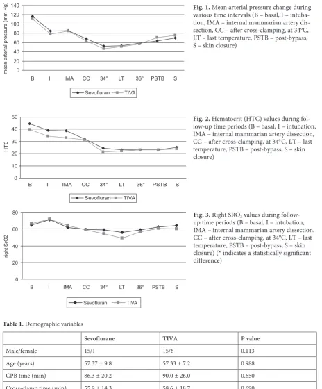
The changes in mean arterial pressure by time interval were similar (p = 0.185). No difference was detected in mean arterial pressure between the groups during any period (p = 0.477) (Fig. 1).
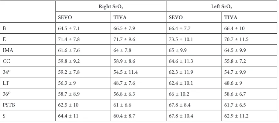
The changes in hematocrit level relative to the time interval were similar in the 2 groups (p = 0.153) (Fig. 2). There were no differences in oxygen saturation (SpO2), PaCO2, glucose or pH
between the groups (p = 0.829, 0.738, 0.150 and 0.837, respectively).
Cerebral oxygen saturation (SRO2) values on
the right side were higher in the sevoflurane group
Fig. 2. Hematocrit (HTC) values during fol-low-up time periods (B – basal, I – intubation, IMA – internal mammarian artery dissection, CC – after cross-clamping, at 34°C, LT – last temperature, PSTB – post-bypass, S – skin closure)
0 10 20 30 40 50
B I IMA CC 34° LT 36° PSTB S
HT
C
Sevofluran TIVA
Table 1. Demographic variables
Sevoflurane TIVA P value
Male/female 15/1 15/6 0.113
Age (years) 57.37 ± 9.8 57.33 ± 7.2 0.988
CPB time (min) 86.3 ± 20.2 90.0 ± 26.0 0.650
Cross-clamp time (min) 55.9 ± 14.3 58.6 ± 18.7 0.690
Other coexisting disease 4/16 5/21 1.0
Fig. 1. Mean arterial pressure change during various time intervals (B – basal, I – intuba-tion, IMA – internal mammarian artery dis-section, CC – after cross-clamping, at 34°C, LT – last temperature, PSTB – post-bypass, S – skin closure)
0 20 40 60 80 100 120 140
B I IMA CC 34° LT 36° PSTB S
mean arterial pressure (mm Hg)
Sevofluran TIVA
Fig. 3. Right SRO2 values during
follow-up time periods (B – basal, I – intubation, IMA – internal mammarian artery dissection, CC – after cross-clamping, at 34°C, LT – last temperature, PSTB – post-bypass, S – skin closure) (* indicates a statistically significant difference)
0 20 40 60 80
B I IMA CC 34° LT 36° PSTB S
right SrO
2
than in the TIVA group, but the only significant difference was determined at lowest temperature (p = 0.012). The values on the left side were also higher in the sevoflurane group than in the TIVA group, and significant differences were seen at cross clamping, at the lowest temperature and at
36°C (p = 0.03, 0.02 and 0.03, respectively) (Ta-ble 2, Figs 3 and 4).
The change in SRO2 on the right side
com-pared with the baseline value, which is termed rO2,
was less in the sevoflurane group than in the TIVA group, with a significant difference at the lowest Fig. 4. Left SRO2 values during follow-up
time periods (B – basal, E – entubation, IMA –internal mammarian artery dissec-tion, CC – after cross-clamping, at 34°C, LT – last temperature, PSTB – post-bypass, S – skin closure) (* indicates a statistically significant difference)
Table 2. SrO2 values according to measurement times. The values are presented as mean ± SD
Right SrO2 Left SrO2
SEVO TIVA SEVO TIVA
B 64.5 ± 7.1 66.5 ± 7.9 66.4 ± 7.7 66.4 ± 10
E 71.4 ± 7.8 71.7 ± 9.6 73.5 ± 10.1 70.7 ± 11.5
IMA 61.6 ± 7.6 64 ± 7.8 65 ± 9.9 64.5 ± 9.9
CC 59.8 ± 9.2 58.9 ± 8.6 64.6 ± 11.3 55.8 ± 7.2
34O 59.2 ± 7.8 54.5 ± 11.4 62.3 ± 11.9 54.7 ± 9.9
LT 56.3 ± 9 48.7 ± 7.6 62.4 ± 10.1 48.6 ± 9
36O 58.7 ± 8.9 56.8 ± 6.3 66 ± 10.2 58.6 ± 6.7
PSTB 62.5 ± 10 61 ± 6.6 67.8 ± 8.4 61.7 ± 6.5
S 64.4 ± 11 60.4 ± 8.7 67.8 ± 10.4 62.9 ± 11.2
B – basal, E – entubation, IMA – internal mammarian artery dissection, CC – after cross-clamping, at 34°C, LT – last tem-perature, PSTB – post-bypass, S – skin closure.
0 20 40 60 80
B E IMA CC 34° LT 36° PSTB S
leftSrO
2
Sevofluran TIVA
*
* *
Fig. 5. Right RO 2 values during follow-up time periods (I – intubation, IMA –internal mammarian artery dissection, CC – after cross-clamping, at 34°C, LT – last temperature, PSTB – post-bypass, S – skin closure) (* indicates a statistically significant differ-ence)
temperature. On the left side, rO2 differed
signifi-cantly at the cross clamp and lowest temperature time intervals (Fig. 5 and 6).
The extubation and intensive care times were similar in both groups (p = 0.212 and 0.296).
Discussion
This study was designed to compare the effects of sevoflurane and TIVA on cerebral oxygen satu-ration in patients undergoing elective cardiac sur-gery. The success of cardiac surgery is related to the neurological outcome of the patients, which can be adversely affected by cerebral oxygen de-saturation [9–11, 15]. During CPB, cerebral oxy-gen desaturation occurs at different time points, such as the beginning of bypass due to hemodilu-tion, during low perfusion and during rewarming. Correcting the cause of desaturation is an impor-tant issue in anesthetic management, and preserv-ing cerebral oxygen saturation is a major concern in cardiac surgery.
Many factors, including PaCO2, pH and
tem-perature, have been used to explain cerebral oxy-gen desaturation during cardiac surgery, and in-flammatory activation has been reported as a main effect of CPB [16–19]. In this study, only the effect of sevoflurane on cerebral oxygen saturation was assessed, and no other regulatory parameters.
Hemodilution at the start of CPB causes a de-cline in regional cerebral oxygen saturation. Oth-er critical times for desaturation are the low pOth-er- per-fusion and early rewarming stages [20]. Cerebral cortical oxygen saturation reflects the dynamic balance between cerebral oxygen supply and con-sumption. When body temperature drops, the ce-rebral metabolic rate and oxygen consumption de-crease, but the cerebral oxygen supply does not change significantly because the amount of arterial blood transported from the artificial pump to the aorta is steady. Thus, saturation can decrease dur-ing the initial part of the CPB procedure as well as the rewarming stage [16, 21].
In the current study desaturation was not-ed at the onset of CPB, which might have been due to blood-free prime-induced hemodilution.
According to the study findings, sevoflurane pre-served cerebral oxygen saturation better than the fentanyl and midazolam combination, as satura-tion was higher in the sevoflurane group.
The effect of CPB temperature on the neuro-logical outcome remains controversial. Regragui et al. reported a worse neurological outcome of normothermic bypass (37°C) compared with that of moderately hypothermic (32°C) perfusion [22]. Similar results were reported by Martin et al. [23]. In the present study, a moderately hypothermic bypass was used.
Piquette et al. administered intravenous ni-troglycerin to prevent the decrease in NIRS asso-ciated with CPB during high-risk cardiac surgery and reported that nitroglycerin may also prevent a decrease in NIRS [24]. Although there are some conflicting results regarding NIRS and outcomes, NIRS monitoring is an easy, non-invasive and helpful technique for detecting cerebral oxygen saturation during cardiac surgery [25, 26].
The difference in oxygen saturation between the right and left sides was a confusing result of the present study. False-negative factors (e.g., skin, hair follicles, forehead shape, and A-V anatom-ic shunts) might have been responsible for the difference.
The results of this study indicate that the use of sevoflurane during CPB maintained cerebral oxy-gen saturation. Sevoflurane has a direct cerebral vasodilatory effect and plays a role in brain protec-tion by reducing CMRO2 [14]. Although there was
desaturation in some patients, cerebral oxygen sat-uration was better in the sevoflurane group than in the TIVA group.
Several limitations of this study must be con-sidered. Cerebral oximetry was not monitored in the critical care unit, and neurocognitive and neu-ropsychological tests must be used after surgery to identify neurological outcomes, as mentioned in other studies.
The present study demonstrated that cerebral oxygen saturation was higher in the sevoflurane group than in the TIVA group. This effect may be useful for preventing desaturation during perfu-sion, but more studies are needed to verify these results.
References
[1] Shroyer AL, Coombs LP, Peterson ED, Eiken MC, DeLong ER, Chen A, Ferguson TB Jr, Grover FL: The Society of Thoracic Surgeons: 30-day operative mortality and morbidity risk models. Ann Thorac Surg 2003, 75, 1856–1864.
[2] Uehara T, Tabuchi M, Kozawa S, Mori E: MR angiographic evaluation of carotid and intracranial arteries in Japanese patients scheduled for coronary artery bypass grafting. Cerebrovasc Dis 2001, 11, 341–345.
[3] Yoon BW, Bae HJ, Kang DW, Lee SH, Hong KS, Kim KB, Park BJ, Roh JK: Intracranial cerebral artery disease as a risk factor for central nervous system complications of coronary artery bypass graft surgery. Stroke 2001, 32, 94–99.
[5] Nollert G, Jonas RA, Reichart B: Optimizing cerebral oxygenation during cardiac surgery: A review of experimen-tal and clinical investigations with near infrared spectrophotometry. Thorac Cardiovasc Surg 2000, 48, 247–253.
[6] Edmonds HL, Ganzel BL, Austin EH: Cerebral oximetry for cardiac and vascular surgery. Semin Cardiothorac Vasc Anesth 2004, 8, 147–166.
[7] Murkin JM, Arango M: Near-infrared spectroscopy as an index of brain and tissue oxygenation, Br J of Anaest 2009, 103, Suppl 1, i3–i13.
[8] Edmonds HL Jr: Multi-modality neurophysiologic monitoring for cardiac surgery. Heart Surg Forum 2002, 5, 225–228.
[9] Goldman S, Sutter F, Ferdinand F, Trace C: Optimizing intraoperative cerebral oxygen delivery using noninva-sive cerebral oximetry decreases the incidence of stroke for cardiac surgical patients. Heart Surg Forum 2004, 7, 376–381.
[10] Slater JP, Guarino T, Stack J, Vinod K, Bustami RT, Brown JM 3rd, Rodriguez AL, Magovern CJ, Zaubler T, Freundlich K, Parr GV: Cerebral oxygen desaturation predicts cognitive decline and longer hospital stay after cardiac surgery. Ann Thorac Surg 2009, 87, 36–44.
[11] Yao FF, Tseng CA, Ho CA, Levin SK, İllner P: Cerebral oxygen desaturations is associated with early postopera-tive neuropsychological dysfunction in patients undergoing cardiac surgery. J Cardiothorac Vasc Anesth 2004, 18, 552–58l.
[12] Young WL, Prohovnik I, Correll JW, Ornstein E, Matteo RS, Ostapkovich N: Cerebral bood flow and metabo-lism in patients undergoing anaesthesia for carotid endarterectomy: a comparison of isoflurane, halothane and fentanyl. Anesth Analg 1999, 68, 712–717.
[13] Mielck F, Stephan H, Weyland A,Sonntag H: Effects of one minimum alveolar anesthetic concentration sevoflu-rane on cerebral metabolism, blood flow, and CO2 reactivity in cardiac patients. Anesth Analg 1999, 89, 364–369.
[14] Malta B, Heath K, Tipping K, Summors AC: Direct cerebral vasodilatory effects of sevoflurane and isoflurane. Anesthesiology 1999, 91, 677.
[15] Gugino LD, Edmonds HL: Neuromonitoring for vascular surgery. Bailliere Clin Anaesthesiol 2000, 14, 17–62.
[16] Lassnigg A, Hismayr M, Keznicki P, Müllner T, Ehrlich M, Grubhofer G: Cerebral oxygenation during car-diopulmonary bypass measured by near-infrared spectroscopy: effects of haemodilution, temperature and flow. J Cardiothorac Vasc Anesth 1999, 13, 544–548.
[17] Harris DN: Cerebral oxygenation during cardiopulmonary bypass. Adv Exp Med Biol 1996, 388, 41–44.
[18] Nollert G, Mohne P, Tassani-Prell P, Reichart B: Determinants of cerebral oxygenation during cardiac surgery. Circulation 1995, 92, 327–333.
[19] Talpahewa SP, Ascione R, Angelini GD, Lovell AT: Cerebral cortical oxygenation changes during OPCAB sur-gery. Ann Thorac Surg 2003, 76, 1516–1522.
[20] Teng Y, Ding H, Gong Q, Jia Z, Huang L: Monitoring cerebral oxygen saturation during cardiopulmonary bypass sing near-infrared spectroscopy: the relationships with body temperature and perfusion rate. J Biomed Opt 11, 024016 (March/April 2006).
[21] Kadoi Y, Kawahara F, Saito S, Morita T, Kunimoto F, Goto F, Fujita N: Effects of hypotermic and normotermic cardiopulmonary bypass on brain oxygenation. Ann Thorac Surg 1999, 68, 34–39.
[22] Regragui I, Birdİ I, İzzat MB, Black AM, Lopatatzidis A, Day CJ, Gardner F, Bryan AJ, Angelini GD: The effects of cardiopulmonary bypass temperature on neurpsychologic outcome after coronary artery operations: a prospective randomized trial. J Thorac Cardiovasc Surg 1996, 112, 1036–1045.
[23] Martin TD, Craver JM, Gott JP, Weintraub WS, Ramsay J, Mora CT, Guyton RA: Prospective, randomized trial of retrograde warm blood cardioplegia: myocardial benefit and neurological treat. Ann Thorac Surg 1994, 57, 298–302.
[24] Piquette D, Deschamps A, Belisle S, Pellerin M, Levesque S, Tardif JC, Denault AY: Effects of intravenous nitroglycerin on cerebral saturation in high risk cardiac surgery. Can J Anaesth 2007 Sep, 54, 718–727.
[25] HongSW, ShimJK, ChoiYS, Kim DH, Chang BC, Kwak YL: Prediction of cognitive dysfunction and patients’ outcome following valvular heart surgery and the role of cerebral oximetry. Eur J Cardiothorac Surg 2008, 33, 4, 560–565.
[26] Reents W, Muellges W, Franke D, Babin-EbellN J, Elert O: Cerebral oxygen saturation assessed by near-infrared spectroscopy during coronary artery bypass grafting and early postoperative cognitive function. Ann Thorac Surg 2002, 74, 109–114.
Address for correspondence:
Çiğdem Y. Güçlü
Sancak Mah. 525 st. 15/7 Yıldız Ankara
Turkey
Tel.: +90 532 457 66 48
E-mail: [email protected]
Conflict of interest: None declared