Bhairav Bhushan A
Poona College of Pharmacy, Bharati Vidyapeeth Deemed University, Erandwane, Pune
Email: [email protected]
Address for correspondence
Access this article online www.japer.in
Detection of Microbial Contamination in quality control of
Ophthalmic Preparations by PCR
INTRODUCTION
Contamination of pharmaceutical preparations by micro-organisms continues to be a matter of concern to the pharmacists. From the beginning it has been an official requirement that parenteral preparations shall be sterile. More recently a number of serious infections in the eye in Britain and elsewhere have resulted in the further requirement that all preparations used in the eye should be sterile. Side by side with this has been a greater appreciation of the need to control microbial contamination and to establish microbial standards for Pharmaceutical products.
Sweden who traced an outbreak of Salmonellosis to tablets containing the drug of natural origin and who also found large number of enterobacteriaceae in number of tablets of synthetic drugs; other reports have also been published in different countries. These undoughtedly laid to standards being set or
recommended in Sweden and other countries of Europe limiting the level of microbial contamination in individual materials or in Pharmaceutical products, similar standards are being proposed in the USA. Thus in Sweden all corticosteroids preparations must be sterile, and with certain qualifications, it is recommended that no preparation should have a viable microbial count greater than 100/gm or ml, in Britain the BPC requires that salmonellae shall be absent from carmine, the USPXVII gave a series of microbial limit test and in the USPXVIII several standards are set, including a modified specification for the total bacteria count in gelatine (not more than 5000/gm) and Escherichia coli absent from 10gm, a limit of 100 bacteria per ml for the various magnesia and alumina suspensions (But not for their dried preparations) Water shall be of the US public health service standard for potable water. Escherichia coli
and salmonellae shall be absent from starch, and Salmonellae absent from powdered Digitalis Thyroid. Pharmaceutical preparations, which are checked for the presence of microbial contamination, are given below;2
The genetic material of each living organism- plant or animal, bacterium or virus possesses sequences of its nucleotide building blocks (usually DNA, sometimes RNA) that are uniquely and specifically present only in its own species. These unique variations make it possible to trace genetic material back to its origin, identifying with precision at least what species of organism it come from, and often which particular member of that species.Such an investigation requires, however, that enough of the DNA understudy is available for analysis which is where PCR (Polymerase Chain Reaction) comes in. PCR exploits the remarkable natural function of the enzymes known as polymerases.PCR is an essential tool for improving human health and human life in areas of detection of variation and mutation in genes, especially human genes. The method is useful for searching out disease organisms that are difficult or impossible to culture, such as many kinds of bacteria, fungi, and viruses, because it can generate analysable quantities of the organism’s genetic material for identification. The aim of this study was to develop a method for detection of microbial contamination in quality control of ophthalmic preparations by PCR.
Keywords: Polymerase Chain Reaction, PCR, quality control, ophthalmic
preparations ABSTRACT Bhairav Bhushan A*1
Dunake Uday U Chaudhary Mohan R
Harsulkar Abhay M Mahadik Kakasaheb R
1Department of Quality assurance Techniques, Poona College of Pharmacy, Bharati Vidyapeeth Deemed University, Erandwane, Pune-411 038
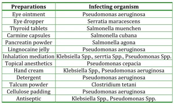
Table 1: Microbial contamination in pharmaceutical preparations
Preparations Infecting organism
Eye ointment Pseudomonas aeruginosa
Eye dropper Serratia maracescens
Thyroid tablets Salmonella muenchen
Carmine capsules Salmonella cubana
Pancreatin powder Salmonella agona
Lingnocaine jelly Pseudomonas aeruginosa
Inhalation mediation Klebsiella Spp., serrtia Spp., Pseudomonas Spp. Topical anesthetics Pseudomonas cepacia
Hand cream Klebsiella Spp., Pseudomonas aeruginosa
Detergent Pseudomonas aeruginosa
Talcum powder Clostridium tetani
Cellulose padding Pseudomonas aeruginosa Antiseptic Klebsiella Spp., Pseudomonas Spp.
IP3 and USP 4 also describe the microbial limits for raw materials given bellow
Table 2: Microbial contamination in pharmaceutical raw materials
Raw material Specifications
Gelatin The microbial count NMT 1000/gm is free from E. coli. Bentonite 10 mg is free from E. coli.
Hard gelatin capsule
shells The microbial count NMT 1000/ gm of the capsule shell, 1 gm is free from E. coli and Salmonellae. Guar gum Total microbial count does not exceed 5000/ gm, 1gm is free from E. coli and 10 gm is free from Salmonellae.
Pectin 10 gm is free from Salmonellae.
Starch 10 gm is free from E. coli and Salmonellae.
Tragacanth 1 gm is free from E. coli and 10gm is free from Salmonellae. Magnesium aluminium
silicate Its total aerobic microbial count does not exceed 1000/ gm and it meets the requirement of the test from absence of E. coli. Acacia It meets the requirement of the test for the absence of Salmonellae species under microbial limit test.
Agar It meets the requirement of the test for the absence of Salmonellae species under microbial limit test. Magnesium silicate Total microbial count does not exceed 1000/ gm, and the test for E. coli is negative.
APLICATIONS OF PCR:
The genetic material of each living organism- plant or animal, bacterium or virus possesses sequences of its nucleotide building blocks (usually DNA, sometimes RNA) that are uniquely and specifically present only in its own species. These unique variations make it possible to trace genetic material back to its origin, identifying with precision at least what species of organism it come from, and often which particular member of that species.5
The term genome refers to the total genetic information contained in a cell. The bacterium Escherichia coli contain about 3000 genes present on a single chromosome. The genome of humans is more
complex, with 23 pairs of (diploid) chromosomes containing 6 billion base pairs of DNA, with an estimated 100000 genes.6
microbes, animals, or plants, some of them may thousands or possibly even millions of year olds. PCR requires a template molecule, the DNA or RNA, and two primer molecules to get the copying process started. The primers are short chain of the four different chemical components that make up any strand of genetic material. These four components are like bricks or building blocks that are used to construct genetic molecules; called nucleotides or bases.
DNA itself is a chain of nucleotides. Under most conditions, DNA is double- stranded, consisting of two such nucleotide chains that wind around each other in the famous shape known as the double helix. Primers are single-stranded. They consist of a string of nucleotides in a specific order that will, under the right condition, bind to a specific complementary sequence of nucleotides in another piece of single-stranded RAN or DNA.
For PCR, primers must be duplicates of nucleotides sequences on either side of the piece of DNA of interest, which means that the exact order of the primers nucleotides must already be known. These flanking sequences can be constructed in the lab, or purchased from commercial suppliers.
PCR is an essential tool for improving human health and human life in areas of detection of variation and mutation in genes, especially human genes. The method is useful for searching out disease organisms that are difficult or impossible to culture, such as many kinds of bacteria, fungi, and viruses, because it can generate analyzable quantities of the organism’s genetic material for identification.3
For example- PCR detect the virus sooner during the first few weeks after infection than the standard ELISA test. PCR looks directly for the virus’s unique DNA, instead of the method employed by the standard test, which looks for indirect evidence that the virus is present by searching for antibodies the body has made against it. PCR technique has detected bacterial DNA in children’s middle ear fluid, signalling an active
infection i.e. otitis media even when culture method fails to detect it.
PCR can detect three different sexually transmitted disease organisms on a single swab (herpes, papilloma virus and Chlamydia) and can even distinguish the particular strain of papilloma virus that predisposes to cancer which other test cannot do.
PCR can easily distinguish among the small variation in DNA that make genetically unique in human being, and thus leading to genetic testing. These tests diagnose not only people with inherited disorders, but also people who carry deleterious variations, known as mutations that could be passed to their children. The task of DNA sequencing can also be assisted by PCR. Known segments of DNA can easily be produced from a patient with a genetic disease mutation. Modifications to the amplification technique can extract segments from a completely unknown genome, or can generate just a single strand of an area of interest.1
A common application of PCR is the study of patterns of gene expression. Tissues (or even individual cells) can be analyzed at different stages to see which genes have become active, or which have been switched off. This application can also use quantitative PCR to quantitate the actual levels of expression.
PCR has numerous applications to the more traditional process of DNA cloning. It can extract segments for insertion into a vector from a larger genome, which may be only available in small quantities. Using a single set of 'vector primers', it can also analyze or extract fragments that have already been inserted into vectors. Some alterations to the PCR protocol can generate mutations (general or site-directed) of an inserted fragment.1
treatments and radiation therapy can be started or stopped as soon as possible.
PCR is an essential tool for the Human Genome Project, as it can quickly and easily generate an unlimited amount of any piece of DNA for the study of mutational changes that produce disease and even death. PCR is an indispensable adjunct to forensic DNA typing – commonly called DNA fingerprinting that has ability to identify and copy the tiniest amounts of even old and damaged DNA which proved exceptionally valuable in the law. For example- hairs are found at scene of crimes. The root of a single shed hair contains enough DNA for typing at PCR.
PCR helps in sorting out relationship among vanished human groups and tracing human migration. PCR can illuminate human practices as well as human biology for Archaeologist.
With PCR scientists can glean genetic information from the rarest animal- urine, faeces, and scent marks, infinitesimal bits of hair or skin rubbed onto a tree as the elusive creature passes by. The Mobile Molecular Laboratory is a professional- quality DNA laboratory in a suitcase. It is appropriate for field investigation, education, or as a quick and inexpensive way to equip a lab just beginning DNA research. Epidemiologist need to test for pathogens directly in the field, rather than just sending samples back to the home lab. This concept has been revolutionized by the PCR.2
ESSENTIAL COMPONENTS OF POLYMERASE CHAIN REACTIONS: 7
A thermostable DNA polymerase to catalyze template-dependent synthesis of DNA
A pair of synthetic oligonucleotides to prime DNA synthesis
Deoxynucleoside triphosphate (dNTPs)
Divalent Cations
Buffer to maintain pH
Monovalent cations
Template DNA
MATERIALS AND METHODS:
Reagents:
Taq Polymerase enzyme and buffer (Bangalore Genei). It is preserved at 20oC in small aliquots to minimize the repeated freezing and thawing. DNTP mix (Bangalore Genei). This mixture is preserved at 20oC in small aliquots to minimize the repeated freezing and thawing. DNA ladder (100 bp) (Bangalore Genei). Gel loading buffer (Bangalore Genei). PA1 – TCC AAA CAA TCG TCG AAA GC and PA2 – CCG AAA ATT CGC GCT TGA AC primers. Primers PA1 and PA2 received from Ocimum Biosolutions (India) Ltd. Hyderabad, are dissolved in triple glass distilled water were used. All primer solutions are preserved at -20oC in small aliquots to minimize the repeated freezing and thawing. DNA GeNeiTM Spin Genomic DNA Prep Kit, Cat No. 107415. (Bangalore Genei).
Strains of Pseudomonas aeruginosa
ATCC No. 19429
ATCC No. 9027
ATCC No. 15442
ATCC No. 27853
ATCC No. 25619
METHODS
Method of isolation of DNA for positive control: For the extraction of the DNA, GeNeiTM Spin Genomic DNA Prep Kit (From Bacteria) from the Bangalore Genei was used.
1. Nutrient broth of five different strains of Pseudomonas Aeruginosa was prepared. 5 ml broth each of five strain of pseudomonas aeruginosa was centrifuge at 5000 rpm to form pellet. To these pellet 1 ml of PBS was added and then spined followed by draining, repeated three times.
2. Bacterial pellet of appropriate culture volume was resuspended in 180 μl of lysis buffer I.
4. The sample was spined at maximum speed in a microcentrifuge for 5 minutes and the supernatant decanted carefully to a fresh vial.
5. To the supernatant 200 μl of Lysis buffer II was added and mixed thoroughly by vortexing and incubating at 70oC for 20 minutes.
6. 4 μl of RNase (100 mg/ml) was added, mixed by vortexing and incubated at room temperature for 5 minutes. The sample was spined at maximum speed in a microcentrifuge for 5 minutes and the supernatant decanted carefully to a fresh vial. 7. To the supernatant 200 μl of distilled ethanol was
added and mixed thoroughly by vortexing.
8. The spin column was kept in a 2 ml collection tube and the sample-ethanol mixture was added. 9. It was then centrifuge at maximum speed for 1
minute and the collection tube with flow through was discarded.
10.The spin column was kept in a fresh 2 ml collection tube and 500 μl of wash buffer I was added and spined at maximum speed for 1 minute and collection tube with wash sample was discarded. 11.The spin column was kept in a fresh 2 ml collection
tube and 500 μl of wash buffer II was added and spined at maximum speed for 3 minute and wash fraction discarded and collection tube retained for step 12.
12.The empty column was spin for two minutes at maximum speed to ensure the removal of wash buffer.
13.The spin column was placed in a fresh 1.5 ml tube and 200 μl of pre warmed elution buffer was
added and incubated at room temperature for 5 minutes and centrifuged for 2 minutes to elute the DNA.
Isolated DNA confirmation by Agarose Gel Electrophoresis:
1 % agarose with ethidium bromide in 0.5 X TAE was prepared and transferred to submerged agarose gel electrophoresis apparatus and allow to cool to form a gel .The comb was removed and then the electrophoresis buffer (0.5 X TAE) was added. Then 10 μl isolated DNA with gel loading buffer was loaded in the well. The gel was run at a constant voltage (100 volt.) for 45 minutes and monitored by the tracer present in the gel loading buffer. The isolated DNA bands were observed in Gel documentation instrument.
Quantitation of DNA by UV-Spectrophotometric method:13
1 μl from each isolated DNA of five different strains was dissolved in 500 μl double distilled water. The OD values on spectrophotometer were taken at 260 nm.The amount of DNA was quantified using the formula:
DNA concentration (ng/ml.) = OD260 x 500 (dilution factor) x 50 μg/ml.
PCR Thermal Cycling:
For positive control: The PCR thermal cycling of each strain which was previously subjected to the DNA extraction procedure and the negative control was done as follows;
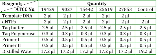
Table 3: A typical 25 μl PCR reaction
Reagents
ATCC No. 19429 9027 15442 25619 27853 Control Quantity Template DNA 2 μl 2 μl 2 μl 2 μl 2 μl -
dNTPs 2 μl 2 μl 2 μl 2 μl 2 μl 2 μl
The above mixture was subjected to the PCR thermal cycling. The above mixture was first of all subjected to 94oC for 5 minutes and then the 30 cycles at different temperatures as shown in the table 6 were run by setting the programme on the PCR thermal cycler.
Table 4: PCR conditions
Process Temperature oC Time Initial denaturation 94 oC 5 minutes
Denaturation 94 oC 30 second
Annealing 57 oC 30 second
Extension 72 oC 30 second
Incubation 72 oC 7 minutes
Cooling 4 oC 5 minutes
For microbial testing in ophthalmic solutions: 0.1 ml nutrient broth of Pseudomonas aeruginosa (ATCC NO. 9027.) was transferred in the conical flask containing 100 ml of saline solution (A). 0.1 ml of solution was transferred from A to the other conical
flask containing 100 ml of saline solution (B -10-3 dilution). Then 0.1 ml of solution was transferred from B to the other conical flask containing 100 ml of saline solution (C -10-6 dilution). Again 0.1 ml of solution was transferred from C to the other conical flask containing 100 ml of saline solution (D -10-9 dilution). In this way the dilutions of Pseudomonas aeruginosa (ATCC NO. 9027.) in the saline solution were made. The 0.1 ml saline solution of D was then transferred to the two different marketed ophthalmic solutions so as to contaminate them with Pseudomonas aeruginosa (ATCC NO. 9027.) and these ophthalmic solutions were then subjected to the PCR based microbial testing.
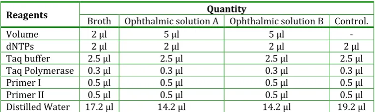
The PCR thermal cycling of ophthalmic solutions prepared as above and the negative control was done as follows;
Table 5: A typical 25 μl PCR reaction
Reagents Quantity
Broth Ophthalmic solution A Ophthalmic solution B Control.
Volume 2 μl 5 μl 5 μl -
dNTPs 2 μl 2 μl 2 μl 2 μl
Taq buffer 2.5 μl 2.5 μl 2.5 μl 2.5 μl
Taq Polymerase 0.3 μl 0.3 μl 0.3 μl 0.3 μl
Primer I 0.5 μl 0.5 μl 0.5 μl 0.5 μl
Primer II 0.5 μl 0.5 μl 0.5 μl 0.5 μl
Distilled Water 17.2 μl 14.2 μl 14.2 μl 19.2 μl
The above mixture was subjected to the PCR thermal cycling.The above mixture was first of all subjected to 94oC for 10 minutes and then the 30 cycles at different temperatures as shown in the table 8 were run by setting the programme on the PCR thermal cycler.
Table 6: PCR conditions
Process Temperature oC Time Initial denaturation 94 oC 10 minutes
Denaturation 94 oC 30 second
Annealing 57 oC 30 second
Extension 72 oC 30 second
Incubation 72 oC 7 minutes
Cooling 4 oC 5 minutes
Visualization of PCR product by agarose gel electrophoresis:
The method adopted for the visualization of PCR product of both i.e. positive control and microbial testing in ophthalmic solutions remains the same, which is carried out as described-
the gel loading buffer. The isolated DNA bands were observed in Gel documentation instrument.
RESULTS AND DISCUSSION: Conventional Microbial Testing
After performing the oxidase test, the deep purple coloure was developed.
This is a positive oxidase test and confirmed the presence of Pseudomonas aeruginosa.
Fig. 1: Oxidase test for Pseudomonas aeruginosa (ATCC No. 9027)
PCR based microbial testing
Isolated DNAs were observed at more than 2000 base pair.
Quantitation of isolated DNA
ATCC No. OD Concentration 19429 0.012 300 ng/μl
9027 0.072 1800 ng/μl 15442 0.006 150 ng/μl 27853 0.020 500 ng/μl 25619 0.005 125 ng/μl
PCR Thermal Cycling for positive control:
The oligonucleotide sequences which are used in the PCR thermal cycling for the amplification of DNA are common to all the five different strains of Pseudomonas aeruginosa, hence PCR based microbial testing developed by us can detect any wild strain of
Pseudomonas aeruginosa in the sample.
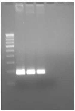
Fig. 2: PCR Thermal Cycling result for DNA isolated from five different strains of Pseudomonas aeruginosa. :Lane 1- DNA ladder, Lane 2- 2 μl DNA ( ATCC.19429), Lane 3 2 μl DNA ( ATCC.9027), Lane 4 -2 μl DNA ( ATCC.1544-2), Lane 5 --2 μl DNA (ATCC.25619), Lane 6 -2 μl DNA ( ATCC.27853), Lane 7 –Control.
PCR Thermal Cycling for microbial testing in ophthalmic solutions:
The different lanes show different bands on the agarose gel as shown in the photograph of agarose gel (fig.3 and fig.4)
The fig. 3 showed different bands of isolated DNA of Pseudomonas aeruginosa (ATCC No.9027.). The first lane on the agarose gel was of 100 bp standard DNA ladder, hence it showed different bands at 100 bp intervals.
The ophthalmic solution samples showed bands corresponding with the 181 bp of standard DNA ladder, hence the samples were PCR positive for the presence of Pseudomonas aeruginosa.
Fig. 3: PCR Thermal Cycling result for DNA isolated from Pseudomonas aeruginosa. ( ATCC.9027)
Lane 1- DNA ladder ,Lane 2- 2 μl DNA, Lane 3 - 2 μl DNA, Lane 4 -2 μl DNA,
Lane 5 – Control.
Fig.4: PCR Thermal cycling result for Microbial testing in ophthalmic solution.
Lane 1- Broth, Lane 2- Control, Lane 3 – Oph. Sol.A, Lane 4 - Oph. Sol.B
Lane 5 - DNA ladder.
CONCLUSION
There are numerous incidences of the Pseudomonas aeruginosa infections associated use of contaminated medicaments.
For ophthalmic formulation all pharmacopoeias specify certain requirements one of them is sterility. The possibility of serious ocular infection resulting from the use of contaminated ophthalmic solutions
has been documented amply in the literature. Such solutions have been the cause of corneal ulcer and the loss of eyesight. Contaminated solutions have been found in use in physician clinics, eye clinics and dispensed on prescription in community and hospital pharmacy.
Pseudomonas aeruginosa is a dangerous and opportunistic organism that grows well on most culture media and produces both toxins and antibacterial products. The latter tend to kill other contaminants and allow the Pseudomonas aeruginosa
to grow in the pure culture. This gram negative bacillus also grows readily in the ophthalmic solutions, which may become the source of extremely serious infection of the cornea. It can cause loss of sight in 24-48 hours. In concentrations tolerated by tissues of the eye, it seems that all the antimicrobial agents discussed may be ineffective against some strains of this organism.
The exotoxins are produced by the bacteria inside the cell and diffuse out into the surrounding environment. Both Gram positive and Gram negative Bacteria produce them. They are extremely toxic and are responsible for serious effects of certain diseases. For example, the toxin produced by Clostridium tetani (the casual organism of tetanus) is neurotoxic and causes severe muscular spasms due to impairment of neural control. Examples of other toxins identified are necrotoxins (causing tissue damage), enterotoxins (causing intestinal damage) and haemolysins (causing haemolysis of erythrocytes). Gram positive bacteria producing exotoxin are certain members of the genera Clostridium, Streptococcus and Staphylococcus whilst an example of a Gram negative bacterium is Vibrio cholerae (the casual organism of cholera).
Rapid detection of microbial contamination in pharmaceutical environments demands highly sensitive and accurate procedures due to the scale of testing, health risk to consumers and heterogeneous distribution of microorganisms in a given pharmaceutical production batch. Standard conventional methods are slow, not always specific, discriminate poorly among species and strains, and were developed for the isolation of microorganisms from clinical samples.
The study was undertaken to introduce the new method for the microbial testing of the pharmaceuticals, which is based on the Polymerase Chain Reaction hence called as the PCR based microbial testing. On comparison of results it was seen that the PCR based microbial testing has following advantages over conventional microbial testing. Conventional microbial testing requires the enrichment of the culture but in case of PCR the enrichment of culture is not required hence the time is saved. Again, the incubation of the culture is not required in case of PCR. There are PCR thermal cyclers available who can amplify the 10 to 3000 samples at a time hence no need of repetitively preparing the different media, sterilization of the media and performing the biochemical tests.
Conventional microbial testing requires the more inventories in the form of different equipments e.g. Autoclave, Hot air oven, Incubator etc. and different media for the culturing of the microorganisms, Petri plates, biochemical reagent kits which are different for different microorganisms but in case of PCR based microbial testing all these equipments are not required. The manpower required is also less in case of PCR because 20-3000 samples can be subjected to the PCR based microbial testing at a time hence the labour cost is also less in case of PCR.
Handling of pathogenic microorganisms is less in case of PCR based microbial testing because few micro liters of the sample is required and we are not enriching the culture but in case of conventional
microbial testing we are required to enrich the culture.
PCR based microbial testing requires less space as compared to the conventional microbial testing. Conventional microbial testing requires the change of reagents, media, chemicals for microbial testing of the different microorganisms but in case of PCR same protocol is followed for the different microorganisms, only change required is the change of primer which is specific for the specific microorganisms hence the PCR based microbial testing is simple as compared to the conventional microbial testing which is very tedious. For PCR based microbial testing the presence of a single DNA, even one single stranded DNA in the sample is sufficient. The sample requirement in case of PCR is 20-100microlitre but in case of conventional microbial testing the sample required is more.
The PCR based microbial testing is very very selective because only that sequence will get amplified for which the primer is added, but in case of conventional microbial testing the selectivity is less.
Conventional microbial testing requires about three to four days to confirm the presence of Pseudomonas aeruginosa but in case of PCR based microbial testing the time required is very less i.e. For the extraction of DNA from the sample, five hours are required, for PCR thermal cycling three hours are required and for the agarose gel electrophoresis two hours are required. Thus it means that we get the confirm result within nine days hence the time required for the PCR based microbial testing is very less.
Although the PCR based microbial testing detects the dead microorganism we can use it as a master check technique for the presence of particular microorganism previously i.e. it give the footprints of the presence of microorganism previously and during that microorganism stay it might have released the toxins (exotoxins and endotoxins).
Hence it can be concluded that there is a need of fast and accurate method to screen microbes from pharmaceuticals; we have developed PCR based method for quicker and selective microbial testing. Also, PCR analysis is selective, simple and cost effective. Quantitative analysis will be able to calculate microbial load, which is usually expressed as colony forming units in conventional microbial testing methods.
ACKNOWLEDGEMENT
The authors wish to thank IRSHA, Bharati Vidyapeeth Deemed University, Pune for providing with facilities for the study.
REFERENCES
1. Pharmaceutical Journal, Oct. 23, 400-402.
2. S.J. Bloomfield, R. Baird, R. E. Leak. R. Leech,
Microbiological Quality assurance in
Pharmaceuticals, Cosmetics and toilatories, Ellis Harwood Ltd. Chichester, 18.
3. The Pharmacopoeia of India, Vol. 1, 4th Edn, Published by the controller of publications of India. 4. The United State Pharmacopoeia, 22nd Edn, United
states pharmacopoeial Convention,
1990,1943,1896,1945,1897,1585.
5. Breakthroughs in bioscience, The Polymerase Chain
Reaction, Tabitha M. Powledge.
6. Dr. U. Satyanarayana, Biochemistry, 2nd edition, Books and Allied (P) Ltd.1999,571.
7. Sambrook and Russell, Molecular Cloning, A
Laboratory Manual, 3rd edition, 2, 8.4-8.9,8.14-8.15.
8. http://www.fermentas.com/index.html (accessed on
10/01/2008)
9. Carl W. Dieffenbach, Gabriela S. Dveksler, PCR Primer- A laboratory manual, 7-16.
10. R. W. Old, S. B. Primrose, Principles of Gene
Manipulation: An introduction to Genetic
engineering, 4th edition, Blackwell Scientific Publications, 5-6.
11. James A. Watson, Nancy H. Hopkins, Jeffery W. Roberts, Joan Argetsinger Steitz, Alan M. Wener, Molecular Biology Of The Gene, 3rd edition, The Benzamin/ Cummings Publishing Company. Inc., 175.
12. http://en.wikipedia.org/wiki/Applications_of_PCR" (accessed on 25/01/2008)
13. Doane A., Standard Operating Procedure: Gel
Documentation system, College of Biological Science, University of Guelph, 2007.
14. http://www.pcrstation.com/pcrtypes (accessed on
25/01/2008)
15. Praful B. Godkar, Textbook of Medical Laboratory Technology, Bhalani Publishing House, 1994, 340. 16. John C. Sherris et.al, Medical Microbiology: An
introduction to infectious diseases, Elsevier, 2nd edition,394.
17. Duguid J. P., Marmion B. P., Swain R. H., Mackie McCartney Medical Microbiology, English Language Book Society,13th edition, vol.I, 1973,341.
18. Tyler S. D., Rozee K.R., Oligonucleotide primer designed to differentiate pathogenic Pseudomonads on the basis of the sequencing of genes coding for 16S-23 S rRNA internal transcribed spacers, American Society for Microbiology, vol.2(4),1995, 448-453.
19. Singh B.; Biotechnology, 1st edition, Kalyani Publishers, Ludhiana, New Delhi. 2002, 32-40. 20. Kohler R., Wheat L., White A.; Detection of
pseudomonas aeruginosa antigenemia in
granulocytopenic rabbits by radiommunoassay. J. Clin. Microbiol. 12(1), 1980, 39–43.
21. Khan A.,Cerniglia C.; Detection of pseudomonas aeruginosa from clinical and environmental samples by amplification of the exotoxin A gene using PCR. Appl. Environ. Microbiol. 60(10), 1994, 3739–3745. 22. Kingsford N., Raadsma H.; Detection of pseudomonas
aeruginosa from ovine fleece washings by PCR amplification of 16S ribosomal RNA. Vet. Microbiol. 47(1-2), 1995, 61-70.
in clinical and municipal wastewater. FEMS Microbiology Ecology.2006, 571(10),158-167. 24. Kevin G., Sarah E., Linda S.; Method for detection of
pseudomonas aeruginosa using polymerase chain reaction. US Patent Issued on April 11, 2000. 25. Brodsky M., Nixon M.; Rapid Method for Detection of
pseudomonas aeruginosa on MacConkey Agar under ultraviolet light. Appl. Microbiol. 26(2), 1973, 219– 220.
26. Remington: The science and the practice of
pharmacy, 19th edition, vol. II, Mack publishing company, 1995, 1569-1570.
27. Fundamentals of microbiology, 6th edition, Edited by Hugo W. B., Russel A. D., Blackwell Science, 283.
28. Rosamund M. Baird, R.A. Shooter, British Medical Journal, 1976, 2,349-350.
29. Richard C.B., John F.P., Medical Microbiology, A guide to microbial infections: pathogenesis, immunity, laboratory diagnosis and control, Churchill Livinnstone, 16th edition, 2002,282.
30. Lubert Stryer, Biochemistry, W. H. Freeman and company, 3rd edition, 1988, 71-88, 118-140.
How to cite this article: Bhairav Bhushan A*1, Dunake Uday U, Chaudhary Mohan R , Harsulkar Abhay M, Mahadik Kakasaheb R; Detection of Microbial Contamination in quality control of Ophthalmic Preparations by PCR; J. Adv. Pharm. Edu. & Res. 2014: 4(3): 319-329.