Dr. Satyabrata Bhanja Associate Professor
Department of Pharmaceutics, Malla Reddy College of Pharmacy, Maisammaguda, Dhulapally
Secunderabad-500014, India
E-mail: [email protected] Address for correspondence
Access this article online www.japer.in
Formulation and Evaluation of Diclofenac Transdermal Gel
INTRODUCTION
The Transdermal drug delivery systems are self-contained, discrete dosage forms which when applied to intact skin deliver the drug through the skin at a controlled rate to the systemic circulation[1].At present, the most common form of delivery of drugs is the oral route. While this has the notable advantages of easy administration, it also has significant drawbacks namely poor bioavailability due to hepatic metabolism (first pass) and the tendency to produce rapid blood level spikes leading to a need for high and /or frequent dosing, which can be both cost prohibitive and involvement[2].To overcome these difficulties there is a need for the development of new drug delivery system; which will improve the therapeutic efficacy and safety of drugs by more precise (i.e. site specific), spatial and temporal placement within the body thereby reducing both size
and number of doses. One of the methods most often utilized has been Transdermal delivery. This delivery transport therapeutic substance through the skin for systemic effect. The success of Transdermal delivery depends on the ability of the drug to permeate the skin in sufficient quantities to achieve its desired therapeutic effects. The skin is very effective as a selective penetration barrier. Percutaneous absorption involves the passage of the drug molecule from the skin surface into the stratum corneum under the influence of a concentration gradient and its subsequent diffusion through the stratum corneum and underlying epidermis through the dermis and into the blood circulation. The skin behaves as a passive barrier to the penetrating molecule. The stratum corneum provides the greatest resistance to penetration and it is the rate-limiting step in percutaneous absorption[3].
Gels are transparent to opaque semisolids containing a high ratio of solvent to gelling agent merge or entangle to form a three-dimensional colloidal network structure. This network limits fluid flow by entrapment and immobilization of the solvent molecules. The network structure is also responsible for a gel resistance to deformation and therefore, its
R R R
Researchesearchesearch ArticleesearchArticleArticleArticle
The present investigation is concerned with formulation and evaluation of Transdermal gels of Diclofenac sodium, anti-inflammatory drug, to circumvent the first pass effect and to improve its bioavailability with reduction in dosing frequency and dose related side effects. Twelve formulations were developed with varying concentrations of polymers like Carbopol 934P, HPMCK4M and Sodium CMC. The gels were tested for clarity, Homogeneity, Spreadability, Extrudability, Viscosity, surface pH, drug Content uniformity, in-vitro drug diffusion study and ex-vivo
permeation study using rat abdominal skin. FTIR studies showed no evidence on interactions between drug, polymers and excipients. The best in-vitro drug release
profile was achieved with the formulation F4 containing 1 gm of Diclofenac sodium exhibited 6 h sustained drug release i.e. 98.68 % with desired therapeutic concentration which contains the drug and Carbopol 934p in the ratio of 1:2. The surface pH, drug content and viscosity of the formulation F4 was found to be 6.27, 101.3% and 3,10,000cps respectively. The drug permeation from formulation F4 was slow and steady and 0.89gm of Diclofenac sodium could permeated through the rat abdominal skin membrane with a flux of 0.071 gm hr-1 cm-2. The in-vitro release
kinetics studies reveal that all formulations fits well with zero order kinetics followed by non-Fickian diffusion mechanism.
Key words: Transdermal gel, Viscosity, In-vitro drug release, In-vitro drug release
kinetics study, Ex-vivo permeation study. ABSTRACT
ABSTRACT ABSTRACT ABSTRACT Satyabrata Bhanja*, P.Kishore
Kumar 1 , Muvvala Sudhakar1,
Arun kumar Das 2
*1Department of Pharmaceutics, Malla Reddy College of Pharmacy, Maisammaguda Secunderabad. Andhra Pradesh. 2Department of Pharmaceutics, Malla Reddy Pharmacy College, Maisammaguda Secunderabad. Andhra Pradesh.
visco-elastic properties. Gels tend to be smooth, elegant, non greasy and produce cooling effect and utilize better drug release as compared to other semi-solid formulation[4-5]. Gels have better potential as a vehicle to administered drug topically in comparison to ointment, because they are non-sticky requires low energy during the formulation are stable and have aesthetic value [6].
Diclofenac sodium (2-{2-[(2,6 dichlorophenyl) amino]phenyl}acetic acid is a selective COX-2 inhibitor used in a variety of inflammatory, pain and fever condition [7]. Diclofenac sodium is an effective as classical Non-steroidal anti-inflammatory drug (NSAID) for the relief of a wide variety of pain and inflammatory conditions, but it is better tolerated than other (NSAID’s). After oral administration the drug is rapidly and extensively absorbed. It is rapidly distributed, extensively bound to albumin and eliminated with a terminal half-life of about 2hr. Molecular Weight is 296.149, Protein binding More than 99%,Metabolism by Hepatic Excretion of the unchanged drug in urine and faeces is negligible. Generally the formulations of Diclofenacsodium commercially available are in oral and rectal form. More recently, a topical gel formulation will be introduced specifically for the treatment of localized painful and inflammatory condition, such as soft tissue musculoskeletal disorders and osteoarthritis. So the present study, formulation and evaluation of Diclofenac sodium transdermal gel will attempt to increase the efficacy of the drug at the site of action.
MATERIALS AND METHODS
Materials:
Diclofenac sodium was a gift sample from Yacht Pharma, Hyderabad. Carbopol 934P and Sodium CMC were purchased from S.D. Fine chem. Ltd, Mumbai. HPMCK4M was purchased from Yarrow chemicals ltd, Mumbai. All other reagents used were of analytical grade.
Preformulation studies:
Characterization of Diclofenac sodium:
Description
The sample of Diclofenac sodium was analysed for its nature, colour and taste.
Melting Point
The melting point was determined by using thiesel’s tube apparatus method.
Drug Excipient compatibility studies:
The drug polymer and polymer-polymer interaction was studied by the FTIR spectrometer using Shimadzu 8400-S, Japan. Two percent (w/w) of the sample with respect to a potassium bromide disc was mixed with dry KBr. The mixture was grind into a fine powder using an agate mortar and then compressed into a KBr disc in a hydraulic press at a pressure of 1000psi. Each KBr disc was scanned 16times at 2 mm/sec at a resolution of 4 cm-1 using cosine apodization. The characteristic peaks were recorded.
Preparation of Transdermal Gels
1% w/w Diclofenac sodium Transdermal gels were prepared by using different Concentrations of polymers such as Carbopol 934P, HPMCK4M and Sodium CMC. The formulation data for the preparation of Diclofenac sodium Transdermal gels using Carbopol 934P, HPMCK4M and Sodium CMC in different ratio’s is shown in [Tables 01]
Procedure:
remaining ingredients were added to obtain a homogeneous dispersion of gel.
Evaluation of Gels
About twelve formulations i.e. F1 to F12 were conducted. Gels were evaluated for their clarity, pH, viscosity, spreadability, extrudability, skin irritation test, percentage drug content, in-vitro diffusion studies, in-vitro drug release kinetic study, ex-vivo permeation studies using rat abdominal skin and stability studies by using standard procedure. All studies were carried out in triplicate and average values were reported.
Clarity
Clarity of various formulation was determined by visual inspection under black and white background and it was graded as follows: turbid +; clear ++; very clear (glassy) +++. The results are shown in [Table 02].
Homogeneity:
All developed gels were tested for homogeneity by visual inspection after the gels have been set in the container. They were tested for their appearance and presence of any aggregates. The results are shown in [Table 02].
Consistency
The estimation of consistency of the prepared gels was done by dropping a cone attached to a holding rod from a fixed distance of 10cm in other way that it should fall down on the centre of the glass cup was filled with the gel. The penetration by the cone was accurately measured from the surface of the gel to the tip of the cone inside of the gel. The distance traveled by cone in the period was noted down after 10sec. The results are shown in [Table 02]
Spreadability:
It was determined by wooden block and glass slide apparatus. For the determination of spreadability, excess of sample was applied in between two glass slides and then was compressed to uniform thickness. The weight (50gm) was added to pan. The time required to separate the two slides i.e., the time in
which upper glass slide moves over the lower plates was taken as a measure of spreadability (S).
It is calculated by using the formula: S = M . L / T
Where M = wt. tied to upper slide L = length of glass slides
T = time taken to separate the slides The results are shown in [Table 02].
Extrudability
Extrudability test was carried out by using Pfizer hardness tester. 15gm of gel was filled in collapsible aluminium tube. The plunger was adjusted to hold the tube properly the pressure of 1kg/cm2 was applied for 30 sec. The quantity of the gel extruded was weighed. The procedure was repeated at three equidistance places of the tube. The test was carried out in triplates. The results are shown in [Table 02].
Surface pH [8]
2.5 gm of gel was accurately weighed and dispersed in 25ml of distilled water. The pH of the dispersion was determined by using digital pH meter. The results are shown in [Table 03].
Viscosity[8]
Viscosity was determined by using brookfield viscometer. Viscosity measurements were carried out at room temperature (25- 27°C) using a Brookfield viscometer (Model RVTDV II, Brookfield Engineering Laboratories, Inc, Stoughton, MA). The results are shown in [Table 03].
Drug content[8]
A specified quantity (100mg) of developed gel and marketed gel were taken and dissolved in 100ml of phosphate buffer of pH 6.8. The volumetric flask containing gel solution was shaken for the period 2hr on mechanical shaker in order to get absolute solubility of drug. This solution was filtered and estimated spectrophotometrically at 285.0nm using phosphate buffer (pH 6.8) as blank. The results are shown in [Table 03].
In vitro diffusion study[8]:
sac (Cellophane membrane) was used in franz diffusion cell. The gel sample was applied on the membrane and then fixed in between donor and receptor compartment of quality diffusion cell. The receptor compartment contained phosphate buffer (100ml) of pH 6.8. The temperature of diffusion medium was thermostatically controlled at 37º ± 0.5ºC by surrounding water in jacketand the medium was continuously stirred by magneticstirrer at speed of 600rpm. The sample at predetermined intervals were withdrawn and replaced by equal volume of freshly prepared fluid. The samples withdrawn were spectrophotometrically measured at 285nm against their blank. The results are shown in [Fig. 07 -10]. Drug release kinetic studies
Various models were tested for explaining the kinetics
of drug release. To analyze the mechanism of the drug
release rate kinetics of the dosage form, the obtained
data was fitted into zero-order, first order, Higuchi
and Korsmeyer-Peppas release model, to study the
drug release from the dosage form. The results are shown in [Table 04].
Zero order release rate kinetics:-
To study the zero-order release kinetics the release
rate data are fitted to the following equation.
F=K0t
Where ‘F’ is the drug release, ‘K’ is the release rate
constant and ‘t’ is the release time. The plot of % drug
release versus time is linear.
First-order release rate kinetics:-
The release rate data are fitted to the following
equation.
Log (100-F) = kt
A plot of log % drug release versus time is linear.
Higuchi release model:-
To study the Higuchi release kinetics, the release data
were fitted to the following equation.
F = kt1/2
Where ‘k’ is the Higuchi constant.
In Higuchi model, a plot of % drug release versus square root of time is linear.
Korsmeyer-Peppas release model:-
The release rate data were fitted to the following equation.
Mt/M∞ = ktn
Where, Mt/M∞ is the fraction of drug released, ‘K’ is the release constant,
‘t’ is the release time. ‘n’ is diffusion exponent.
If n = 0.89, the release is zero order. If n = 0.45 the release is best explained by Fickian diffusion, and if 0.45 < n < 0.89 then the release is through anomalous diffusion or non fickian diffusion (Swellable & Cylindrical Matrix). In this model, a plot of log (Mt/M∞) versus log (time) is linear.
The drug release data of optimised tablet were fitted to Zero-order, First-order, Higuchi and Korsmeyer-Peppas model to study the kinetics of drug release. Ex vivo permeation studies[9]
Tissue Isolation
Rats weighing 135-160 gm were used to obtain freshly excised full thickness skin. Animal was sacrificed by spinal dislocation. Hairs from abdominal regions was removed by means of surgical and razor taking care not to damage the epidermal surface, Subcutaneous fats was removed carefully without damaging to the skin.
In vitrodrug permeation through rat abdominal
skin membrane
In vitro permeation of Diclofenac sodium transdermal
gel was studied through the rat abdominal skin
membrane. The skin membrane was mounted
between the donor and receptor compartment of the standard Franz diffusion cell with a diffusion area of 2.1 cm2 and the acceptor compartment volume of 21ml.The two chambers were tied with the help of springs so that the skin membrane did not move from its place. The phosphate buffer pH 6.8 in the acceptor compartment was continuously stirred at 600rpm using a magnetic stirrer. The entire setup was placed over a magnetic stirrer and the temperature was
maintained at 37˚±0.5°C by placing the diffusion cell in
compartment. The amount of drug permeated through the membrane was determined by removing aliquots from the receptor compartment and by replacing the same volume of buffer. The amount of Diclofenac sodium in the diffusion samples was estimated by the
HPLC method.
The flux (J) through the membrane was calculated by using the equation.
J = dQ / A dt
Where J is flux (mg h-1cm-2);
dQ/dt is the slope obtained from the steady-state portion of the curve and
A is the area of diffusion (cm2)
HPLCanalysis:
Instrument: youngling instrument Software: Autochro 3000+ Column: C18, 5µm Lambda max: 285nm Temp: 35˚C
Injection volume: 20µl Time -10min
Mobile phase: Methanol: Phosphate buffer (4:1 ratio)
pH: adjusted to 2 with HCl
The results are shown in [Table 05] and [Fig 11] Skin Irritation Test
The hair on the dorsal side of Wister albino rats was removed by clipping 1 day before the experiment. The rabbits were divided into 3 groups. Group 1 served as control; group 2 received optimized formulation; group 3 received 0.8% v/v aqueous solution of formalin as a standard irritant. Finally, the application sites were graded according to visual scoring scale. Stability studies
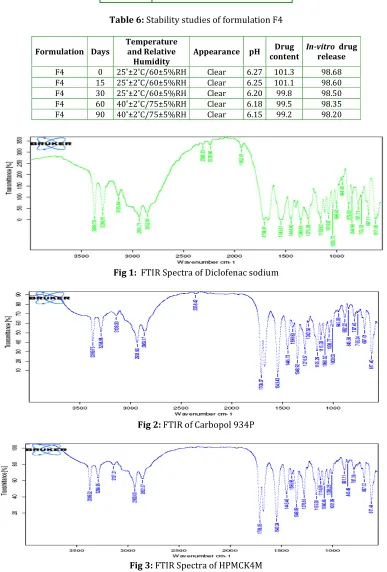
The optimized formulation F4 was subjected to a stability testing for the period of three months as per ICH norms at a temperature of 25˚±2°C with relative humidity RH= 60±5% and 40º ± 2°C with relative humidity RH= 75±5%. The optimized formulation F4 was analyzed for the changes in appearance, pH, percentage of drug content and in-vitro diffusion study
by procedure stated earlier. The results are shown in [Table 06]
RESULTS AND DISCUSSIONS
The objective of the present study was to formulate Transdermal gels of Diclofenac sodium. Total twelve different Diclofenac sodium transdermal gels with different polymer ratios were prepared. In order to select the optimized formulation, various evaluation parameters were checked and subjected to in-vitro diffusion study and their release kinetic study were observed. The optimized formulation was further studied for ex-vivo permeation using rat abdominal skin.
Preformulation studies
Characterization of Diclofenac sodium:
The following tests were performed according to British Pharmacopoeia.
Description: A white or almost white powder Solubility: Methanol and Ethanol
Melting Point: 296.149˚C
From these tests it was confirmed that the sample complies with the monograph.
Compatibility studies
The incompatibility between the drug and excipients were studied by FTIR spectroscopy. The results indicate that there was no chemical incompatibility between drug and excipients used in the formulation. The results are shown in [Fig 01- 05].
Evaluation of Transdermal gels:
Clarity:
Carbopol 934P gels were found to be sparkling and transparent, HPMC K4M gels were found to be translucent. All gels were free from presence of particles. The results are shown in [Table 02].
Homogeneity:
Spreadability:
The value of spreadability indicates that the gel is easily spreadable by small amount of shear. In formulations F1 to F4, Spreadability of Carbopol 934P gel was in the range 18.75- 27.39 g.cm/sec. In formulations F5 to F8, Spreadability of HPMCK4M gel was in the range 20.06- 24.27 g.cm/sec. In formulations F9 to F12, Spreadability of Na CMC was in the range of 19.07- 24.57 g.cm/sec, indicating Spreadability of Carbopol 934P containing Diclofenac sodium gel i.e. F4 was good i.e. 27.39 g. cm/sec as compared to HPMC K4M gel and Na CMC gel. The results are shown in [Table 02].
Extrudability:
The extrusion of the gel from the tube is an important during its application and in patient acceptance. Gels with high consistency may not extrude from tube whereas, low viscous gels may flow quickly, and hence suitable consistency is required in order to extrude the gel from the tube. Extrudability of Carbopol 934P gel i.e. F4 formulation was found to be Excellent when compared to other formulations. The results are shown below in [Table 02].
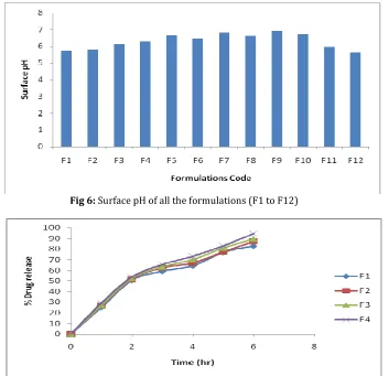
Surface pH:
The pH value of all developed formulations of Carbopol gels (F1-F4) were in the range of 5.71- 6.27, HPMC gels (F5-F8) were in the range of 6.45- 6.82 and Na CMC gels (F9-F12) were in the range of 5.65- 6.91 which is well within the limits of skin pH i.e. 5.6-7.5. Hence, it was concluded that all the formulations could not produce any local irritation to the skin. The results are shown in [Table 03].
Viscosity Measurement:
The Viscosity of the formulations i.e. F1-F4 containing drug and Carbopol 934P were in the range of 1,92,000 -3,10,000 cps, whereas the formulations i.e F5-F8 containing drug and HPMC K4M were in the range of 1,36,000 – 1,47,000 cps, whereas formulations i.e F9-F12 containing drug and Sodium CMC were in the range of 1,52,000- 1,80,000 cps. From the results it was found that the formulation F1 showed maximum
viscosity i.e. 3,20,000 cps and formulation F8 showed minimum viscosity i.e. 1,36,000 cps.
The results are shown in[Table 03]. Drug Content:
The percentage drug content of all prepared gel formulations i.e. F1 to F12 were found to be in the range of 97.21±0.18 to 101.46±0.26%. The percentage drug content of formulations was found to be within the I.P limits. Hence methods adopted for gels formulations were found suitable. The results are shown in [Table 03].
In-vitro drug diffusion studies:
In-vitro drug release study of different gel formulations i.e. F1 to F12 were carried out through dialysis sac (cellophane membrane) and are plotted. The percentage drug release for the formulations containing drug and carbopol 934P i.e. F1 to F4 were found to be in the range of 82.88% to 98.68% in 6 hours. Among these formulations, formulation F4 containing drug and carbopol 934P in the ratio1:2 showed high percentage of drug release i.e. 98.68% in 6 hours. The results indicate that increase in the concentration of Carbopol 934p, increases the drug release.
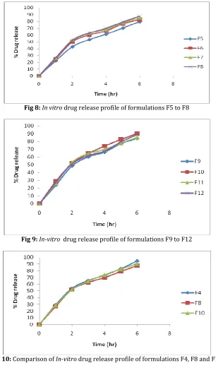
The percentage drug release for the formulations containing drug and HPMC K4M i.e. F5-F8 were in the range of 79.59 – 87.72% in 6 hours. Among these, formulation F8 containing drug and HPMCK4M in the ratio 1:4 showed highest percentage of drug release i.e. 87.72% in 6 hours. From the above it was observed that increase in the concentration of HPMC K4M, increases the drug release.
conducted for the formulations F4, F8 and F10. The results are shown in [Fig 07-10].
From the above result it is observed that the formulation F4 containing drug and Carbopol 934P in the ratio 1:2 showed highest percentage drug release i.e. 98.68% in 6 hours.
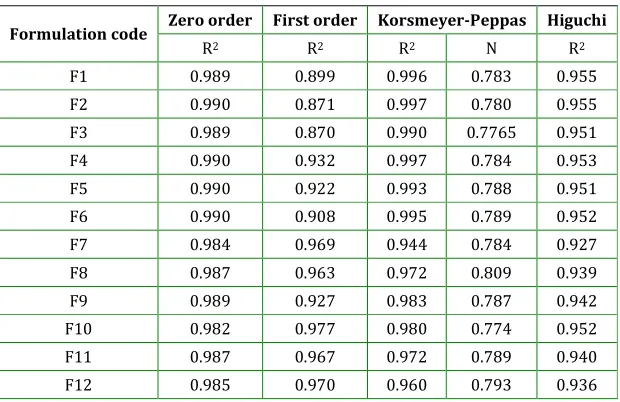
Drug release kinetics:
In-vitro drug release data of F1 to F12 were fitted to zero order, first order, Higuchi and Korsmeyer-Peppas equations to ascertain the pattern of drug release. The results are shown in [Table 04]. In-vitro drug release data for all the formulations F1 to F12 were subjected to release kinetic study according to Zero order, First order, Higuchi and Korsemeyer-Peppas equation to ascertain the mechanism of drug release. Among the zero-order and first-order, the R2 values were found to be higher in zero-order. So, all the formulations followed zero-order kinetics. But in case of mechanism of drug release, between Higuchi and Korsemeyer-Peppas equation, the R2 value were found to be higher in Korsemeyer-Peppas equation and release exponent “n” value less than 1 i.e. (n > 0.5). This indicates that all the formulations followed non-Fickian diffusion. Hence it was concluded that all the formulations followed zero-order drug release with non-Fickian diffusion.
Ex-vivo permeation studies:
It was concluded that the formulation F4 containing drug, carbopol 934P in the ratio 1:2, showed good spreadability, extrudability and invitro drug release.
On the basis of above results formulation F4 was studied for ex-vivo permeation using rat abdominal skin. The optimized formulation was analyzed by HPLC method at 285nm for 6hrs release through rat abdominal skin. The flux was calculated. The results of drug permeation from optimized formulation through the rat abdominal skin revealed that Diclofenac sodium was released from the optimized formulation and permeate through the rat abdominal membrane and could possibly permeate through the human abdominal membrane. The drug permeation from F4 was slow and steady and 0.89gm of Diclofenac sodium could permeate through the skin membrane with a flux of 0.071 gm hr-1 cm-2. The results are shown in [Table 05] and [Fig.11].
Skin irritation test:
Based on in-vitro diffusion study formulation F4 containing drug and Carbopol 934P in the ratio 1:2 was optimized. Furthur, Skin irritation test was performed with optimized formulation F4 in white rabbits divided in 3 groups. It was found that the gel F4 causes no irritation or erythema.
Stability Studies:
Accelerated stability studies was conducted in best formulation F4, according to ICH guidelines i.e. 25˚±2˚C/60±5%RH for first 30 days and 40˚±2˚C/75±5%RH upto 90 days. The results indicate that there was no so much change in appearance, pH, drug content and in-vitro drug release studies. The results are shown in [Table 06].
Table 1: Formula for the preparation of Diclofenac sodium Transdermal gels using Carbopol 934P, HPMCK4M and Sodium CMC
Ingredients F1 F2 F3 F4 F5 F6 F7 F8 F9 F10 F11 F12
Diclofenac sodium (gm) 1 1 1 1 1 1 1 1 1 1 1 1
Carbopol 934P (gm) 0.5 1 1.5 2 - - - -
HPMC K4M (gm) - - - - 2.5 3 3.5 4 - - - -
Sodium CMC - - - 1 1.5 2 2.5
Triethanolamine (ml) 0.4 0.6 0.8 1.0 - - - -
Alcohol (ml) 20 20 20 20 - - - --
Propylene glycol (ml) 10 10 10 10 30 30 30 30 30 30 30 30
PEG 400 (ml) - - - - 7 7 7 7 7 7 7 7
Table 2: Clarity, Homogeneity, Spreadability, Extrudability Parameters
Formulation
Code Clarity Homogeneity Spreadability Extrudability
F1 + Satisfactory 18.75 +
F2 ++ Good 19.85 ++
F3 ++ Good 22.55 ++
F4 +++ Excellent 27.39 +++
F5 ++ Good 20.06 +
F6 ++ Good 21.08 ++
F7 ++ Good 23.54 ++
F8 ++ Good 24.27 + +
F9 ++ Good 19.07 ++
F10 +++ Excellent 21.81 +++
F11 ++ Good 24.57 ++
F12 ++ Good 23.25 ++
Table 3: pH, Viscosity and Drug Content (%)
Formulation code pH Viscosity (cps) Drug Content (%)
F1 5.71±0.05 3,20,000 98.53±0.21
F2 5.79±0.15 1,92,000 97.21±0.18
F3 6.12±0.02 2,40,000 98.92±0.27
F4 6.27±0.03 3,10,000 101.3±0.22
F5 6.64±0.02 1,44,000 101.46±0.26
F6 6.45±0.07 1,47,000 98.92±0.25
F7 6.82±0.05 1,38,000 98.82±0.31
F8 6.60±0.04 1,36,000 99.95±0.18
F9 6.91±0.02 1,52,000 97.94±0.33
F10 6.73±0.09 1,60,000 98.08±0.40
F11 5.98±0.12 1,70,000 97.24±0.38
F12 5.65±0.14 1,80,000 99.13±0.19
For all n=3±S.D.
Table 4: Drug release kinetics of all the formulations (F1 - F12)
Formulation code Zero order First order Korsmeyer-Peppas Higuchi
R2 R2 R2 N R2
F1 0.989 0.899 0.996 0.783 0.955
F2 0.990 0.871 0.997 0.780 0.955
F3 0.989 0.870 0.990 0.7765 0.951
F4 0.990 0.932 0.997 0.784 0.953
F5 0.990 0.922 0.993 0.788 0.951
F6 0.990 0.908 0.995 0.789 0.952
F7 0.984 0.969 0.944 0.784 0.927
F8 0.987 0.963 0.972 0.809 0.939
F9 0.989 0.927 0.983 0.787 0.942
F10 0.982 0.977 0.980 0.774 0.952
F11 0.987 0.967 0.972 0.789 0.940
Table 5: Ex-vivo drugpermeation of optimized Formulation F4
Time (h) Cumulative drug permeated (gm)
0 0
1 0.17
2 0.31
3 0.49
4 0.62
5 0.77
6 0.89
Table 6: Stability studies of formulation F4
Formulation Days
Temperature and Relative Humidity
Appearance pH Drug
content
In-vitro drug
release
F4 0 25˚±2˚C/60±5%RH Clear 6.27 101.3 98.68
F4 15 25˚±2˚C/60±5%RH Clear 6.25 101.1 98.60
F4 30 25˚±2˚C/60±5%RH Clear 6.20 99.8 98.50
F4 60 40˚±2˚C/75±5%RH Clear 6.18 99.5 98.35
F4 90 40˚±2˚C/75±5%RH Clear 6.15 99.2 98.20
Fig 1: FTIR Spectra of Diclofenac sodium
Fig 2: FTIR of Carbopol 934P
Fig 4: FTIR Spectra of Sodium CMC
Fig 5: FTIR Spectra of Diclofenac sodium + Carbopol 934P + HPMCK4M + Sodium CMC
Fig 6: Surface pH of all the formulations (F1 to F12)
Fig 8: In vitro drug release profile of formulations F5 to F8
Fig 9: In-vitro drug release profile of formulations F9 to F12
Fig 10: Comparison of In-vitro drug release profile of formulations F4, F8 and F10
CONCLUSION
It was observed that Carbopol 934P gel containing Diclofenac sodium in 1:2 ratio (F4) produced better spreadability and consistency as compared to other formulations. The developed F4 gel showed good homogeneity, suitable pH, no skin irritation and good stability. The maximum percentage of drug release was found to be 98.68% in 6 hours in formulation F4. The drug permeation from optimized formulation i.e. F4 was slow and steady and 0.89 gm of Diclofenac sodium could permeated through rat abdominal skin membrane with a flux 0.071 gm hr-1 cm-2 and could possibly permeate through human abdominal membrane. The Carbopol 934P forms water washable gel because of its water solubility and has wider prospects to be used as a topical drug delivery system.
REFERENCES
1. Hsieh D.,Drug Permeation Enhancement-Theory and
Applications. In Drug and the Pharmaceutical Sciences,
New York, Marcel Dekker, 1987; 11-13
2. Langer R.,Transdermal Drug Delivery: Past progress,
current status and future prospects. Adv Drug Deliv
Rev. 2004; 56: 557-558.
3. Barry B.,Transdermal Drug Delivery. In: Aulton ME,
Pharmaceutics. The science of dosage form design. 2nd
ed. Churchill, Livingstone, 2002; 499-543.
4. Idson B.,Jack L.,Semisolids. In: Lachmann L,
Liebermann HA and Kanig JL. The Theory and Practice
of Industrial Pharmacy, 3rd ed. Bombay: Varghese
Publishing House, 1990; 534-563.
5. Pena LE.,Gel dosage form: Theory, Formulation and
Processing. In: Osborne DW, Amann AH. Topical drug
delivery formulation. New York, Marcel Dekker; 1990;
381-388.
6. Alberto B.,Clinical pharmacokinetics and
metabolism of Nimesulide in flammopharmacology.
2001; 9: 81-89.
7. Sankar S V.,Chandrasekharan AK.,Durga S., Prasanth
KG., Nilani P., Formulation and stability evaluation of
diclofenac sodium ophthalmic gels. Ind. J. Pharm. Sci.
2005; 67(4): 473-476.
8. Lakshmi P K.,Marka K K.,Aishwarya S., Shyamala B.,
Formulation and evaluation of Ibuprofen Topical gel: A
Novel approach for penetration enhancement. Int.J.
Applied Pharm. 2011; 3 (3): 25-30.
9. Swamy N.G.N., Mazhar P., Zaheer A., Formulation and
evaluation of Diclofenac sodium gels using Sodium
carboxymethyl Hydroxypropyl Guar and
Hydroxypropyl methylcellulose. Indian J. Pharm. Educ.
Res. 2010; 44 (4): 310-314.
How to cite this article: Satyabrata Bhanja*, P. Kishore
Kumar, Muvvala Sudhakar, Arun Kumar Das; Formulation and Evaluation of Diclofenac Transdermal Gel; J. Adv. Pharm. Edu. & Res. 2013: 3(3): 248-259.