I
WONAB
EDNARZ−M
ISA1, J
ADWIGAP
IETKIEWICZ1, T
ERESAB
ANAŚ1, A
NDRZEJG
AMIAN1, 2Enolase from
Klebsiella pneumoniae
and Human Muscle Cells.
I. Purification and Comparative Molecular Studies
Enolaza z komórek
Klebsiella pneumoniae
i mięśniowa enolaza ludzka.
I. Oczyszczanie i porównawcze badania właściwości molekularnych
1 Department of Medical Biochemistry, Wroclaw Medical University, Poland
2 Institute of Immunology and Experimental Therapy, Polish Academy of Sciences, Wrocław, Poland
Adv Clin Exp Med 2009, 18, 1, 71–78 ISSN 1230−025X
ORIGINAL PAPERS
Abstract
Background. This report concerns the glycolytic enzyme enolase from the cytoplasm of Klebsiella pneumoniae
bacterial cells and its similarity with human muscle−specific enzyme.
Material and Methods. Human muscle−specific enolase was purified from crude extract using standard chro− matographic techniques, but this procedure was unsuccessful for isolation of bacterial enzyme. Gel filtration on a Sephadex G−100, anion−exchange chromatography on a DEAE−Sephadex A−50 column, and preparative elec− trophoresis were applied to obtain an electrophoretically homogenous cytosolic protein from K. pneumoniaecells. Results.Human muscle−specific enolase was purified 83−fold with a specific activity of 75 U/mg. The new proce− dure resulted in a 76−fold purification of bacterial enolase, with a recovery rate of 14% and specific activity of 31 U/mg. The purified protein analyzed in SDS−PAGE appeared as a single band with a molecular mass of 47 kDa. A similar molecular weight (45 kDa) for the human enolase monomer was obtained. The molecular mass of the native K. pneumoniaeenolase was estimated to be 94 kDa.
Conclusions.Although the specific activity of purified K. pneumoniaeenolase is half that observed for human muscle−specific enzyme, the application of preparative gel electrophoresis for the purification of the bacterial eno− lase permits obtaining a homogenous enzyme with relative good recovery. The results presented in this report indi− cate that both enzymes have a dimeric native structure, comparable to enolases from other sources. In the second part of these investigations the kinetic differences between the bacterial and human muscle−specific enolase will be presented (Adv Clin Exp Med 2009, 18, 1, 71–78).
Key words: enolase, enzyme purification, preparative gel electrophoresis, molecular properties, Klebsiella pneu− moniae.
Streszczenie
Wprowadzenie. Doniesienie dotyczy glikolitycznego enzymu enolazy z cytoplazmy komórek bakteryjnych
K. pneumoniaei jego podobieństwa z ludzką enolazą mięśniowoswoistą.
Materiał i metody. Enolazę mięśniowoswoistą człowieka otrzymywano, stosując standardowe techniki chromato− graficzne, ale były one nieskuteczne w wydzielaniu enzymu bakteryjnego. W celu uzyskania homogennego białka cytozolowego z komórek K. pneumoniaewykorzystano filtrację żelową na Sephadex G−100, chromatografię jono− wymienną w żelu DEAE Sephadex A−50 i elektroforezę preparatywną
Wyniki. W procedurze wydzielania enolazy mięśniowoswoistej człowieka otrzymano enzym jednorodny, o aktyw− ności specyficznej 75 U/mg i 83−krotnym stopniu oczyszczenia. W opracowanej nowej metodzie izolowania enzy− mu bakteryjnego uzyskano 76−krotne oczyszczenie, z wydajnością 14% i aktywnością specyficzną 31 U/mg. Ana− lizowane metodą SDS−PAGE oczyszczone białko ujawniło się jako pojedynczy prążek o ciężarze cząsteczkowym 47 kDa. Dla monomeru enolazy ludzkiej określono niższy ciężar cząsteczkowy – 45 kDa. Masę cząsteczkową na− tywnej enolazy K. pneumoniaeokreślono w warunkach elektroforezy niedenaturującej jako 94 kDa, co wskazuje na dimeryczną strukturę cząsteczki.
Enolase (2−phosphopyruvate hydrolyase – EC
4.2.1.11) catalyses the Mg2+−dependent conversion
of 2−PGA (2−phospho−D−glycerate) to PEP (phos− phoenolpyruvate) (Fig. 1). It is essential for the degradation of carbohydrates along the glycolysis pathway as well as for glucose synthesis via glu− coneogenesis. As this reaction occupies a key position in the metabolic pathway of fermentation, enolase is ubiquitously present in abundance in the biological world [1]. Enolase has been found in almost all organisms in several isoforms. In verte− brates, among them mammalian and human cells, it is active as a homo− or heterodimer. Tissue−spe− cific isoforms of the enzyme are formed by two of
the three types of subunits, α, β, and γ. Each sub−
unit is encoded by a distinct gene whose expres− sion is regulated in a tissue−specific and develop−
ment−specific manner. The ααembryonic form is
widely distributed in most adult tissues. During development, the accumulation of specific iso− forms accompanies the differentiation of two tis−
sues with high energy demands: αβand ββin stri−
ated muscles and αγ and γγ in the brain [1, 2].
Alpha−enolase is present in most vertebrate tis− sues, including liver, kidney, lung, spleen, and adi−
pose tissue, whereas βis located in the heart and
skeletal muscles and the γ form is found only in
neurons and neuroendocrine cells [3]. An approx− imately 82% amino−acid sequence identity
between the three types of subunits in mammalian and human enolases was observed [3, 4].
Enolase has been isolated and characterized from a broad spectrum of sources. The enzyme has been found in organisms from eubacteria to mam− mals and has maintained a highly conservative pri− mary and tertiary structure throughout evolution [5, 6]. Comparison of the amino−acid sequences determined for about 80 enolases from different species demonstrated a high degree of identity in evolutionarily distant species. Between primates and lower organisms an about 50% sequence homology of this protein was observed [3, 6].
Although enolase from all eukaryotes and many prokaryotic species appears as a dimer, an octamer− ic enzyme has been reported in some bacterial strains, for example the hyperthermophilic
Thermotoga maritima and the Gram−positive
pathogen Streptococcus pneumoniae [7, 8]. The
subunit molecular weight of the most isolated eno− lases is in the range of 40–50 kDa, but for the
monomer from Streptococcus rattusit is 22 kDa [9].
Enolase has been isolated from many different organisms in this department [10–12]. The subject of this investigation is a bacterial enolase from
cytoplasm fromKlebsiella pneumoniaecells.The
cell surface enolase like protein of K. pneumoniae
has been described in prerias report. This protein showed some epitope similarity with human mus− cle−specific enolase and maintained residual enzy− matic activity in the inner−membrane fraction [13]. In the present study a simple method for the purifi−
cation of enolase from the cytosolic fraction of K.
pneumoniae cells is presented. The essential mol− ecular properties of this enzyme are described and compared with those of human muscle enolase for a better understanding of some differences be− tween the bacterial and human enzymes.
Material and Methods
All chemicals used were of analytical grade. 2−Phospho−D−glycerate was purchased from Fluka. The kit of molecular mass protein markers for SDS− PAGE (sodium dodecyl sulfate) was from Bio−Rad. Native molecular mass protein standards and other
jak enolazy z innych źródeł. W drugiej części niniejszych badań zostaną przedstawione różnice kinetyczne między bakteryjną i mięśniowoswoistą enolazą człowieka (Adv Clin Exp Med 2009, 18, 1, 71–78).
Słowa kluczowe: enolaza, oczyszczanie enzymów, elektroforeza preparatywna, właściwości cząsteczkowe, Kleb− siella pneumoniae.
Fig. 1. The reversible dehydration of 2−phospho−D− glycerate (2−PGA) to phoshoenolpyruvate (PEP) cat− alyzed by enolase
reagents were purchased from Sigma−Aldrich.
K. pneumoniae strain 21 was obtained from the Department of Microbiology of Wroclaw Medical University. Bacterial cells were cultivated in TSB (Tripticase Soy Broth) (BIOCORP). Tissue samples
of human tibialis anterior muscle were obtained
from postoperative material from the Department of Vascular, General, and Transplantation Surgery of Wroclaw Medical University in accordance with the Polish legal requirements under a license issued by the Commission of Bioethics of Wroclaw Medical University.
Purification of Human
Muscle−Specific Enolase
Human β−enolase was isolated according to the
method of Witkowska et al. [13] with some modifi− cations. Briefly, frozen human striated muscle was homogenized with deionized water containing 3 mM
MgSO4and the protease inhibitors PMSF (phenyl−
methylsulfonyl fluoride) and aprotinin (2 µg/ml).
The homogenate was centrifuged at 4500 × gfor 30
min. and the supernatant was filtered through gauze
and heated to 53–54°C for 3 min, cooled to 4°C,
and centrifuged at 9000 × gfor 45 min. The super−
natant was treated with 60–80% saturated ammoni− um sulfate and the precipitated proteins were cen−
trifuged at 9000 × gat 4°C for 45 min. The pellet
was dissolved in buffer A (20 mM Tris−HCl buffer (2−amino−2(hydroxymethyl)−1,3−propanediol), pH
9.0, containing 3 mM MgSO4 and 1 mM β−mercap−
toethanol (β−ME)), dialyzed overnight against the
same buffer, and applied to a DEAE−Sephadex A−50 column (30 × 3 cm) equilibrated with buffer A, which was also used for elution. Enolase was not retained under these conditions and fractions con− taining enolase activity were collected and precipi− tated with ammonium sulfate. The pellet was dis−
solved in buffer B (10 mM phosphate (Na+) buffer,
pH 6.4, with 3 mM MgSO4) and, after dialysis
against buffer B, was run on a CM−Sephadex C−50 column (10 × 3 cm) equilibrated with the same buffer. The protein was eluted with a pH gradient of 6.4–9.0. Fractions with enolase activity were pooled, concentrated and, after dialysis against buffer A, fractionated on a QAE−Sephadex column (5 × 1.6 cm) in buffer A. The main peak, containing about 90% enolase activity, was collected and pre− cipitated by dialysis in 80% ammonium sulfate. The pellet was dissolved in 7.5 mM imidazole−HCl
buffer, pH 6.8, containing 2.5 mM MgSO4, 50 mM
NaCl, and 50% glycerol and stored at 4°C for sev−
eral months without loss of activity.
Purification
of
K. pneumoniae
Enolase
The purification of enolase from K. pneumoni−
aebacterial cells required a simplification of the
procedure used for human muscle−specific
enzyme. Bacterial cells were grown at 37°C for 24
h in TSB without shaking, then centrifuged and washed with PBS (phosphate buffered saline). In the next step the cells were resuspended in 10 mM
Tris−HCl buffer, pH 7.8, containing 1 mM MgSO4,
0.5 mM β−ME, 1% glycerol, and the protease
inhibitors 4 mM PMSF and aprotinin 2 µl/10−ml. The suspension was treated with ultrasound for 30 min at 0°C with a Vibra−Cell YC−130PB (Labo− Plus). The disrupted cell suspension was cen−
trifuged at 4000 × gfor 45 min at 4°C to remove
cell debris. The supernatant, obtained by low−
speed centrifugation, was centrifuged at 100,000 × g
for 1 h to separate the envelope fraction and the supernatant was used as the crude extract for fur− ther purification.
Bacterial enolase from the crude extract was precipitated by dialysis against an 80–100% sat−
urated AS (ammonium sulfate) solution at 4°C.
The pellet was collected after centrifugation at
8000 × gat 4°C for 30 min, dialyzed against 20
mM Tris−HCl buffer, pH 7.8, containing 1 mM
MgSO4 and 1 mM β−ME (buffer C), and fraction−
ated on a Sephadex G−100 column (100 × 1.8 cm)
at 4°C. Fractions with enolase activity were elut−
ed with buffer C, collected, and precipitated overnight by dialysis against an 80–100% satu−
rated AS solution, pH 7.0, at 4°C. The pellet was
centrifuged at 8000 × gfor 30 min at 4°C, resus−
pended in 20 mM Tris−HCl buffer, pH 9.0, con−
taining 1 mM MgSO4 and 1 mM β−ME (buffer
D), and dialyzed overnight at 4°C against large
containing 70–80 mg of protein in 1.5 ml of elec− trode buffer was applied for resolution and elec− trophoresis was run at 100 V. The elution of pro− teins with the electrode buffer was started when the bromophenol blue indicator band reached the base of the separating gel. Fractions with enolase activity were pooled and concentrated using a 10− kDa cut−off membrane (Amicon, Millipore). The homogeneity of the enzyme was determined by SDS−PAGE. The enolase preparation was dia− lyzed against 20 mM Tris−HCl buffer, pH 7.8,
with 1 mM MgSO4 and stored at –80°C.
SDS−Polyacrylamide
Gel Electrophoresis
SDS−PAGE was performed using a mini gel apparatus (Biometra). Samples of protein were applied to 10% acrylamide resolving gels [14] using an electrode buffer containing 25 mM Tris, 192 mM glycine, and 0.1% SDS for 45 min at 200 V. After electrophoresis, the gels were stained with 0.25% Coomassie Brilliant Blue R 250 in 10% acetic acid and 40% methanol and destained using a solution of 5% methanol and 7.5% acetic acid in water. Under these conditions the molecu− lar weight of the enolase subunit was estimated using the Vilber Lourmat System and BIO 1D+ software.
Molecular Weight
Determination of Native
K. pneumoniae
Enolase and
Human
ββ
−enolase
by Polyacrylamide Gel
Electrophoresis
in Native Conditions
The molecular weight of native enolase was determined by electrophoresis in non−denaturing systems according to the Sigma manual bulletin. A sample of 10 µg of enolase and non−denatured protein molecular−weight markers were charac− terized by 7%, 8%, 9%, and 10% polyacrylamide gels. Resolution was performed in 5 mM Tris− HCl buffer, pH 8.3, containing 38 mM glycine.
After determining the Rfof the protein in each gel
relative to the tracking dye, values of 100×[log−
(Rf × 100)] were plotted against the percent gel
concentration for each protein (Rf is the protein
electrophoretic mobility). From these plots, indi−
molecular mass of the native enolase was deter− mined.
Protein Concentration
Determination
Enolase concentration was determined spec− trophotometrically at 280 nm using the absorption
coefficient A0.1%= 0.89 established for 1 mg/ml of
rabbit muscle enolase [1].
Enolase Activity Assay
Bacterial enolase activity was assayed spec− trophotometrically at 240 nm at room temperature as the increase in PEP concentration in a standard assay containing 50 mM imidazole−HCl buffer, pH
7.8, with 1 mM MgSO4, 0.4 M KCl, and 1 mM
2−PGA as a substrate. For measuring human eno− lase activity, the pH of the assay medium was 6.8
and contained 3 mM MgSO4. One unit of enolase
activity was defined as the amount of protein which catalyses the synthesis of 1 µmol of PEP from 2−PGA in 1 min under these conditions. The molar absorption coefficient for PEP was taken as
1.52 M–1cm–1[11].
Results
Purification of Enolases
The purification of bacterial and human mus− cle−specific enolases is summarized in Table 1. Enolase from human striated muscle was purified from crude extract using standard chromatographic techniques. Nine mg of 83−fold purified enolase with a specific activity of 75 U/mg and 15% recov− ery was obtained from 150 g of muscle tissue. Such a procedure applied to bacterial cells gave inhomo− geneous enolase of low activity. Therefore, the heat−treatment step was omitted and the fractiona− tion on CM−Sephadex and QAE−Sephadex columns was replaced by preparative gel elec− trophoresis for bacterial proteins. The procedure for the isolation and purification of enolase from bacterial cells is demonstrated in Fig. 2. The eno−
lase from the cytosol of K. pneumoniae was puri−
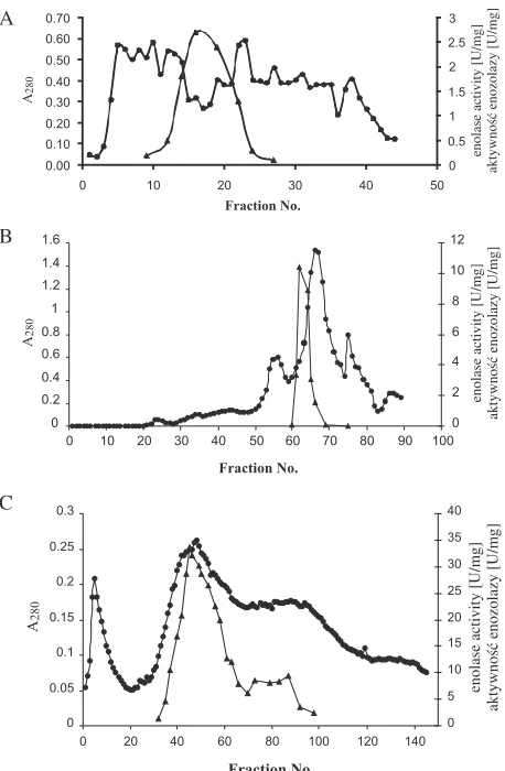
K. pneumoniae crude extract by Sephadex G−100 gel chromatography is presented in Fig. 3A. In this step, substantial amounts of ballast proteins were removed. Fractions with enolase activity were applied to the DEAE−Sephadex A−50 column. The
adsorbed enolase on the anion exchanger was elut− ed with a linear gradient of 0–0.5 M NaCl (Fig. 3B). Fractions with significant enolase activity were pooled, concentrated, and, after dialysis, continu− ous preparative electrophoresis in non−denaturing conditions on a Prep−Cell 491 system followed. The elution profile from the preparative elec− trophoresis is presented in Fig. 3C. The fractions with enolase activity formed major and minor peaks of activity, but only the main peak contained electrophoretically homogenous protein (Fig. 4, lane 3). This pure enzyme preparation was used for
molecular studies. The enolase from K. pneumoni−
ae was purified 76−fold with a specific activity of
31.2 U/mg and has been stored at –80°C for one
year with no apparent loss of activity (multiple freezing and thawing steps were avoided).
Molecular Properties
The molecular mass of native K. pneumoniae
enolase was determined by electrophoresis in a non−denaturing system using molecular mass markers and various percentages of polyacry− lamide gels. The molecular mass of the native bac− terial enolase was found to be 94 kDa (Fig. 5). The
molecular weights of the monomers of K. pneu−
moniae enolase and human β−enolase determined by electrophoresis in SDS−PAGE were 47 kDa and 45 kDa, respectively (Fig. 4). Because the molec− ular mass of the native bacterial enolase was 94 kDa,
the enolase from K. pneumoniae has a dimeric
structure. The single band of protein observed Table 1. Purification of K. pneumoniae enolase and human muscle−specific enolase
Tabela 1. Bilans oczyszczania enolazy cytosolowej z komórek bakteryjnych K. pneumoniae i mięśniowoswoistej enolazy ludzkiej
Step Total protein Total activity Specific activity Purification Yield
(Etap) [mg] [U] [U/mg] (fold) [%]
(Całkowita (Aktywność (Aktywność (Stopień (Wydajność) ilość białka) całkowita) specyficzna) oczyszczenia)
Human β−enolase
Crude extract 5000 4500 0.9 1 100
Heat treatment 3000 4200 1.4 1.6 93
60–80% (NH4)2SO4 750 3225 4.3 4.8 72
precipitation
DEAE−Sephadex A−50 108 2270 21 23 50
CM−Sephadex C−50 48 1440 30 33 32
QAE−Sephadex 9 675 75 83 15
K. pneumoniae enolase
Crude sonic extract 3276 1441 0.41 1 100
80–100% (NH4)2SO4 790 987.5 1.25 3.05 68.5
precipitation
Sephadex G−100 280 588 2.1 5.15 40.8
DEAE−Sephadex A−50 33 293.7 8.9 21.7 20.4
Preparative electrophoresis 6.5 202.8 31.2 76.1 14.1
bacterial cell mass sonication centrifugation 4000×g, 4°C, 50 min
supernatant pellet
(undissrupted cells)
centrifugation 100,000×g, 4°C, 1 h
centrifugation 8000×g, 4°C, 30 min. pellet supernatant
AS precipitation
pellet supernatant
Sephadex G-100
centrifugation 8000×g, 4°C, 30 min pellet supernatant
DEAE-Sephadex A-50
Preparative gel electrophoresis (Prep Cell 491)
enolase (store at –80°C)
AS precipitation
Fig. 2.Schedule of enolase preparation from the cytosol of K. pneumoniaecells
Ryc. 2.Schemat izolacji enolazy z cytozolu komórek
after SDS−PAGE analysis (Fig. 4, lane 3) suggest− ed that identical subunits formed a dimeric mole− cule. Similar properties were observed for human
β−enolase (Fig. 4, lane 4).
Discussion
The method of enolase purification from human muscle summarized in Table 1 involves thermal denaturation of the crude protein extract at
a temperature of 53–54°C, precipitation of the pro−
teins with 60–80% saturated AS, and ion− exchange chromatography [12, 15–17]. The condi− tions for ion−exchange chromatography in our experiments were established on the basis of the pI
7.72 value for human β−enolase [18].
The method used to obtain homogenous
human β−enolase had low efficiency when applied
to the cytosolic enzyme from K. pneumoniaecells.
Therefore, in the first steps of bacterial enzyme purification (Table 1), the heat treatment was omit− ted because enzyme activity was lost. For the pre− cipitation of bacterial enolase from the protein mixture, AS was needed in a degree of saturation of 80–100%, higher than for the human muscle enzyme (60–80%). After precipitation, similar efficiency of purity was obtained, namely about 70% in both cases. The ion−exchange chromatog− raphy on the DEAE−Sephadex column resulted in a significant amount of colorful protein adsorption under equilibration conditions of the column with
0 0.5 1
0.00 0.10 0.20
0 10 20 30 40 50
enolase activity [U/mg]
aktywność enozolazy [U/mg]
Fraction No.
0 0.2 0.4 0.6 0.8 1 1.2 1.4 1.6
0 10 20 30 40 50 60 70 80 90 100
Fraction No.
0 2 4 6 8 10 12
enolase activity [U/mg]
aktywność enozolazy [U/mg]
A280
0 0.05 0.1 0.15 0.2 0.25 0.3
0 20 40 60 80 100 120 140
Fraction No.
0 5 10 15 20 25 30 35 40
enolase activity [U/mg]
aktywność enozolazy
[U/mg]
A280
Fig. 3. Purification of K. pneumoniae enolase.
(A) Sephadex G−100 gel chromatography of K. pneumo− niae. Enolase. About 700 mg of protein sample was applied to the column. (B) Elution profile of 270 mg of protein sample fractioned on DEAE Sephadex A−50 col− umn. (C) K. pneumoniaeenolase purification by continu− ous preparative non−denaturing electrophoresis using a Prep Cell apparatus model 491 (Bio−Rad). 70–80 mg sample was applied on top of the stacking gel. (z) protein profile determined at 280 nm, (c) enolase specific acti− vity
Ryc. 3. Oczyszczanie enolazy z komórek bakteryjnych
K. pneumoniae. (A) Rozdział ok. 700 mg białek wyi− zolowanych z cytosolu pałeczek K. pneumoniae
w kolumnie z żelem Sephadex G−100.(B) Profil elucji bakteryjnych białek cytosolowych (ok. 270 mg) po rozdziale w kolumnie z żelem DEAE Sephadex A−50. (C) Rozdział białek metodą elektroforezy preparaty− wnej w warunkach natywnych. Próbki białek ok. 70 mg rozdzielano z użyciem aparatu Prep Cell model 491 (Bio−Rad). (z) profil elucji białek, (c) aktywność specyficzna enolazy
B
C
Fig. 4. SDS−PAGE analysis of K. pneumoniaeenolase purification: lane 1) protein Mwstandards; lane 2) 25 µg of partially purified bacterial enolase after DEAE−Sephadex fractionation; lane 3) 8−µg sample of bacterial enolase after preparative electrophoresis; lane 4) 8−µg sample of human muscle enolase after the QAE−Sephadex step
Ryc. 4.Analiza oczyszczania enolazy bakterii
a Tris−HCl buffer of low ionic strength and high alkaline pH, similar to the case for human muscle−
specific enolase purification. Human β−enolase
was eluted under these conditions with elution buffer, but bacterial enolase was adsorbed on the DEAE−Sephadex column and was subsequently eluted using a gradient of NaCl. This stage of the purification of bacterial enolase permitted obtain− ing a degree of purity similar to that obtained from human muscle, i.e. 21.7 and 23, respectively. The yield of this process was lower by about half for
the enolase from K. pneumoniae(20.4%) than for
human β−enolase (50%). The ion−exchange chro−
matography step on the CM−Sephadex column was omitted in the bacterial enzyme purification because the yield of the procedure was decreased due to the instability of the catalytic activity. In the last step of purification, preparative electrophore− sis was performed. This procedure can be espe−
cially useful in the isolation of enolase from bac− terial cells for a good efficiency of the purification process. We obtained homogeneous protein with 76−fold purity and with a final yield of 14%. Similar results were reported for the purification
of enolase from Escherichia coli [19] and Strep−
tococus mutanscells [20]. The specific activity of
homogenous enolase from K. pneumoniae was
about 50% lower than that of the human muscle enzyme, but similar values were obtained for the enolases from other bacterial and fungal pathogens [8, 12].
Similarly to other glycolysis enzymes, enolase has been shown to have been highly conservative during evolution [5]. Comparison of the amino− acid composition and sequences shows 40–90% identity among enolases from different species [3]; therefore, most eukaryotic enolases have similar subunit molecular weights, ranging from 82 to 100 kDa. The native molecule is usually a homo− or heterodimer [1, 16, 21], but octameric forms have been reported for enolases from a variety of
bacterial strains, such as Streptococcus mutans, S.
pneumoniae, Bacillus subtilis, and Thermotoga maritima [7, 8, 20, 22]. The monomeric molecular
mass of K. pneumoniae enolase determined by
SDS−PAGE was 47 kDa. This is consistent with earlier findings for the enzyme from other sources and is in agreement with the subunit size found for other prokaryotic enolases obtained from various bacterial [22, 19, 24] and fungal strains [25, 15]. The subunit molecular mass established for human
β−enolase was 45 kDa, this result being similar to
that reported by Cali et al. [32]. The molecular
masses of native K. pneumoniae and human β−eno−
lase were estimated to be 94 kDa and 90 kDa, respectively, suggesting that both enzymes are composed of two identical subunits.
In conclusion, the results reported in this paper represent a starting point for investigating the kinetic properties of the obtained bacterial enolase
for a better knowledge of glycolysis in K. pneu−
moniaecells. These results will be presented in the second part of these studies.
Fig. 5. Determination of the native molecular mass of
K. pneumoniaeenolase by polyacrylamide gel elec− trophoresis in nondenaturing conditions. Protein stan− dards: (1) bovine lactate albumin 14.2 kDA, (2) car− bonic anhydrase 29 kDa, (3) chicken eggs albumin 45 kDa, (4) monomer BSA 66 kDa, (6) dimer BSA 132 kDa(5) enolase from the cytosol fraction of K. pneu− monie cells
Ryc. 5. Wyznaczanie masy cząsteczkowej natywnego enzymu metodą elektroforezy w warunkach niedenatu− rujących. Białka standardowe: (1) albumina z mleka wołowego 14.2 kDa, (2) anhydraza węglanowa 29 kDa, (3) albumina z jaja kurzego 45 kDa, (4) monomer BSA 66 kDa, (6) dimer BSA 132 kDa, (5) enolaza cytosolowa komórek K. pneumoniae
Acknowledgments
We thank Katarzyna Jermakow of the Department of Microbiology, Wroclaw Medical University, for the cultivation of the Klebsiella pneumoniae strain. This work was supported in part by grant no. 2 P05A 101 26 from the Ministry of Science and Higher Education in Warsaw and by grant no. 1004 from Wroclaw Medical University, Poland.
References
[1] Wold F:Enolase. In: The Enzymes, Boyer, P.D. (ed), Acad. Press, New York 1971, 5, 499–538.
[2] Rider CC, Taylor TB:Enolase isoenzymes in rat tissues. Electrophoretic, chromatographic, immunological and kinetic properties. Biochim Biophys Acta 1974, 365, 285–300.
[3] Pancholi V:Multifunctional alpha−enolase: its role in diseases. Cell Mol Life Sci 2001, 58, 902–920.
Termotoga maritima: Purification, characterization, and image processing. Protein Sci 1995, 4, 228–236. [8] Ehinger S, Schubert WD, Bergmann S, Hammerschmidt S, Heinz DW: Plasmin(ogen)−binding α−enolase
form Streptococcus pneumoniae: crystal structure and evaluation of plasmin(ogen)−binding sites. J Mol Biol 2004, 343, 997–1005.
[9] Hüther FJ, Psarros N, Duschner H: Isolation, characterization and inhibition kinetics of enolase from
Streptococcus rattus FA−1. Infect Immun 1990, 58, 1043–1047.
[10] Pietkiewicz J, Kustrzeba−Wójcicka I, Wolna E:Purification and properties of enolase from carp (Cyprinus Carpio). Comparison with enolases from mammals’ muscles and yeast. Comp Biochem Physiol B 1983, 75, 693–698.
[11] Baranowski T, Wolna, E:Enolase from human muscle. In: Methods in Enzymology. Eds.: Colowick SP, Kaplan NO, Acad. Press, New York 1975, Vol. XLII, pp. 335–338.
[12] Kustrzeba−Wójcicka I, Golczak M:Enolase from Candida albicans – purification and characterization. Comp Biochem Physiol B 2000, 126, 109–120.
[13] Witkowska D, Pietkiewicz J, Szostko B, Danielewicz R, Masłowski L, Gamian A:Antibodies against human beta−enolase recognize a 45−kDa bacterial cell wall outer membrane protein. FEMS Immunol Med Microbiol 2005, 45, 53–56.
[14] Laemmli UK:Cleavage of structural proteins during the assembly of the head of bacteriophage T4. Nature 1970, 227, 680–685.
[15] Kustrzeba−Wójcicka I, Pietkiewicz J, Wolna E:Studies on immunological properties of enolase from carp mus− cles after chemical modification of some aminoacid residues. Arch Immunol Ther Exp 1986, 34, 93–99. [16] Merkulova T, Lucas M, Jabet C, Lamandé N, Rouzeau J−D, Gros F, Lazar M, Keller A:Biochemical char−
acterization of the mouse muscle−specific enolase: developmental changes in electrophoretic variants and selec− tive binding to other proteins. Biochem J 1997, 323, 791–800.
[17] Kornblatt MJ, Zheng SX, Lamande N, Lazar M:Cloning, expression and mutagenesis of a subunit contact of rabbit muscle−specific (ββ) enolase. Biochim Biophys Acta 2002, 1597, 311–319.
[18] Chai G, Brewer JW, Lovelace LL, Aoki T, Minor W, Lebioda L:Expression, purification and the 1.8 Åreso− lution crystal structure of human neuron specific enolase. J Mol Biol 2004, 341, 1015–1021.
[19] Dannelly HK, Reeves HC:Purification and characterization of enolase from Escherichia coli. Curr Microbiol 1988, 17, 265–268.
[20] Kaufmann M, Bartholmes P: Purification, characterization and inhibition by fluoride of enolase from
Streptococcus mutans DSM 320523. Caries Res 1992, 26, 110–116.
[21] Marangos JP, Zis AP, Clark RL, Goodrin FK: Neuronal, non−neuronal and hybrid forms of enolase in brain: structural immunobiological and functional comparision. Brain Res 1978, 150, 117–33.
[22] Brown CK, Kuhlman PL, Mattingly S, Slates K, Calie JP, Farrar WW:A model of the quaternary structure of enolases, based on structural and evolutionary analysis of the octameric enolase from Bacillus subtilis. J Protein Chem 1998, 17, 855–866.
[23] Sijbradi R, Blaauwen TD, Tame JR, Oudega B, Luirnh J, Otto BR:Characterization of an iron−regulated alpha−enolase of Bacteroides fragilis. Microb Infect 2005, 7, 9–18.
[24] Bergmann S, Rhode M, Chhatwal GS, Hammerschmidt S:α−Enolase of Streptococcus pneumoniae is a plas− min(ogen)−binding protein displayed on the bacterial cell surface. Mol Microbiol 2001, 40, 1273–1287.
[25] Polidori E, Saltarelli R, Ceccaroli P, Buffalini M, Pierleoni R, Palma F, Bonfante P, Stocchi V:Enolase from the ectomycorrhizal fungus Tuber borchii Vittad.: biochemical characterization, molecular cloning, and localiza− tion. Fungal Genet Biol 2004, 41, 157–167.
[26] Cali L, Feo S, Oliva D, Giallongo A:Nucleotide sequence of a cDNA encoding the human muscle−specific eno− lase (MSE). Nucleic Acids Res 1990, 18, 1893.
Address for correspondence:
Jadwiga Pietkiewicz
Department of Medical Biochemistry Wroclaw Medical University Chałubińskiego 10
50−368 Wrocław Poland
Tel.: +48 71 784 13 78
E−mail: [email protected]
Conflict of interest: None declared