Witold Szaflarski
1, Patrycja Sujka-Kordowska
1, Radosław Januchowski
1,
Karolina Jaszczyńska-Nowinka
2, Małgorzata Andrzejewska
1, Adnan Savan
3,
Piotr Dzięgiel
1, 4, Michał Nowicki
1, Maciej Zabel
1, 4Expression of Cell Vault Components MVP, TEP1
and vPARP in Cancerous Ovarian Tissues*
Ekspresja składników krypt komórkowych MVP, TEP1 oraz vPARP
w tkankach nowotworowych z jajnika
1 Department of Histology and Embryology, Poznań University of Medical Sciences, Poland 2 Department of Gynecological Oncology, Poznań University of Medical Sciences, Poland
3 Department of Biomaterials and Experimental Dentistry, Poznań University of Medical Sciences, Poland 4 Department of Histology and Embryology, Wroclaw Medical University, Poland
Abstract
Background. Cell vaults are composed of three proteins (MVP, TEP1, vPARP) and vRNA. They are known to be involved in the development of resistance mechanisms in cancer cells. There is currently a great deal of controversy over how vaults and their components participate in increasing resistance.
Objectives. The absolute quantification of mRNA molecules for MVP, TEP1 and vPARP genes in normal and can-cerous cells, and investigation of the quantitative relations between their levels of expression in cells.
Material and Methods. The study involved 18 surgical specimens from patients diagnosed with ovarian cancer. After RNA isolation from the specimens, the expression of genes encoding MVP, TEP1 and vPARP was deter-mined by quantitative reverse-transcription polymerase chain reaction (RT-QPCR) analysis. In some cases gene expression was confirmed by standard immunohistochemistry. The cancer cells in the specimens were identified by standard H+E staining, correlations with the analyzed gene expression levels were calculated using Spearman’s rank correlation technique and the statistical significance was determined.
Results. The absolute quantification of MVP, TEP1 and vPARP mRNA demonstrated a strong correlation between the expression of MVP and TEP1. To some extent, vPARP acts as a housekeeping gene. In the specimen collection used in the current study, the higher level of MVP expression was not associated with a larger number of ovarian cancer cells.
Conclusions. The RT-QPCR analysis revealed that MVP, TEP1 and vPARP genes are widely expressed in normal and cancerous human tissues; however, MVP and TEP1 might associate with each other in order to perform a still- -undefined vault function. The high expression of MVP, TEP1 and vPARP also suggests their ubiquitous role in both normal and malignant cells (Adv Clin Exp Med 2011, 20, 3, 285–294).
Key words: cancer, cell vaults, MVP/LRP, TEP1, vPARP.
Streszczenie
Wprowadzenie. Krypty komórkowe są zbudowane z 3 białek (MVP, TEP1 i vPARP) oraz vRNA. Struktury te uzna-je się za czynniki uczestniczące w rozwoju mechanizmów lekooporności komórek nowotworowych. Dotychczas funkcjonowanie krypt wzbudzało wiele kontrowersji, nie poznano, w jaki sposób krypty oraz ich składniki odpo-wiadają za zwiększenie odporności.
Cel pracy. Bezwzględne określenie liczby cząsteczek mRNA dla genów MVP, TEP1 oraz vPARP w normalnej i nowotworowej komórce oraz określenie relacji między poziomami ich ekspresji w komórce.
Materiał i metody. Zanalizowano 18 próbek chirurgicznych od pacjentów ze zdiagnozowanym rakiem jajnika. Określono ekspresję genów kodujących MVP, TEP1 oraz vPARP za pomocą ilościowej reakcji PCR w czasie
rze-Adv Clin Exp Med 2011, 20, 3, 285–294 ISSN 1230-025X
ORIGINAL PAPERS
© Copyright by Wroclaw Medical University
Cell vaults were described for the first time in 1986 as ribonucleoprotein particles found in the cytoplasm of eukaryotes [1]. Such a late date of dis-covery was due to vaults’ low visibilityin standard electron microscopy (EM). The EM technique is suitable for revealing membranes and nucleic ac-ids, while the protein-rich vaults were misidentified as clathrin-coated vesicle contaminants. Finally, Kedersha and Rome correctly interpreted negative stain EM results based on liver tissue photographs. They termed these new structures “vaults” because of their specific morphology resembling the ceil-ings of Gothic cathedrals [2]. Vaults were charac-terized as the largest ribonucleoprotein particles (RNP) ever described. Approximate calculations made at that time showed their mass was equal to 12.9 MDa and dimensions around 41 × 41 × 72 nm. Interestingly, this was significantly larger than other RNPs and also larger than well-known ribo-somes. Detailed biochemical studies revealed that vaults are composed of three proteins: the 100 kDa major vault protein MVP [1], the 193 kDa vault poly(ADP-ribose) polymerase vPARP [3] and the 240 kDa telomerase-associated protein TEP1 [4]. Furthermore, at least one copy of untranslated RNA was found in vault fractions [5].
Vaults have been identified in many different species. In addition to mammals, they have been observed in such evolutionarily diverse entities as slime mold (Dictyostelium discoideum), sea urchins (Strongylocentrotus pupuratus), amphibians (Rana catesbeiana and Xenopus laevis), birds (Gallus gal-lus) and fish (electric ray Torpedo marmorata and
Discopyge ommata) [6–9]. The high evolutionary conservation suggests that vaults have a funda-mental role in the living cell. However, the ques-tion of which role is mediated by vaults is still open and many theories have been suggested. One of the most essential roles that vaults may theoretically play would be maintenance of transport between cytoplasm and nucleus. They may also participate in the formation of nuclear membrane and/or
serve as the scaffold for constructing the nuclear pore complex (NPC) [10].
MVP is the main component of vaults, com-prising approximately 75% of the total mass [11]. Interestingly, in insect cells MVP alone is able to create vault-like particles (VLPs) by self-polymer-ization, without the need for the other components [12]. The self-interactions of MVP molecules are possible due to a coiled-coil domain located at the C-terminal half of the MVP molecule, which was previously found to be essential for vault forma-tion [13]. MVP does not contain endogenous en-zymatic activity. The N-terminal region of MVP interacts with vPARP [3, 13]. vPARP can be also found in non-vault-associated cytoplasmatic frac-tions as well as in the nucleus [3]; it belongs to the PARP (poly-ADP ribose polimerase) family of proteins, which is associated with DNA repair mechanisms [14]. vPARP knockout mice are vi-able, but they are more sensitive to gamma irra-diation and treatment with some alkylating agents [15]. vPARP is able to ADP-rybosylate itself and MVP in purified vaults [3]. Since the precise func-tion of vaults is still unknown, it is difficult to assess the impact of poly(ADP)-rybosylation on vault function. However, it may enhance or ne-gate vault interaction(s) with other vault compo-nents or proteins in the cell. It may also allow for changes in vault conformation, i.e., the opening and closing of vaults. The 240 kDa vault protein, another minor vault protein, has been found to be identical with the already known telomerase-binding protein TEP1 [4]. However, TEP1 is not necessary for adequate telomerase action, nor is it a component of the telomerase core complex. Fur-thermore, TEP1 does not demonstrate telomerase activity. However, TEP1 is able to interact with both telomerase RNA [16] and several human vR-NAs [4]. Co-immunoprecipitation and subcellular fractionation have demonstrated that TEP1 is as-sociated with purified vault particles [4]. Vaults of TEP1 (–/–) mice, viewed by cryo-EM, contain less czywistym z zastosowaniem w odwrotnej transkrypcji (RT-QPCR) po wcześniejszym wyizolowaniu RNA z pró-bek poddanych analizie. Potwierdzenie ekspresji genów w pewnych przypadkach uzyskano za pomocą tradycyjnej immunohistochemii. Przeprowadzono ponadto standardowe barwienie H+E w celu oszacowania liczby komórek nowotworowych w próbkach przeznaczonych do izolacji RNA. Korelacje między zanalizowanymi poziomami eks-presji genów obliczono, stosując korelację rang Spearmana oraz wyznaczając istotność statystyczną dla uzyskanych wyników.
Wyniki. Bezwzględna ocena liczby cząsteczek mRNA dla MVP, TEP1 oraz vPARP wykazała silną korelację między ekspresją MVP i TEP1. vPARP wykazuje pewne cechy genu podstawowego (housekeeping gene). W naszym zbiorze próbek wyższa ekspresja MVP nie jest skorelowana z większą liczbą komórek raka jajnika.
Wnioski. Analiza za pomocą RT-QPCR wykazała, że geny MVP, TEP1 oraz vPARP ulegają szerokiej ekspresji w ludzkich tkankach zmienionych nowotworowo oraz kontrolnych, a białka MVP i TEP1 mogą oddziaływać ze sobą i spełniać nieokreśloną jeszcze funkcję krypty komórkowej. Duża ekspresja MVP, TEP1 oraz vPARP wskazuje również na ich powszechną rolę w każdej komórce, normalnej i zmienionej nowotworowo (Adv Clin Exp Med 2011, 20, 3, 285–294).
density within the very ends of the cap regions, suggesting that that may be its localization [17]. This finding has recently been confirmed by the crystal structure of rat vaults, where TEP1 appears to be in the cap region [18]. Interestingly, several PARP proteins have a telomere length-regulating function. For example, PARP5a and 5b are known to poly-(ADP)-ribosylate and thus inhibit the te-lomere-binding protein 1 (TRF1) [19]. This may suggest that vPARP can modulate vault function, either by direct modification of MVP or indirectly, by modifying TEP1.
Structural research has been performed using standard EM, cryo-EM and X-ray diffraction, and the data obtained were supported by biochemi-cal analysis. The same overall structure of a single vault was observed in all the techniques, but with some differences. Vaults have a barrel-like struc-ture with dimensions of 75 × 42 nm and an esti-mated molecular weight of 13 MDa, which makes them the largest ribonucleoprotein complex now known. A vault has two protruding polar caps, an invaginated waist and a large internal cavity that can encompass several fully assembled 80S ribo-somes. Vaults are organized in two identical moi-eties, each of which consists of 39 copies of MVP [18]. Although the pure fraction of vaults isolated from different eukaryotic organisms contains three proteins (MVP, TEP1, vPARP) and vRNA, the crystal structure based on vaults isolated from rat liver contains only MVP protein [18]. TEP1 is probably localized in the cap regions because of the higher electron density there [18].
An essential breakthrough was finding that MVP has a high homology to LRP (lung resistant protein), which was already known as one of the multidrug resistance inducers [20]. MVP/LRP (MVP and LRP are currently used as synonyms) are abundant in tissues that are chronically ex-posed to the external environment, such as the epithelium of the lungs and the digestive tract [21]. In the process of evolutionary adaptation, MVP became involved in cancer resistant mechanisms. Thus, MVP is highly overexpressed in drug resis-tant human cancer cells selected against a wide array of chemotherapeutic drugs [22]. In order to confirm that up-regulation of MVP in cancer cells is really responsible for the resistant phenotype, Akiyama et al. used a specific ribozyme targeting MVP mRNA [23, 24]. They demonstrated that in the resistant cell line SW-650, elimination of the MVP protein caused higher sensitivity to doxoru-bicin. Several other studies, however, exclude any direct role of MVP in drug resistance. The typical upregulation of vaults was not enough to induce an MDR phenotype [25]. Similarly, the inhibition of MVP expression in SW1573-2R120 cell did not
result in chemosensitivity, nor in the accumula-tion of doxorubicin in nucleoli [26]. Furthermore, based on the mouse model, restoring MVP expres-sion in knockout animals did not restore the resis-tant phenotype even though it caused formation of intact vaults [27, 28].
Material and Methods
Clinical Material
Eighteen ovarian post-surgery specimens were provided by the Department of Gynecological On-cology, Poznań University of Medical Sciences. The Ethics Committee of Poznań University of Medical Sciences approved the study. The specimens used were all from patients who had received no treat-ment before surgery, except for patients p3 and p7, who had had cisplatin- and paclitaxel-based adjuvant treatment. The patientdetails including histopathology profile are presented in Table 1.
Isolation of Total RNA
and Synthesis of cDNA
The post-surgery tissue samples were stored in RNA Stabilization Solution (RNAlater®, Applied Biosystems) at −80°C. The total RNA fraction was prepared using TRI Reagent® Solution (Applied Biosystems) and then purified using a GeneMA-TRIX Universal RNA Purification Kit (EURx). Total RNA was determined by measuring optical density at 260 nm, and purity was verified by the 260/280 nm absorption ratio, which was consis-tently > 1.8 (NanoDrop® ND-1000, ThermoScien-tific). RNA integrity was assessed by electrophore-sis in 1% agarose gel with ethidium bromide. All RNA samples were stored in H2O at −80°C until
used.
immediately used for subsequent amplification re-actions was stored in H2O at −20°C.
Absolute Quantification
of mRNA Copies
Construction of Standard Curves
to Determine Copy Number
The cDNA of MVP, TEP1 and vPARP was amplified using standard PCR protocols. Prim-ers were used as indicated in Table 2. The PCR products were analyzed in 2% agarose gel in order to confirm their specificity, and then all the PCR products were purified separately using a Gen-eMATRIX PCR/DNA Clean-Up Purification Kit (EURx). The concentration of each DNA sample was estimated by measuring optical density at 260 nm (NanoDrop® ND-1000, ThermoScientif-ic). The weight concentrations were converted to the corresponding DNA copy numbers using the Avogadro constant:DNA (copy) = (6.02 × [10] 23 (copies [mol] (–1) × DNA amount (g))/(DNA lenght (bp)
× 660 (g [mol] (–1) [bp] ↑ (–1)).
A ten-fold serial dilution series of known amounts of DNA corresponding to MVP, TEP1 or vPARP and specific primers, ranging from 1 × 10⁷
to 10 copies per µl, was used to construct standard curves. The threshold cycle (Ct) values of each
di-lution were measured in duplicate and were plot-ted against the logarithm of their initial template copy numbers. Each standard curve was generated by a linear regression of the plotted points. From the slope of each curve, the PCR amplification ef-ficiency (E) was calculated according to the follow-ing equation:
E = 10 1/slope – 1.
Real-Time PCR Reaction
One µl of a given cDNA or DNA solution was added to a reaction mixture composed of 12.5 µl 2× Maxima® SYBR Green/ROX qPCR Maxter Mix (Fermentas), 1 µl of a specific pair of prim-ers (f.c. 0.3 µM) and 10.5 µl H2O. The reactions
were carried out in twin.tec real-time PCR plates with PCR Film (Eppendorf) using a Mastercycler ep-realplex2 (Eppendorf). The PCR program was
as follows:
1) initial denaturation, 95°C, 10 minutes;
2) denaturation, 95°C, 15 seconds; 3) annealing 60°C, 30 seconds; 4) extension 72°C, 30 seconds.
The number of cycles was 40–50. Melting curves were obtained and 2% agarose gel electro-phoresis was used to verify the amplification prod-uct specificity and size. All the samples were am-plified in duplicate or triplicate, and in cases where the results varied by more than 15% the reactions were repeated.
Absolute Quantification
in Real-Time PCR
The absolute quantification method was used to quantify the MVP, TEP1 and vPARP mRNA copy numbers. As Lee et al. described it: “Absolute quantification determines the exact copy concen-tration of a target gene by relating the Ct value to
a standard curve” [29]. Prior to absolute quantifi-cation, the Ct values were normalized by
compari-son to the average of the Ct values obtained from
five housekeeping genes (β-actin, B2M, GAPDH, HPRT1, MRLP19; for details, see Table 2). Prim-ers for studying the expression of vault-related genes and reference genes (Table 2) were pur-chased from the Laboratory of DNA Sequenc-ing and Oligonucleotide Synthesis, Institute of Biochemistry and Biophysics, Polish Academy of Sciences, Warsaw.
Immunohistochemistry
Imunohistochemistry was performed on trans-verse 5 µm formalin-fixed paraffin embedded sec-tions from human ovarian carcinoma. The slides were dewaxed with xylene and gradually hydrated. The activity of endogenous peroxidase was blocked by a 30-minute exposure to 1% H2O2. Monoclonal
mouse anti-human MVP (LRP-56, 1 : 20 dilution, Abcam) was used as the primary antibody. The tested sections were incubated overnight with the primary antibody at 4°C, followed by incubation with the EnVision Detection System Peroxidase/ DAB, Rabbit/Mouse (Dako) for 30 minutes. The sections were then reacted with 3,3-diaminoben-zidine, counterstained with hematoxylin, dehy-drated and mounted.
Statistical analysis
Spearman’s Rank Correlation
Spearman’s rank correlation coefficient was calculated using the following equation:R = 1 – 6 ∑d2
i/n(n2 – 1).
All of the calculations were done using Micro-soft Office 2007. The statistical significance was as-sessed using Statistica 7.1 software (StatSoft).
Results
Determination of the Amount
of Cancer Cells in Ovarian
Surgical Specimens
The 18 surgical specimens were carefully ana-lyzed by the naked eye and under a microscope,
to determine the precise number of cancer cells in each sample. Much of the literature data concern-ing RNA isolation from tumors in situ might be uncertain because the ratio between normal cells and cancer cells in the sample was unknown, and the total RNA fraction is therefore represented by two types of cells. In order to diminish this error, one of the current authors identified the most can-cer-cell-prone regions in our specimens, and then two samples from nearby regions weretaken for RNA analysis and for control H+E staining. Fig-ure 1 shows two example specimens (p5 and p16). Figure 1A shows the diagnosed high-prone frag-ments of the tumors, with the parts taken for RNA isolation and for H+E staining marked with red circles. The results of the H+E staining for p5 and p16 is presented in Fig. 1B, with estimations of the number of cancer cells. The estimations for all the tumors in the study are presented in Fig. 2C. The results represent a wide spectrum of cancer cells in tissues, from a lack of cancer cells (p1 and p2) to over 90% cancerous tissue (p16, p17 and p18).
Table 1. Clinical features of the patients in the study
Tabela 1. Dane kliniczne pacjentów objętych badaniami Sample
(Próbka) Diagnosis(Diagnoza) Stage(Zaawansowanie) Grade(Stopień) Age(Wiek)
p1 serous adenocarcinoma III C G1 76
p2 clear cell adenocarcinoma III C G3 69 p3 papillary serous adenocarcinoma II C G3 47 p4 mucinous adenoma (borderline) I C G1 56
p5 serous adenocarcinoma III C G2 60
p6 serous adenocarcinoma III C G2 53
p7 papillary serous adenocarcinoma III B G3 61
p8 serous adenocarcinoma NS G2 57
p9 papillary serous adenocarcinoma III C G3 47 p10 papillary serous adenocarcinoma III C G2 74
p11 serous adenocarcinoma IIIC G2 44
p12 endometrioid adenocarcinoma III B G2 71
p13 serous adenocarcinoma IV G2 50
p14 serous adenocarcinoma IV G2 50
p15 serous adenocarcinoma II C G2 55
p16 serous adenocarcinoma II C G3 71
p17 serous adenocarcinoma III C G3 59 p18 endometrioid adenocarcinoma II C G1 72 NS – non staging.
The Absolute Quantification
of MVP, TEP1 and vPARP
Expression in Cancerous-
-Derived Ovarian Tissues
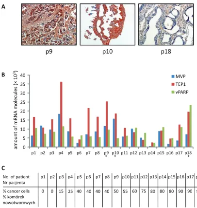
The 18 surgical specimens from patients suffer-ing from ovarian cancer were examined. The histo-pathologic features of the cases are summarized in Table 1. The expression of MVP, TEP1 and vPARP was analyzed using absolute RT-QPCR quantifica-tion. A broad spectrum of expression of MVP, TEP1 and vPARP was observed in the analyzed specimens (Fig. 2A and 2B). MVP expression ranged from 0.5 to 15.8 × 10³ mRNA molecules (p14 and p10,
re-spectively); TEP1 expression was between 2.5–36.3 × 10³ mRNA molecules (p14 and p4, respectively);
and vPARP expression ranged from 2.3 to 23.4 × × 103 mRNA molecules (p14 and p18, respectively).
The lowest expression of all the genes was observed in p16, where 90% of the tissues were cancerous.
The highest average expression was observed in p4, where 25% of the tissues were cancerous (Fig. 1C).
For three samples (p9, p10 and p18) a MVP assessment using classical immunohistochemis-try was performed in order to demonstrate a di-rect correlation between the mRNA copy number and the level of protein expression. The results are shown in Figure 1A. The samples were assessed as (+), (++) and (+++) for p18, p9 and p10, respec-tively. This revealed a gradual increase in MVP expression, which confirmed similar observations obtained using the RT-QPCR method.
Spearman’s rank correlation was done for the expression of MVP, TEP1 and vPARP, as well as for the number of cancer cells in the analyzed specimens. The only statistically significant corre-lation (p < 0.05) observed was for MVP and TEP1 expression (R = 0.613 – see Table 3). Other cor-relations that were found (also shown in Table 3) exhibit no statistical significance and therefore cannot be taken into consideration.
Table 2. Sequences of RT-QPCR primers
Tabela 2. Sekwencje starterów dla RT-QPCR Gene
(Gen) NCBI accesion number(Numer NCBI) Product length(Długość produktu) Sequence(Sekwencja)
MVP NM_017458 135 bp 5’-TGAGGAGGTTCTGGATTTGG (F) 5’-TGCACTGTTACCAGCCACTC (R) TEP1 NM_007110 186 bp 5’-GCTCAAGAGGGAGAAGCTGA (F)
5’-GGACAGAGCCTGTCTGCATT (R) vPARP NM_006437 207 bp 5’-GCCAAATACCGAGCTTTGAG (F) 5’-AGGAGAACCATGCAACAAGG (R) β-ACT NM_001101 169 bp 5’-TCTGGCACCACACCTTCTAC (F)
5’-GATAGCACAGCCTGGATAGC (R) B2M NM_004048 137 bp 5’-CTCACGTCATCCAGCAGAGA (F) 5’-AAGACAAGTCTGAATGCTCCA (R) GAPDH NM_002046 199 bp 5’-GAAGGTGAAGGTCGGAGTCA (F)
5’-CTGAGAACGGGAAGCTTGTC (R) HPRT1 NM_000194 156 bp 5’-CTGAGGATTTGGAAAGGGTG (F) 5’-AATCCAGCAGGTCAGCAAAG (R) MRPL19 NM_014763 171 bp 5’-ACTTTATAATCCTCGGGTC (F)
5’-ACTTTCAGCTCATTAACAG (R)
β-ACT – β-actin.
B2M – β-2-microglobulin.
GAPDH – glyceraldehyde-3-phosphate dehydrogenase.
HPRT1 – hypoxanthine phosphoribosyltransferase 1.
MRPL19 – mitochondrial ribosomal protein L19. F – forward primer.
R – reverse primer.
β-ACT – β-aktyna.
B2M – β-2 mikroglobulina.
GAPDH – dehydrogenaza glyceroaldehydo-3-fosforanowa.
HPRT1 – fosforybozylotransferaza hipoksytanowa 1.
MRPL19 – mitochondrialne białko rybosomalne L19. F – starter wprost.
Interestingly, vPARP behaves like a house-keeping gene, with relatively high expression in all the specimens analyzed (see Fig. 2B). This is supported by the observation that the deviation in vPARP expression in all the samples is lower than that of MVP and TEP1 (Fig. 2B).
Discussion
Multidrug resistance is a major obstacle in attempts to improve clinical outcome in ovarian
Table 3. Spearman’s rank correlation for the mRNA copy number of MVP, TEP1, vPARP and the cancer cell con-tent in cancer-derived tissues
Tabela 3. Korelacja rang Spearman’a dla liczby kopii mRNA MVP, TEP1, vPARP, a także zawartości komórek nowotworowych w tkankach pochodzenia nowotworowego
MVP TEP1 vPARP % cancer cells MVP 1 0.613* (0.325) (0.111) TEP1 1 (0.199) (0.304)
vPARP 1 (0.438)
% cancer
cells 1
% cancer cells – the cancer cell content in cancer-derived tissue (as percentages).
* – p < 0.05 the numbers in parentheses are without statis-tical significance.
% komórek nowotworowych – zawartość komórek nowo-tworowych w tkance nowotworowej (w %).
* – p < 0,05; liczby w nawiasach są nieistotne statystycznie.
Fig. 1. Preparation of the surgical speci-mens for total RNA isolation. A – exam-ples of tumor material from two patients, with the fragments taken for RNA isola-tion and control H+E staining marked (red circles), B – the control H+E stain-ing with an assessment of the number of cancer cells
Ryc. 1. Przygotowanie fragmentów ope-racyjnych do izolacji całkowitego RNA. A – przykład materiału uzyskanego z 2 guzów od pacjentów z zaznaczeniem fragmentów przeznaczonych do izolacji RNA oraz kontrolnego barwienia H+E (czerwona obwódka), B – kontrolne barwienie H+E z oceną liczby komórek nowotworowych
carcinoma patients [30]. The main mechanisms of tumor cells’ resistance to chemotherapeutic drugs are associated with activation of transporter pro-teins that increase drug efflux from cancer cells, or with activation of anti-apoptotic mechanisms [31]. The action of MVP and whole vaults is unclear: These structure do not work as transmembrane proteins causing drug efflux (e.g., P-glycoprotein), but are found frequently in ovarian carcinoma cell lines and tissues [32]. However, MVP is thought to be involved in the nuclear elimination of drugs by an unknown mechanism [33]. Vaults contribute heavily to the multidrug resistance of cancer cells because the range of drugs eliminated by MVP is even broader than those associated with P-gly-coprotein and includes alkylating agents such as melphalan and cyclophosphamide and platinum compounds [21].
activator might be is unknown. It may be directly associated with the vault particle, or it may be any protein located in the cell. The primary can-didates are TEP1 and vPARP. The present study demonstrated a wide expression of both of them in normal cells and in cancer cells. Interestingly, the only statistically significant correlation found was between TEP1 expression and MVP expression. In contrast, vPARP expression is not correlated with either MVP or TEP1 expression. Moreover, none of the analyzed proteins correlate with the number of cancer cells. It seems probable that this means that none of these proteins is a potential activator
of resistance associated with cell vaults. However, the strong correlation between MVP expression and TEP1 expression suggests that the latter may have an important role in the vault function.
The cap region has been proposed as the loca-tion of TEP1 in the vault [17, 18]. It has previously been proposed that cancer resistance may hypo-thetically be caused by vaults docking to the nu-clear pore complex (NPC) [34], which could block the transfer of drugs to the nucleus. A comparison of vault and NPC structures may support this idea, because a vault fits perfectly into the central ca-nal of NPC [35]. This suggestion is also supported
0 5 10 15 20 25 30 35 40
p1 p2 p3 p4 p5 p6 p7 p8 p9 p10 p11 p12 p13 p14 p15 p16 p17 p18
MVP TEP1 vPARP
No. of paent Nr pacjenta
A
B
p18
p9
p10
p1
0 % cancer cells % komórek nowotworowych
C
p2 p3 p4 p5 p6 p7 p8 p9 p10 p11 p12 p13 p14 p15 p16 p17 p18
0 15 25 40 40 40 40 50 55 60 75 80 80 80 90 90 90
* * *
am
ou
nt
o
f m
RN
A
m
ol
ec
ul
es
(×
1
0
3
)
Fig. 2. Quantification of the levels of expression of MVP, TEP1 and vPARP, and assessment of the number of cancer-ous cells in the surgical specimens used in the study. A – Immunohistochemistry with anti-LRP-56 antibodies (MVP) for patients p9, p10 and p18, magnification ×20, B – absolute quantification of the amount of mRNA for MVP, TEP1 and vPARP, C – assessment of the number of cancer cells in the specimens used in the study
by the finding that vaults serve as scaffolds for the correct formation of NPC [10]. Here, TEP1 pro-teins, which are probably located in the two pole caps of the vault [18] may potentially activate the binding of the vault to NPC, causing the closing of the central NPC canal and preventing drug trans-fer to the nucleus.
The current study proposed a new approach to the selection of a tumor fragment for RNA isolation.
In order to determine the number of cancer cells in the tumor, a cancer-prone fragment was divided into two parts, one for isolation of total RNA and the sec-ond for control H+E staining (Fig. 1). In the authors’ opinion this eliminates bias in the results previously caused by normal cells “contaminating” the tumor sample. As shown in Fig. 1B, the number of cancer cells can vary significantly in tumors that have been identified by the naked eye as cancer prone.
Acknowledgments. The authors wish to thank Prof. Dan W. Urry and Kelley D. Urry for helpful discussions.
References
[1] Kedersha NL, Rome LH: Isolation and characterization of a novel ribonucleoprotein particle: large structures contain a single species of small RNA. J Cell Biol 1986, 103, 699–709.
[2] Rome L, Kedersha N, Chugani D: Unlocking vaults: organelles in search of a function. Trends Cell Biol 1991, 1, 47–50.
[3] Kickhoefer VA, Siva AC, Kedersha NL, Inman EM, Ruland C, Streuli M, Rome LH: The 193-kD vault protein, VPARP, is a novel poly(ADP-ribose) polymerase. J Cell Biol 1999, 146, 917–928.
[4] Kickhoefer VA, Stephen AG, Harrington L, Robinson MO, Rome LH: Vaults and telomerase share a common subunit, TEP1. J Biol Chem 1999, 274, 32712–32717.
[5] Kickhoefer VA, Searles RP, Kedersha NL, Garber ME, Johnson DL, Rome LH: Vault ribonucleoprotein particles from rat and bullfrog contain a related small RNA that is transcribed by RNA polymerase III. J Biol Chem 1993, 268, 7868–7873.
[6] Kedersha NL, Miquel MC, Bittner D, Rome LH: Vaults. II. Ribonucleoprotein structures are highly conserved among higher and lower eukaryotes. J Cell Biol 1990, 110, 895–901.
[7] Vasu SK, Kedersha NL, Rome LH: cDNA cloning and disruption of the major vault protein alpha gene (mvpA) in Dictyostelium discoideum. J Biol Chem 1993, 268, 15356–15360.
[8] Vasu SK, Rome LH: Dictyostelium vaults: disruption of the major proteins reveals growth and morphological defects and uncovers a new associated protein. J Biol Chem 1995, 270, 16588–16594.
[9] Herrmann C, Zimmermann H, Volknandt W: Analysis of a cDNA encoding the major vault protein from the electric ray Discopyge ommata. Gene 1997, 188, 85–90.
[10] Vollmar F, Hacker C, Zahedi RP, Sickmann A, Ewald A, Scheer U, Dabauvalle MC: Assembly of nuclear pore complexes mediated by major vault protein. J Cell Sci 2009, 122, 780–786.
[11] Kedersha NL, Heuser JE, Chugani DC, Rome LH: Vaults. III. Vault ribonucleoprotein particles open into flower- -like structures with octagonal symmetry. J Cell Biol 1991, 112, 225–235.
[12] Stephen AG, Raval-Fernandes S, Huynh T, Torres M, Kickhoefer VA, Rome LH: Assembly of vault-like particles in insect cells expressing only the major vault protein. J Biol Chem 2001, 276, 23217–23220.
[13] van Zon A, Mossink MH, Schoester M, Scheffer GL, Scheper RJ, Sonneveld P, Wiemer EA: Structural domains of vault proteins: a role for the coiled coil domain in vault assembly. Biochem Biophys Res Commun 2002, 291, 535–541.
[14] de Murcia G, Menissier-de Murcia J, Schreiber V: Poly(ADP-ribose) polymerase: molecular biological aspects. Bioessays 1991, 13, 455–462.
[15] de Murcia JM, Niedergang C, Trucco C, Ricoul M, Dutrillaux B, Mark M, Oliver FJ, Masson M, Dierich A, LeMeur M, Walztinger C, Chambon P, de Murcia G: Requirement of poly(ADP-ribose) polymerase in recovery from DNA damage in mice and in cells. Proc Natl Acad Sci U S A 1997, 94, 7303–7307.
[16] Harrington L, McPhail T, Mar V, Zhou W, Oulton R, Bass MB, Arruda I, Robinson MO: A mammalian telom-erase-associated protein. Science 1997, 275, 973–977.
[17] Mikyas Y, Makabi M, Raval-Fernandes S, Harrington L, Kickhoefer VA, Rome LH, Stewart PL: Cryoelectron microscopy imaging of recombinant and tissue derived vaults: localization of the MVP N termini and VPARP. J Mol Biol 2004, 344, 91–105.
[18] Tanaka H, Kato K, Yamashita E, Sumizawa T, Zhou Y, Yao M, Iwasaki K, Yoshimura M, Tsukihara T: The structure of rat liver vault at 3.5 angstrom resolution. Science 2009, 323, 384–388.
[19] Weinrich SL, Pruzan R, Ma L, Ouellette M, Tesmer VM, Holt SE, Bodnar AG, Lichtsteiner S, Kim NW, Trager JB, Taylor RD, Carlos R, Andrews WH, Wright WE, Shay JW, Harley CB, Morin GB: Reconstitution of human telom-erase with the template RNA component hTR and the catalytic protein subunit hTRT. Nat Genet 1997, 17, 498–502. [20] Scheffer GL, Wijngaard PL, Flens MJ, Izquierdo MA, Slovak ML, Pinedo HM, Meijer CJ, Clevers HC, Scheper RJ:
The drug resistance-related protein LRP is the human major vault protein. Nat Med 1995, 1, 578–582.
[21] Izquierdo MA, Scheffer GL, Flens MJ, Giaccone G, Broxterman HJ, Meijer CJ, van der Valk P, Scheper RJ: Broad distribution of the multidrug resistance-related vault lung resistance protein in normal human tissues and tumors. Am J Pathol 1996, 148, 877–887.
[23] Kitazono M, Okumura H, Ikeda R, Sumizawa T, Furukawa T, Nagayama S, Seto K, Aikou T, Akiyama S: Reversal of LRP-associated drug resistance in colon carcinoma SW-620 cells. Int J Cancer 2001, 91, 126–131. [24] Kitazono M, Sumizawa T, Takebayashi Y, Chen ZS, Furukawa T, Nagayama S, Tani A, Takao S, Aikou T,
Akiyama S: Multidrug resistance and the lung resistance-related protein in human colon carcinoma SW-620 cells. J Natl Cancer Inst 1999, 91, 1647–1653.
[25] Siva AC, Raval-Fernandes S, Stephen AG, LaFemina MJ, Scheper RJ, Kickhoefer VA, Rome LH: Up-regulation of vaults may be necessary but not sufficient for multidrug resistance. Int J Cancer 2001, 92, 195–202.
[26] Huffman KE, Corey DR: Major vault protein does not play a role in chemoresistance or drug localization in a non-small cell lung cancer cell line. Biochemistry 2005, 44, 2253–2261.
[27] Mossink MH, van Zon A, Franzel-Luiten E, Schoester M, Kickhoefer VA, Scheffer GL, Scheper RJ, Sonneveld P, Wiemer EA: Disruption of the murine major vault protein (MVP/LRP) gene does not induce hypersensitivity to cytostatics. Cancer Res 2002, 62, 7298–7304.
[28] van Zon A, Mossink MH, Schoester M, Scheper RJ, Sonneveld P, Wiemer EA: Efflux kinetics and intracellular distribution of daunorubicin are not affected by major vault protein/lung resistance-related protein (vault) expres-sion. Cancer Res 2004, 64, 4887–4892.
[29] Lee C, Lee S, Shin SG, Hwang S: Real-time PCR determination of rRNA gene copy number: absolute and relative quantification assays with Escherichia coli. Appl Microbiol Biotechnol 2008, 78, 371–376.
[30] Holschneider CH, Berek JS: Ovarian cancer: epidemiology, biology, and prognostic factors. Semin Surg Oncol 2000, 19, 3–10.
[31] Szakacs G, Paterson JK, Ludwig JA, Booth-Genthe C, Gottesman MM: Targeting multidrug resistance in can-cer. Nat Rev Drug Discov 2006, 5, 219–234.
[32] Meijer GA, Schroeijers AB, Flens MJ, Meuwissen SG, van der Valk P, Baak JP, Scheper RJ: Increased expres-sion of multidrug resistance related proteins Pgp, MRP1, and LRP/MVP occurs early in colorectal carcinogenesis. J Clin Pathol 1999, 52, 450–454.
[33] Bouhamyia L, Chantot-Bastaraud S, Zaidi S, Roynard P, Prengel C, Bernaudin JF, Fleury-Feith J: Immunolocalization and cell expression of lung resistance-related protein (LRP) in normal and tumoral human respiratory cells. J Histochem Cytochem 2007, 55, 773–782.
[34] Chugani DC, Rome LH, Kedersha NL: Evidence that vault ribonucleoprotein particles localize to the nuclear pore complex. J Cell Sci 1993, 106, 23–29.
[35] Dickenson NE, Moore D, Suprenant KA, Dunn RC: Vault ribonucleoprotein particles and the central mass of the nuclear pore complex. Photochem Photobiol 2007, 83, 686–691.
Address for correspondence:
Witold Szaflarski
E-mail: [email protected] Tel.: +48 602 311 066
Conflict of interest: None declared