Małgorzata A. Gacka
1, Rajmund Adamiec
1, Paweł Kowal
2Trophic Lesions of the Lower Limbs As Exponents
of Vascular Complications in Essential Thrombocythemia
Zmiany troficzne kończyn dolnych jako wyraz powikłań naczyniowych
w przebiegu samoistnej nadpłytkowości
1 Department of Angiology, Hypertension and Diabetology, Wroclaw Medical University, Poland 2 Students’ Study Group at the Department of Angiology, Hypertension and Diabetology,
Wroclaw Medical University, Poland
Abstract
Background. The etiology of lower limb ulcer formation is highly diversified. Venous and/or arterial insufficiency associated with essential thrombocythemia is one of the possible causes. The available literature offers scant data on the coexistence of essential thrombocythemia and necrotic lower limb lesions resulting from arterial and/or venous perfusion disturbances.
Objectives. The aim of the study was to conduct a retrospective analysis of patients with idiopathic thrombocy-themia treated for trophic lesions of the lower limbs at the Wrocław Medical University Angiology Department between 2002 and 2010.
Material and Methods. Between 2002 and 2010 only 11 of the patients who were treated at the Wroclaw Medical University Angiology Department for trophic limbs lesions also had thrombocythemia. This group consisted of 6 women and 5 men aged 27–85 years. The case histories of these patients were submitted to retrospective analysis.
Results. Trophic lesions coexisted with essential thrombocythemia in only 11 patients (aged 27–85) during this period. Critical ischemia of the toes with the presence of necrotic lesions was found in six study subjects, while ulcers located on the lateral side of the leg were observed in two patients. A futher three patients presented simul-taneous foot and lateral calf ischemic lesions.
Conclusions. Despite advances in vascular diagnostics, the pathomechanism of calf ulcers has proved difficult to explain. Although idiopathic thrombocythemia is not common cause of trophic lesions of the lower limbs, it should be considered in the differential diagnosis. At the same time, patients with idiopathic thrombocythemia should always be monitored for signs of disturbances in lower extremity perfusion and for coexisting risk factors for cardiovascular complications (Adv Clin Exp Med 2011, 20, 4, 455–460).
Key words: vascular complications, ulcers, essential thrombocythemia.
Streszczenie
Wprowadzenie. Etiologia owrzodzeń kończyn dolnych jest różnorodna. Jedną z przyczyn pozostaje niewydolność tętnicza i/lub żylna, która może wystąpić w przebiegu nadpłytkowości samoistnej. W dostępnym piśmiennictwie dane na ten temat są skromne.
Cel pracy. Retrospektywna analiza pacjentów chorych na nadpłytkowość samoistną leczonych z powodu zmian troficznych kończyn dolnych w Klinice Angiologii, Nadciśnienia Tętniczego i Diabetologii AM we Wrocławiu w latach 2002–2010.
Materiał i metody. Grupę badaną stanowiło 11 osób chorych na nadpłytkowość samoistną leczonych z powo-du zmian troficznych kończyn dolnych w Klinice Angiologii, Nadciśnienia Tętniczego i Diabetologii AM we Wrocławiu w latach 2002–2010. Wśród pacjentów, których w czasie choroby poddano analizie retrospektywnej, było 6 kobiet i 5 mężczyzn w wieku 27–85 lat.
Wyniki. U 11 pacjentów (w wieku 27–85 lat) rozpoznano nadpłytkowość samoistną. Wśród 6 chorych występo-wały objawy typowego, krytycznego niedokrwienia palców stóp ze zmianami martwiczymi, u 2 pacjentów obser-wowano duże, płytkie, bardzo bolesne oraz niegojące się owrzodzenie podudzia umiejscowione po stronie bocznej kończyny. U 3 chorych występowały jednoczasowo zmiany niedokrwienne w obrębie stopy i zmiany troficzne na bocznej stronie podudzia. Na podstawie wykonanych badań nie ustalono jednoznacznie patomechanizmu owrzo-dzeń na podudziach.
Adv Clin Exp Med 2011, 20, 4, 455–460 ISSN 1230-025X
oRIGINAl PAPERS
Blood platelets play a significant role in the coagulation process. In essential thrombocythe-mia (ET), elevated platelet counts as well as al-tered platelet activity are noted. This is conducive to the development of thrombosis in venous and arterial vessels as well as in microcirculation. The clinical manifestations of these complications are thus extremely varied. In the literature there are numerous reports of central nervous system and coronary afflictions in patients with ET [1, 2, 3]. Thrombotic lower limb lesions, however, are how-ever rarely mentioned, although they may have significant negative impact on the quality of life, and may lead to a risk of limb loss.
The aim of the study was to conduct a retro-spective analysis of patients with idiopathic throm-bocythemia treated for trophic lesions of the lower limbs at the Angiology, Arterial Hypertension and Diabetology Department of Wroclaw Medical University (Poland) from 2002 to 2010.
Material and Methods
The Wroclaw Medical University Angiology Department admits 15–30 patients with trophic lesions every month. Between 2002 and 2010 only 11 of the patients who were treated there for trophic limb lesions also had thrombocythemia; this group consisted of 6 women and 5 men, from 27 to 85 years old. The case histories of these pa-tients were submitted to retrospective analysis. It is worth noting that the two youngest patients (aged 27 and 47 years) were admitted to the De-partment for investigation of foot necrosis. Suspi-cion of ET arose following routine blood counts (platelet counts at 1,234,000 and 923,000/μl re-spectively), and was confirmed by hematological tests. The remaining patients had been diagnosed with ET a few years before in hematology units. Current criteria for a diagnosis of ET are shown in Table 1 [4]. Unfortunately, a lack of full hemato-logical documentation for the Angiology Depart-ment patients prevented the authors from estab-lishing all the grounds on which the ET diagnoses had been based. Establishing initial platelet counts at the time of diagnosis proved possible in only a few cases (Table 2). only five cases included
re-sults of genetic testing for aberrations in JAK ki-nase activity (the JAK2 V617F mutation was found in only two instances).
Results
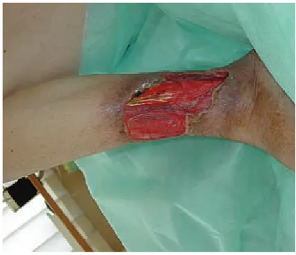
Cases of critical toe ischemia with necrotic le-sions were observed in six study subjects (Fig. 1). Among those, one patient manifested typical chronic lower limb ischemia, with disease progres-sion from Fontaine stage IIb to IV over a two-year observation period; it was possibly accelerated by the patient’s coexistent diabetes. large shallow non-healing and highly painful ulcerations on the lateral side of the calves were observed in a fur-ther two patients (Fig. 2). Anofur-ther three patients presented simultaneous foot ischemia and trophic lesions on the lateral calf.
The study group profile is presented in Tables 2 and 3. Changes in the proximal arterial system were noted in 4 study subjects (3 cases of femoro-popliteal occlusion and 1 case of aorto-iliac occlu-sion). The remaining patients showed distal lesions only, including two subjects with normal ankle-brachial indices. The arteries were checked using color Doppler ultrasonography (Aloka 5500 or Vivid 7) and the ankle-brachial index (Vaskuläres Diagnosesystem).
Thrombosis was found in only 2 study sub-jects (using color Doppler ultrasonography – Alo-ka 5500 or Vivid 7).
The medical histories of 5 patients revealed ischemic heart disease, with myocardial infarctions in 3 subjects; in 1 patient’s coronarography there were no signs of arteriosclerosis, which suggests that the myocardial infarction was of thrombotic origin. 2 patients had undergone ischemic strokes and 1 had upper limb ischemia.
Besides thrombocythemia, other risk factors for vascular complications noted in the study group include arterial hypertension (9/11), type 2 diabetes (2/11), overweight (3/11), obesity (2/11) and dyslipidemia (3/11).
Six patients were treated with hydroxyurea monotherapy, 2 with additional anagrelide, and 3 with anagrelide only (among those 3, 2 also had sulphonylurea intake in their histories). All the Wnioski. Pomimo rzadkiego występowania, w diagnostyce różnicowej zmian troficznych kończyn dolnych należy zawsze rozważyć nadpłytkowość samoistną. Chory z nadpłytkowością samoistną powinien być zawsze oceniany pod kątem zaburzeń krążenia w kończynach oraz współistniejących dodatkowych czynników ryzyka powikłań ser-cowo-naczyniowych. Patomechanizm owrzodzeń podudzi jest nadal niejasny (Adv Clin Exp Med 2011, 20, 4, 455–460).
patients described in the study were administered acetylsalicylic acid; in 4 cases ticlopidine was ad-ministered in addition.
In the patients with critical toe ischemia, phar-macological treatment (cytoreductive, anti-plate-let, anticoagulant and vasoactive agents) resulted in evident local improvement. Additional opioid analegesic administration proved necessary in nu-merous instances. Recurrence of trophic lesions was noted in 4 cases.
Trophic lesions of the calves, on the other hand, showed poor prognosis. During several months of observation, no significant progress in the healing of wounds was noted. limb amputa-tion was necessary in one instance due to second-ary infection and the subsequent development of an extensive phlegmon.
Hemorrhagic complications were observed in 2 patients, with a fatal outcome in one instance (internal bleeding).
Discussion
Vascular complications associated with essen-tial thrombocythemia do not frequently occur in the lower limbs. There is scant data in the available literature on the coexistence of ET and necrotic lower limb lesions resulting from arterial and/ or venous perfusion disturbances. Single reports touch on trophic lesions resulting from arterial ischemia (e.g., three patients out of an observation group of 132), calf ulcerations or peripheral em-bolism [5].
In most cases, signs of peripheral vascular insufficiency manifest many years after an ET di-agnosis. Unfortunately, the Polish public medical care system is not adequately prepared to transfer data in cases of long-term multi-center medical care, so it is sometimes difficult to obtain infor-mation about a patient from another center from previous years. Additionally, some new risk fac-tors for vascular complications in ET have been identified in recent years, but non-hematologists are not likely to be familiar with them. In light of two recent meta-analyses [6, 7], the JAK V617F mutation should also be taken into account when estimating the risk of vascular complications. In the current study, a lack of complete data as to the relationship between the JAK V617F mutation and the observed vascular complications makes it difficult to reach conclusions.
A small number of patients undergo urgent hospitalization in angiology/vascular surgery units due to lower limb necrosis. In these cases essential thrombocythemia should be considered as one of the possible etiological factors, and elevated platelet counts should not be deemed merely a part of the inflammatory response. This can be of crucial im-portance, since thrombocythemia may occur in rel-atively young patients, as seen in the current study. It is still too early to determine whether tro-phic lesions result from arterial thrombosis or from a latent “overlap” of thrombotic processes on existing arteriosclerotic lesions. While the first pathomechanism is more probable in young pa-tients, the second one should be considered in old-er patients. In the future it will be useful to carry out a prospective study to assess patients’ vascular state at the time of diagnosis and after few years.
In the present study, as many as 36% of the patients manifested proximal arterial lesions. The possibility of large vessel involvement in the course of ET was emphasized by Johnson et al. [1]. It is Fig. 1. Critical toe ischemia in essential thrombocythemia
Ryc. 1. Krytyczne niedokrwienie palca w nadpłytko- wości samoistnej
Fig. 2. Calf ulceration in essential thrombocythemia
Table 1. Diagnostic criteria for essential thrombocythemia (WHo) [4]
Tabela 1. Kryteria diagnostyczne nadpłytkowości samoistnej (WHo)
• platelet count ≥ 450 × 103/μl
• megakaryocyte proliferation with large and mature morphology
• not meeting WHo criteria for polycythemia vera, primary myelofibrosis, chronic myelogenous leukemia, myelodys-plastic syndrome or other myeloid neoplasm
• demonstration of JAK2V617F or other clonal marker or no evidence of reactive thrombocytosis
Table 2. Group profile
Table 2. Profil grupy
No
(Nr) Age(Wiek) Years(lata) PlT 1 PlT 2 leu Cholesterol lDl [mg/dl] 1 2 3 4 5 6 7 8 9 10 11 43 27 47 79 74 71 69 54 85 67 82 12 0 0 4 10 n.d. n.d. 9 n.d. n.d. 5 1260 1234 921 402 870 n.d. n.d. n.d. n.d. n.d. 700 615 1234 921 407 368 351 419 836 613 872 684 19.65 14.2 13.2 6.4 28 8.5 7.7 15.7 12.5 7.67 15.7 138 69 135 97 43 232 118 124 80 74 53 Years – time elapsed since ET diagnosis to trophic lesion occurrence.
PlT 1 – platelet count at ET diagnosis.
PlT 2 – platelet count at time of trophic lesion occurrence. leu – leukocyte count at time of trophic lesion occurrence.
Age – patient age at time of trophic lesion occurrence in lower limbs. n.d. – no data.
lata – czas od rozpoznania ET (nadpłytkowości samoistnej) do wystąpienia zmian troficznych. PlT 1 – liczba płytek w okresie rozpoznania ET.
PlT 2 – liczna płytek w chwili pojawienia się zmian troficznych. leu – liczba leukocytów w chwili pojawienia się zmian troficznych.
Wiek – wiek pacjenta w chwili pojawienia się zmian troficznych w kończynach. n.d. – brak danych.
worth noting that ET may also occur in patients with chronic lower limb ischemia at the claudica-tion stage, without pain at rest or trophic lesions. Screening tests for peripheral vascular involvement in patients with ET are therefore needed in order to reveal early perfusion disturbances. This would optimize the selection of treatments to prevent the progression of ischemia.
It is worth noting that most of the patients in the current study presented additional cardiovas-cular risk factors: arterial hypertension and dyslip-idemia. Radaelli et al. demonstrated the occurrence of more than one risk factor for cardiovascular complications in as many as 43% of patients with ET [2]. Hence there is a need for emphasis on more conscientious primary and secondary prevention of cardiovascular disease [8].
An analysis of blood counts among the pa-tients in the current study revealed that in some instances vascular complications occurred in spite of effective cytoreductive treatment maintaining
platelet numbers at an appropriate level. This in-dicates a more complex pathomechanism under-lying the vascular disorder, where both absolute platelet counts and platelet dysfunction play a key role. A similar conclusion was drawn by Regev et al., who noted that reductions in vascular incidents are related not to any arbitrarily-set target platelet count, bur rather to gradual decreases in platelet count relative to each individual patient’s count at the start of therapy [9].
The role of platelet-leukocyte aggregates also needs to be considered in thrombotic complica-tions: Elevated leukocyte counts may result from essential thrombocythemia as well as from inflam-mation. Elevated leukocyte count should also be regarded as factors for a poor prognosis [10, 11].
long-term anticoagulant therapy is applied, some-times resulting in severe bleeding.
The potential influence of cytoreductive agents – hydroxyurea in particular – on the devel-opment of trophic lesions, remains an important issue. Most of the patients in the current study had been treated with hydroxyurea, either during hos-pitalization in the authors’ department or at some point in the past, and the possibility of this drug having negative effects on lesion healing cannot be excluded. Calf ulcerations during hydroxyu-rea therapy are observed in 8.5–30% of patients, a mean three years after the drug’s implementa-tion at 16–50 mg/kg t.d.d. [12–15]. Such lesions are typically located in the lateral malleolus area, are round in shape, highly tender and painful and slow to heal. The mechanism of drug-related ul-ceration has not yet been fully elucidated. A cu-mulation of cytotoxic keratinocyte damage, colla-gen synthesis inhibition, microinjury and essential thrombocythemia has been suggested [16]. In the available literature, in most instances large ves-sel structure as visualized using ultrasound tech-niques remains unaffected. Coexistent venous or
arterial dysfunction is seldom described. In some patients, histopathological analysis reveals vascu-litis [15, 17]. Immunohistochemistry, on the other hand, indicates disturbances in microcirculation connected with platelet-leukocyte aggregate for-mation. Platelet accumulation may promote the secretion of 5-hydroxytryptamine, prostaglandins and platelet-activating factor. Vascular permeabil-ity, proinflammatory cytokine activity (Il-1, Il-8) and growth factor activity are increased. Exces-sive P-selectin expression induces lymphocyte-endothelium interactions leading to vessel de-struction [18]. The most common response to the development of ulceration is the discontinuation of the drug [19], although there are single reports of continued treatment [20]. Positive effects have been achieved with local use of modern dressings such as Promogran (55% collagen and 45% oxi-dized regenerated cellulose) [16].
Although idiopathic thrombocythemia is not common cause of trophic lesions of the lower limbs, it should be considered in the differential diagno-sis. At the same time, every patient with throm-bocytosis should be screened for disturbances of
Table 3. Vascular anamnesis
Tabela 3. Wywiad naczyniowy
No
(Nr) Vascular anamnesis in the past(Wywiad naczyniowy w przeszłości) ABI Present trophic changes in legs (obecne zmiany w nogach) arterial system venous system ulcer location 1 *Negative 1,15 below-ankle artery toe
2 *Negative 1,15 below-ankle artery toes 3 *Negative 1,03 below-ankle artery toe 4 *Revascularisation of a superficial
femoralis and popliteal (twice) *Myocardial infarction
0,38 superficial femoral and
popliteal artery toe
5 *Ischaemic heart disease
*Pulmonary embolism n.d. deep vein throm-bosis calf 6 *Deep thrombosis of subclavian vein
*Stroke
*Ischaemic heart disease
1,6 recurrence deep
and superficial vein thromboses
calf
7 *Venous ulcer 1,16 below-ankle artery toes 8 *Myocardial infarction
*Stroke 0,6 superficial femoral and popliteal artery and below-ankle artery
toes + calf
9 * Negative 0,6 below-ankle artery calf 10 *Sympathectomy
*Subclavian steal syndrome n.d. superficial femoral, pop-liteal and calf arteries foot+calf 11 * Myocardial infarction 1,5 aorta and iliac artery foot ABI – ankle brachial pressure index.
n.d. – no data.
the peripheral circulation. Patients with thrombo-cythemia should be examined for additional car-diovascular risk factors in order to optimize treat-ment programs through close cooperation among hematologists, angiologists and vascular surgeons.
Further investigation of the pathomechanism underlying the development of calf ulceration in essential thrombocythemia is needed in order to elaborate effective treatment methods, considering the poor prognosis in this patient group.
References
[1] Johnson M, Gernsheimer T, Johansen K: Essential thrombocytosis: underemphasized cause of large-vessel throm-bosis. J Vasc Surg 1995, 22, 443–447.
[2] Radaelli F, Colombi M, Calori R, Zilioli VR, Bramanti S, Iurlo A, Zanella A: Analysis of risk factors predicting thrombotic and/or haemorrhagic complications in 306 patients with essential thrombocythemia. Hematol oncol 2007, 25, 115–120.
[3] Tomczykiewicz K, Staszewski J, Wajs J, Hałka J: Nadpłytkowość samoistna istotnym czynnikiem ryzyka udaru mózgowego – opis przypadku. Pol Merk lek 2008, 25, 158–160.
[4] Tefferi A, Thiele J, Vardiman JW: order out of Chaos. The 2008 World Health organization classification sys-tem for myeloproliferative neoplasms. Cancer 2009, 115, 3842–3847.
[5] Arellano-Rodrigo E, Alvarez-Larra´n A, Reverter JC, Villamor N, Colomer D, Cervantes F: Increased platelet and leukocyte activation as contributing mechanisms for thrombosis in essential thrombocythemia and correlation with the JAK2 mutational status. Haematologica 2006, 91, 169–175.
[6] Lussana F, Caberlona S, Paganib C: Association of V617F Jak2 mutation with the risk of thrombosis among patients with essential thrombocythaemia or idiopathic myelofibrosis: A systematic review. Thromb Res 2009, 124, 409–417.
[7] Ziakas PD: Effect of JAK2 V617F on thrombotic risk in patients with essential thrombocythemia: measuring the uncertain. Haematologica 2008, 93, 1412–1414.
[8] O’Keefe J, Carter M, Lavie C: Primary and secondary prevention of cardiovascular diseases: A practical evidence-based approach. Mayo Clin Proc 2009, 84, 741–757.
[9] Regev A, Stark P, Blickstein D, Lahav M: Thrombotic complications in essential thrombocythemia with relatively low platelet counts. Am J Hematol 1997, 56, 168–172.
[10] Barbui T, Carobbio A, Rambaldi A, Finazzi G: Perspectives on thrombosis in essential thrombocythemia and polycythemia vera: is leukocytosis a causative factor? Blood 2009, 114, 759–763.
[11] Carobbio A, Finazzi G, Antonioli E: Thrombocytosis and leukocytosis interaction in vascular complications of essential thrombocythemia. Blood 2008, 112, 3135–3137.
[12] Barbui T, Finazzi G: Management of essential thrombocythemia. Crit Rev oncol Hematol 1999, 29, 257–266.
[13] Birgegård G: long-term management of thrombocytosis in essential thrombocythaemia. Ann Hematol 2009, 88, 1–10.
[14] Vilez A, Garcia-Aranda JM, Moreno JC: Hydroxyurea-induced leg ulcers: is macroerythrocytosis a pathogenic factor? J Eur Acad Dermatol Venereol 1999, 12, 243–244.
[15] Weinlich G, Schuler G, Greil R: leg ulcers associated with long-term hydroxyurea therapy. J Am Acad Dermatol 1999, 39, 372–374.
[16] Romanelli M, Dini V, Romanelli P: Hydroxyurea-induced leg ulcers with a protease-modulating matrix. Arch Dermatol 2007, 143, 1310–1313.
[17] Young HS, Kirby B, Stewart EJC: Aggressive, extensive, vasculitic leg ulceration associated with hydroxyurea therapy and a fatal outcome. Clin Exp Dermatol 2007, 26, 664–667.
[18] Wirth K, Schoepf E, Mertelsmann R, Lindemann A: leg ulceration with associated thrombocytosis: healing of ulceration associated with treatment of the raised platelet count. Br J Dermatol 1998, 138, 533–535.
[19] Daoud MS, Gibson LE, Pittelkow MR: Hydroxyurea dermopathy: A unique lichenoid eruption complicating long-term therapy with hydroxyurea. J Am Acad Dermatol 1997, 36, 178–182.
[20] Dissemond J, Hoeft D, Knab J, Franckson T, Franckson T, Kroger K, Goos M: leg ulcer in a patient associated with hydroxyurea therapy. Int J Dermatol 2006, 45, 158–160.
Address for correspondence:
Małgorzata Gacka
Department of Angiology, Hypertension and Diabetology Wroclaw Medical University
Borowska 213 50-556 Wrocław Poland
Tel.: +48 71 733 2200
E-mail: [email protected]
Conflict of interest: None declared