the presence of a large mediastinal mass of at least 10 cm in width, or a mass occupying at least one-third of the internal transverse diameter of the tho-rax. It is known to be an adverse prognostic fac-tor [3]. The treatment of patients suffering from HL, based on either chemotherapy alone or in com-bination with radiotherapy, depends on the clinical stage of the disease and prognostic factors [4].
Appropriate imaging is of essential impor-tance throughout the diagnostic and therapeu-tic process. Computed tomography (CT) remains a standard imaging study in HL because it is gen-Hodgkin lymphoma (HL) is a malignancy
orig-inating from the B lymphocytes of lymph nodes’ germinal centers. It accounts for approximately 11% of all lymphomas [1]. In developed countries there are two peaks of HL prevalence: in young adults (20–30 years old) and in the older population (around 60 years old). Asymptomatic localized lymphadenopathy, particularly involving the cer-vical, supraclavicular and mediastinal regions, is known to be the major clinical presentation of HL [2]. In some cases bulky HL is diagnosed on the basis of imaging studies. Bulky disease refers to
Ewa Zabrocka
A–D, F, Ewa Sierko
A, B, E, F, Marek Z. Wojtukiewicz
E, FPositron Emission Tomography Scanning
in the Management of Hodgkin Lymphoma Patients:
A Single-institution Experience
Department of Oncology, Medical University of Białystok, Poland
A – research concept and design; B – collection and/or assembly of data; C – data analysis and interpretation;
D – writing the article; E – critical revision of the article; F – final approval of article
Abstract
Background. Positron emission tomography (PET-CT) has become a valuable implement in the management of Hodgkin lymphoma (HL). However, since PET-CT is a relatively new imaging method, its capabilities have not been fully explored.
Objectives. The aim of the study was to evaluate the role of PET-CT at different points in HL management.
Material and Methods. The medical documentation of 47 HL patients treated at the Comprehensive Cancer Center in Białystok, Poland, was analyzed retrospectively. The study group consisted of 15 men and 32 women, aged 18–59 years, in HL clinical stages II–IV, treated either with chemotherapy or sequential chemoradiotherapy.
Results. In 65.2% of the patients who underwent post-chemotherapy PET-CT scanning before their qualification for adjuvant radiotherapy (RT), PET-CT was decisive in qualifying them for RT, by establishing whether or not metabolic partial remission (PR) had occurred. With regard to the achievement of partial or complete response (CR), computed tomography (CT) and PET-CT results correlated in 45.5% of the patients after the completion of chemotherapy, and in 18.7% after the completion of the entire treatment (chemotherapy or chemoradiotherapy). Among the patients from the advanced-stage group (stages III/IV stage and/or bulky HL), morphological PR in CT scans after two to three courses of chemotherapy was more often associated with a lack of metabolic CR in post-treatment PET-CT scanning (p = 0.022) than in other patients. Post-post-treatment PET-CT scanning was shown to be highly prognostic of a relapse-free follow-up (p < 0.0001) and superior to post-treatment CT imaging in relapse prediction (p < 0.0001).
Conclusions. Compared to CT, PET-CT was more accurate in residual masses assessment. Notable ability of PET-CT in relapse-free follow-up prediction encourages to more common use of PET-CT in clinical practice. Further clinical research on the need for RT in patients with PR in CT parallel to CR in PET-CT is required (Adv Clin Exp Med 2016, 25, 6, 1185–1192).
Key words: computed tomography, Hodgkin lymphoma, positron emission tomography, residual masses.
ORIGINAL PAPERS
Adv Clin Exp Med 2016, 25, 6, 1185–1192
erally available, reproducible and easy to perform, and there is credible evidence of its diagnostic sig-nificance [5]. It is used in the assessment of the ex-tent of the primary disease, in monitoring disease regression during therapy and in determining the final response to the administered treatment (re-staging) [6, 7]. Nonetheless, some restrictions of CT usage in HL management are known. Firstly, minor lesions (less than 1.5 cm in diameter) are likely to be missed [7]. Moreover, determining the character of residual masses (i.e., differentiating between fibronecrotic scar tissue and a persistent lymphoma mass) is extremely difficult [8, 9].
By detecting cells’ metabolic function, posi-tron emission tomography-computed tomography (PET-CT) makes it possible to address the imper-fections of CT scanning [8]. Owing to enhance-ment of the uptake of the radiotracer 18
F-fluoro-deoxyglucose (18F-FDG), not only is it feasible to
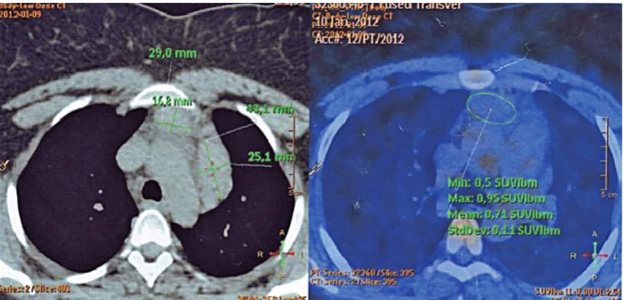
detect even slight neoplastic areas that are imper-ceptible on CT scans, but residual masses can also be investigated (Fig. 1) [10]. An interim PET-CT carried out after two or three cycles of chemother-apy helps to evaluate the response to current treat-ment, and therefore allows for adjustment of the therapy to the individual case, if required. Like-wise, the prognostic value of interim PET-CT has been demonstrated [11].
Currently, the assessment of HL therapy out-comes on the basis of anatomical imaging studies is done in accordance with revised Response Eval-uation CriteriaIn Solid Tumors (RECIST) guide-lines (v. 1.1) [12]. Complete response (CR) refers to the disappearance of all target lesions, with a si-multaneous decrease in the short axis of any
path-ological lymph node to < 10 mm. Partial response (PR) is a 30% decrease (minimum) in the sum of the target lesions’ diameters. Progressive disease (PD) is the designation when the sum increas-es by at least 20% or when a new lincreas-esion appears. A reduction or increase in diameter that cannot be classified either as PR nor as PD is designated as stable disease (SD). As pointed out in the revised RECIST guidelines, there is no undeniable data that discredits the substantial role of morphologi-cal assessment of tumor burden [12]. PET-CT im-aging, however, could be a useful approach in the assessment process, provided further evidence for its role is obtained [12].
In the assessment of treatment response on the base of PET-CT imaging, the Deauville five-point scale is used. A visual interpretation of 18F-FDG
uptake is compared to uptake in the mediastinal blood pool and liver. Scores 1, 2 and 3 stand for CR; 4 and 5 are considered PR, unless there has been no significant change in 18F-FDG uptake
from baseline (SD), or if there is an increasing in-tensity or new malignant focus (PD).
So far the official worldwide guidelines regard-ing HL have referred to PET-CT use rather cau-tiously. However, a more and more appreciable role of PET-CT is being emphasized [13]. In its current recommendations of diagnostic and ther-apeutic proceedings in malignant tumors, the Pol-ish Society for Medical Oncology encourages the use of PET-CT scanning at different points in HL management, particularly supporting it in the as-sessment of final post-therapy outcome [14].
Due to the relatively short history of its clinical usage in Hodgkin disease, the full capabilities and
limitations of PET-CT still have been not thorough-ly explored. Therefore, in order to unequivocalthorough-ly de-fine the exact role of PET-CT in HL management, further clinical research should be performed.
The aim of the present study was to analyze current usage of PET-CT and its clinical usefulness in HL patients at one of the comprehensive cancer centers in Eastern Poland.
Material and Methods
A retrospective analysis of the medical docu-mentation of 67 classical Hodgkin lymphoma pa-tients treated at the Comprehensive Cancer Center in Białystok, Poland, between May 2007 and Feb-ruary 2014 was performed. In 47 cases, PET-CT scanning with 2-[F-18]-fluoro-2-deoxy-d-glucose (18F-FDG) was carried out at least once during HL
management, therefore only those patients were included in the study group. The group was com-posed of 32 women and 15 men, aged 18–59 years at the time of diagnosis (mean age 33.38 ± 10.08) (Table 1). The extent of the disease, determined on the basis of the Ann Arbor staging system with the Cotswolds modifications, ranged between stages II and IV. Advanced-stage HL, defined as stages III/ IV and/or the presence of bulky HL, was found in 25 cases (53.2%). The remaining patients (stages IIA and IIB) were included in the early-stage HL group. All the patients were treated with the doxoru-bicin/bleomycin/vinblastine/dacarbazine (ABVD) chemotherapy regimen, which was followed by ra-diotherapy (RT) in 80.9% of cases. Furthermore, in
cases of progression (found in 5 patients), auto-he-matopoietic stem cell transplantation (auto-HCT) was performed, preceded by salvage chemothera-py regimens: dexamethasone/cytarabine/cisplatin (DHAP) in three patients, and ifosfamide/carbopl-atin/etoposide (ICE) in two patients. Radiological studies performed at different points in HL man-agement, included CT and PET-CT.
The initial CT result was considered positive when a nodal mass or extranodal lesion was found with the longest diameter greater than 1.5 cm and 1.0, respectively. RECIST guidelines (v. 1.1) were used to assess subsequent CT results and assign them to either the CT-positive group (PR, PD) or the CT-negative group (CR). Each PET-CT result was assigned to a particular group on the Deau-ville five-point scale. PR, SD or PD were designat-ed as PET-CT positive, whereas CR was consid-ered PET-CT negative.
A statistical analysis was performed using GraphPad Prism 5 software (GraphPad Software Inc., La Jolla, USA). The c2 test was used and
a p-value less than 0.05 was considered statistical-ly significant.
Privacy protection policies were abided by throughout the study and ethical approval was ob-tained from the Ethics Committee of the Medical University of Bialystok, Poland.
Results
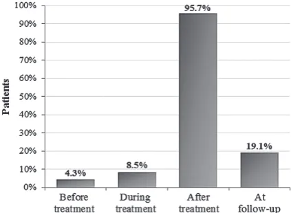
PET-CT scanning was performed once in 66% of the patients, twice in 21.2%, three times in 6.4% and four times in 6.4%. The various time points when PET-CT scans were performed are juxtaposed in Fig. 2. In the vast majority of cases (95.7%), PET-CT was used to evaluate the final re-sponse to the applied treatment (chemotherapy or chemotherapy followed by RT). The use of interim Table 1. Study group characteristics
Characteristics Patients
n (%)
Sex male
female 1532 (31.9)(68.1) Stage (Ann Arbor scale)
II III IV
28 13 6
(59.6) (27.6) (12.8) CS (clinical stage)
early-stage HL
advanced-stage HL 2225 (46.8)(53.2) Treatment modality
CTh
CTh-RT 938 (19.1)(80.9) HL – Hodgkin lymphoma; CTh – chemotherapy;
man-PET-CT was noted in four patients (8.5%). In nine cases (19.1%), PET-CT was used at a follow-up evaluation, either routinely or to detect a recur-rence suspected in clinical examination. This im-aging method was found to be least frequently em-ployed in initial staging process, being used for this purpose in only two patients (4.3%).
In 23 patients (48.9%) PET-CT was ordered af-ter chemotherapy and before they were qualified for RT. For the majority of these patients (15 cas-es, 65.2%), the PET-CT result was decisive either for foregoing RT (when complete metabolic remis-sion was observed) or administering (when com-plete metabolic response had not been achieved). There were eight patients (34.8%) who were qual-ified for RT irrespective of their PET-CT results. Among them, seven were administered RT despite PET-CT negativity (complete response), primarily due to either bulky disease at the baseline or con-siderable residual masses in CT scans after chemo-therapy. All patients diagnosed with bulky disease (10, or 21.3%) were qualified for RT. Post-chemo-therapy PET-CT was carried out in only 4 of them, and metabolic CR did not change the decision re-garding RT administration in any of these patients. Residual masses were revealed in 17 of the 19 patients in whom post-chemotherapy CT was performed. Subsequently, 10 of them underwent PET-CT examinations and CR was found in five patients. Two of them were spared irradiation due to PET-CT negativity.
Both CT and PET-CT were performed af-ter completion of the planned chemotherapy in 11 patients (23.4%); on average, the CT scans were performed four days after the completion of chemotherapy and the PET-CT scans 5.9 weeks af-ter. A discrepancy between the results of the two tests was observed in 54.5% of the patients: most-ly, while the CT scans revealed morphological PR, metabolic complete remission (CR) was shown in the PET-CT scans (Table 2a). One case of progres-sive disease was also detected by PET-CT where CT indicated morphological PR. In the remaining cases the PET-CT and CT results were concordant.
Completion of the planned therapy (chemo-therapy or chemoradio(chemo-therapy) was followed by both CT and PET-CT in 16 patients (34%); on av-erage, the CT scans were performed 6.4 weeks af-ter the completion of treatment and the PET-CT scans 12.25 weeks after. PET-CT and CT results were dissonant in 13 cases (81.3%) (Table 2b), and again, the most prevalent variance was CT positiv-ity (PR) while the PET-CT showed that metabolic CR had been achieved.
There were 30 patients (19 from the ad-vanced-stage group and 11 from the early-stage group) in whom CT scans were performed after
two or three cycles of chemotherapy, as well as PET-CT following the completion of the planned treatment. It was found that among the ad-vanced-stage HL patients, partial morphological response in CT was significantly more often asso-ciated with the absence of complete metabolic re-mission in PET-CT (p = 0.022) (Fig. 3), compared to early-stage patients.
Table 2. Comparison of PET and CT results (a) after chemotherapy and (b) after completion of the entire treat-ment (chemotherapy or chemoradiotherapy) in Hodgkin lymphoma patients
(a)
CT
CR PR
PET CR 1 5
PR – 4
PD – 1
(b)
CT
CR PR PD
PET CR 2 9 1
PR – 1 –
PD – 3 –
PET – positron emission tomography; CT – computed tomography; CR – complete remission; PR – partial remission; PD – progressive disease.
Fig. 3. A juxtaposition of results of computed tomog-raphy (CT) after 2–3 cycles of chemotherapy and positron emission tomography (PET-CT) after the completion of treatment (chemotherapy or chemora-diotherapy) in Hodgkin lymphoma patients. CT(+) –
This study also sought to determine whether there is any correlation between PET-CT results after the completion of therapy and subsequent re-lapse occurrence. There were 30 cases in which it was possible to investigate both PET-CT scans fol-lowing the completion of treatment and follow-up data (Table 3). The average time of observation was 19.9 months (3–53 months). In 26 patients no re-lapse was observed. Among them, CR in PET-CT was achieved in 25 cases and PR in one case. Con-versely, in one patient who experienced disease re-currence, the post-treatment PET-CT scan showed PR. PET-CT scans performed as part of post-treat-ment evaluation showed high predictive value re-garding possible relapse occurrence (p < 0.0001).
For comparison, the results of CT scans fol-lowing therapy were available in 16 cases (mean time of observation: 17.4 months; 3–52 months).
Although in 11 cases (69%) morphological CR was not achieved, no relapses were noted in the follow-up. In another three patients (18.75%), relapse-free follow-up was preceded by CR in post-treatment CT imaging; recurrence was experienced by the remaining two patients (12.5%) whose post-treat-ment CT scans were positive.
A comparison of positive post-treatment CT and PET-CT results in relation to future relapse occurrence (Fig. 4) showed the superior predictive value of PET-CT in this respect (p < 0.0001).
A general increase in the number of PET-CT examinations was observed in the time period re-flected in the study. In 2008 only five PET-CT stud-ies were performed, whereas in 2010 the number rose to 14. In 2013 PET-CT was performed over four times more often than in 2008 (21 studies).
Discussion
In this study CT was the imaging method of choice during the initial HL staging process. The very rare implementation of PET-CT in stag-ing (only 4.3% of the patients) is conspicuous. This probably results from the higher costs of PET-CT compared to CT, the small number of PET-CT centers in the region, and the lack of well-defined recommendations concerning PET-CT implemen-tation in routine practice. It is noteworthy that in one recent study PET-CT imaging – in compari-son with CT alone – was shown to upstage the dis-ease among patients initially diagnosed as stage I or II [15]. However, the official recommenda-tions concerning the use of PET-CT in initial HL staging are slightly inconsistent. The National Comprehensive Cancer Network (NCCN) defines PET-CT as an integral part of this procedure [13], whereas in the European Society for Medical On-cology (ESMO) guidelines PET-CT is considered only an additional tool that may be included in the staging process [16].
Interim PET-CT is a promising implement that could make the treatment of HL patients more individualized. It has been reported to pre-dict treatment failure in advanced-stage Hodgkin disease [11]. An early PET-CT examination and its role in further treatment stratification are be-ing investigated by the ongobe-ing HD18 trial [17]. However, since evidence from randomized trials is still being collected, interim PET-CT has not been established as a standard yet. The Consen-sus of the Imaging Subcommittee of the Interna-tional Harmonization Project in Lymphoma stat-ed that for now the use of this imaging method to monitor treatment during therapy should be con-fined to clinical trials or prospective registries [9]. Table 3. Post-treatment (chemotherapy or
chemoradio-therapy) PET results in relation to relapse occurrence in Hodgkin lymphoma patients
Relapse PET result
n = 30
Relapse n (%) Relapse-free n (%)
PET(+) 4 (13.3) 1 (3.3) PET(–) 0 25 (83.3)
PET – positron emission tomography; PET(+) – partial remission in PET; PET(–) – complete remission in PET.
It was difficult to investigate the usefulness of in-terim PET-CT in the present study, since its imple-mentation was rare.
The need for RT use among advanced-stage HL patients with initial bulky disease and/or resid-ual lesions in post-treatment imaging studies still remains unclear. According to the current ESMO guidelines, the standard treatment for advanced-stage HL comprises chemotherapy followed by RT only when residual post-chemotherapy mass-es are found [16]. In the prmass-esent study this stan-dard scheme was implemented in the vast major-ity of advanced-stage HL patients. However, as the German Hodgkin Study Group (GHSG) HD15 tri-al has recently showed, RT could be omitted – ir-respective of residual masses – when PET-CT neg-ativity is demonstrated in patients treated with the BEACOPP chemotherapy regimen [18]. Similarly, Savage et al. reported that omitting consolidative RT did not increase the risk of relapse in advanced-stage HL patients who had received ABVD-based chemotherapy, provided PET-CT scans showed complete response [19]. Furthermore – unlike the GHSG HD15 trial – the study by Savage et al. re-ferred to cases with bulky HL at the baseline, and found that in cases of post-chemotherapy PET-CT negativity there was no statistically significant dif-ference between irradiated bulky and non-bulky patients in terms of three-year time-to-progression [19]. Although these results seem promising, and avoiding RT-related toxicity would be definitely very beneficial, further PET-CT-ex-ploiting research on the need for RT among ad-vanced-stage HL patients is still required. In cases where post-chemotherapy RT is considered inev-itable, PET-CT has been found essential in plan-ning radiotherapy [20]. It could decrease both the irradiation volume and target misses.
As far as the role of PET-CT in surveillance is concerned, it has been recognized in the NCCN guidelines as unnecessary [13]. Likewise, recent research is providing a growing body of evidence of the limitations in PET-CT usage in follow-up. For instance, a substantial number of false-positive PET-CT results was revealed in routine surveillance among patients who had achieved remission after first-line therapy, which is particularly unfortunate considering the high costs involved [21]. Maeda et al.also reported that PET-CT has low positive predictive value in surveillance [22]. In contrast, in an investigation of clinically suspected relapse Hutchings reported that PET-CT has high
nega-tive predicnega-tive value and considered it eligible for this purpose [23]. The present study seems to re-flect Hutching’s results, since among five patients who were clinically suspected of relapse at follow-up (mean 7.2 months after completing treatment) PET-CT accurately determined the character of the suspicious lesions, whereas none of the patients in whom PET-CT was implemented in routine sur-veillance was shown to relapse (mean 25 months of observation).
The present study revealed a striking discrep-ancy between morphological and functional re-sponse to chemotherapy and chemoradiotherapy. The most prominent discordance was a complete metabolic response in PET-CT parallel to a par-tial morphological remission in CT. Although the mean time after treatment completion differed be-tween the two types of scan, these results seem to point to the need for combined interpretation of PET-CT and CT in the assessment of treatment outcome. This is of particular importance since most evidence-based recommendations concern-ing the treatment of HL patients are derived from randomized trials based on CT assessment.
The present study showed that post-treatment PET-CT results are highly predictive of relapse oc-currence in the future, which is in line with previ-ous reports [24, 25]. Moreover, it was found to be incomparably superior to CT imaging as a predic-tor of relapse.
Undoubtedly, the high cost of PET-CT exam-inations is a drawback that stands in the way of broader usage of this imaging method in clinical practice. Furthermore, although the availability of PET-CT is improving, it is still less accessible than CT scanning. Since the results of clinical studies on the role of PET-CT in HL are very promising and the use of PET-CT is expected to increase, steps should be undertaken to address the prob-lems with costs and accessibility.
In conclusion, in the present study PET-CT re-sults were decisive for further management deci-sions. PET-CT was found to be superior to CT in determining the character of residual masses. Un-like CT, post-therapy PET-CT imaging was found to be a good predictor of relapse. A number of is-sues concerning PET-CT, such as the need for RT in patients with positive CT scans parallel to negative PET-CT results, necessitate thorough investigation.
References
[1] Jemal A, Siegel R, Ward E, Hao Y, Xu J, Thun MJ: Cancer Statistics 2009. CA Cancer J Clin 2009, 59, 225–249.
[2] Mauch PM, Kalish LA, Kadin M, Coleman CN, Osteen R, Hellman S: Patterns of presentation of Hodgkin dis-ease. Implications for etiology and pathogenesis. Cancer 1993, 71, 2062–2071.
[3] Gobbi PG, Broglia C, Di Giulio G, Mantelli M, Anselmo P, Merli F, Zinzani PL, Rossi G, Callea V, Iannitto E, Paulli M, Garioni L, Ascari E: The clinical value of tumor burden at diagnosis in Hodgkin lymphoma. Cancer 2004, 101, 1824–1834.
[4] Connors JM: State-of-the-art therapeutics: Hodgkin’s lymphoma. J Clin Oncol 2005, 23, 6400–6408.
[5] Vinnicombe SJ, Reznek RH: Computerised tomography in the staging of Hodgkin’s disease and non-Hodgkin’s lymphoma. Eur J Nucl Med Mol Imaging 2003, 30, 42–55.
[6] Juweid ME: Utility of positron emission tomography (PET) scanning in managing patients with Hodgkin lym-phoma. Hematology Am Soc Hematol Educ Program 2006, 2006, 259–265.
[7] Connors JM: Positron emission tomography in the management of Hodgkin lymphoma. Hematology Am Soc Hematol Educ Program 2011, 2011, 317–322.
[8] Juweid ME, Cheson BD: Positron emission tomography and assessment of cancer therapy. N Engl J Med 2006, 354, 496–507.
[9] Juweid ME, Stroobants S, Hoekstra OS, Mottaghy FM, Dietlein M, Guermazi A, Wiseman GA, Kostakoglu L, Scheidhauer K, Buck A, Naumann R, Spaepen K, Hicks RJ, Weber WA, Reske SN, Schwaiger M, Schwartz LH, Zijlstra JM, Siegel BA, Cheson BD: Use of positron emission tomography for response assessment of lymphoma: Consensus of the Imaging Subcommittee of International Harmonization Project in Lymphoma. J Clin Oncol 2007, 25, 571–578.
[10] Hueltenschmidt B, Sautter-Bihl ML, Lang O, Maul FD, Fischer J, Mergenthaler HG, Bihl H: Whole body posi-tron emission tomography in the treatment of Hodgkin disease. Cancer 2001, 91, 302–310.
[11] Hutchings M, Loft A, Hansen M, Pedersen LM, Buhl T, Jurlander J, Buus S, Keiding S, D’Amore F, Boesen AM, Berthelsen AK, Specht L: FDG-PET after two cycles of chemotherapy predicts treatment failure and progression-free survival in Hodgkin lymphoma. Blood 2006, 107, 52–59.
[12] Eisenhauer EA, Therasse P, Bogaerts J, Schwartz LH, Sargent D, Ford R, Dancey J, Arbuck S, Gwyther S, Mooney M, Rubinstein L, Shankar L, Dodd L, Kaplan R, Lacombe D, Verweij J: New response evaluation crite-ria in solid tumours: revised RECIST guideline (version 1.1). Eur J Cancer 2009, 45, 228–247.
[13] Hoppe RT, Advani RH, Ai WZ, Ambinder RF, Aoun P, Bello CM, Benitez CM, Bernat K, Bierman PJ, Blum KA, Chen R, Dabaja B, Forero A, Gordon LI, Hernandez-Ilizaliturri FJ, Hochberg EP, Huang J, Johnston PB, Kaminski MS, Khan N, Maloney DG, Mauch PM, Metzger M, Moore JO, Morgan D, Moskowitz CH, Mulroney C, Poppe M, Rabinovitch R, Seropian S, Smith M, Winter JN, Yahalom J, Burns J, Sundar H: NCCN Clinical Practice Guidelines in Oncology (NCCN Guidelines). Hodgkin lymphoma. http://www.nccn.org/professionals/ physician_gls/f_guidelines.asp#hodgkin. (Access date: 23.04.2014).
[14] Krzakowski M, Warzocha K: Zalecenia postepowania diagnostyczno-terapeutycznego w nowotworach złośliwych – 2013 rok. Via Medica: Gdańsk 2013, 981–990 [in Polish].
[15] Raanani P, Shasha Y, Perry C, Metser U, Naparstek E, Apter S, Nagler A, Polliack A, Ben-Bassat I, Even-Sapir E: Is CT scan still necessary for staging in Hodgkin and non-Hodgkin lymphoma patients in the PET/CT era? Ann Oncol 2006, 17, 117–122.
[16] Eichenauer DA, Engert A, Dreyling M: Hodgkin’s lymphoma: ESMO Clinical Practice Guidelines for diagnosis, treatment and follow-up. Ann Oncol 2011, 22, 55–58.
[17] Eich HT, Kriz J, Schmidberger H, Böll B, Klimm B, Rancea M, Müller RP, Engert A: The German evidence-based guidelines for Hodgkin’s lymphoma. Aspects for radiation oncologists. Strahlenther Onkol 2013, 189, 445–447.
[18] Engert A, Kobe C, Markova J, Haverkamp H, Borchmann P, Hitz F, Zijlstra J, Eich H, Mueller RP, Dietlein M, Diehl V: Assessment of residual bulky tumor using FDG-PET in patients with advanced-stage Hodgkin Lymphoma after completion of chemotherapy: Final report of the GHSG HD15 trial. ASH annual meeting abstracts 2010, 116, 764.
[19] Savage KJ, Connors JM, Klasa RJ, Hoskins P, Shenkier TN, Gascoyne RD, Bhimji S, Pickles T, Benard F, Wilson D, Sehn LH: The use of FDG-PET to guide consolidative radiotherapy in patients with advanced-stage Hodgkin lymphoma with residual abnormalities on CT scan following ABVD chemotherapy. J Clin Oncol 2011, 29 (Suppl; abstr 8034).
[20] Lammering G, De Ruysscher D, van Baardwijk A, Baumert BG, Borger J, Lutgens L, van den Ende P, Ollers M, Lambin P: The use of FDG-PET to target tumors by radiotherapy. Strahlenther Onkol 2010, 186, 471–481.
[21] El-Galaly TC, Mylam KJ, Brown P, Specht L, Christiansen I, Munksgaard L, Johnsen HE, Loft A, Bukh A, Iyer V, Nielsen AL, Hutchings M: Positron emission tomography/computed tomography surveillance in patients with Hodgkin lymphoma in first remission has a low positive predictive value and high costs. Haematologica 2012, 97, 931–936.
[22] Maeda LS, Horning SJ, Iagaru AH, Lin FI, Hoppe RT, Rosenberg SA, Advani RH: Role of FDG-PET/CT surveil-lance for patients with classical Hodgkin’s disease in first complete response: The Stanford University experience [Abstract]. Blood 2009, 114, 1563.
[23] Hutchings M: How does PET/CT help in selecting therapy for patients with Hodgkin lymphoma? Hematology Am Soc Hematol Educ Program 2012, 2012, 322–327.
[24] de Wit M, Bohuslavizki KH, Buchert R, Bumann D, Clausen M, Hossfeld DK:18FDG-PET following treatment
as valid predictor for disease-free survival in Hodgkin’s lymphoma. Ann Oncol 2001, 12, 29–37.
Address for correspondence:
Ewa Sierko
Department of Oncology Medical University of Białystok ul. Ogrodowa 12
15-027 Białystok Poland
Tel.: +48 85 664 67 53 E-mail: [email protected]
Conflict of interest: None declared Received: 13.03.2015